Understanding the Differential Diagnosis
Cervical radiculopathy typically presents as neck and arm pain secondary to nerve root compression or inflammation. This most frequently involves the C6 and C7 nerve roots due to spondylotic foraminal stenosis or acute disc herniation. Epidemiological data suggest an annual incidence of approximately 83 per 100,000 individuals, though this may be an underestimation due to varying diagnostic criteria. Differentiating true radiculopathy from Myofascial Pain Syndrome (MPS) is a common and critical clinical challenge, as trigger points in the cervical and shoulder musculature frequently refer pain in pseudo-radicular patterns. Accurate diagnosis is essential, as observational studies note that the natural history of cervical radiculopathy is generally favorable, with a vast majority of patients improving without surgical intervention.
The relationship between cervical radiculopathy and myofascial pain is complex and often bidirectional. Nerve root irritation can lead to secondary protective muscle spasm, facilitating the development of satellite trigger points in the myotome supplied by the affected nerve. Conversely, the "double-crush" phenomenon postulates that proximal compression—such as a taut band in the anterior scalene entrapping the brachial plexus—may render the distal nerve more susceptible to minor compressive forces at the intervertebral foramen or carpal tunnel. Furthermore, even after the primary radicular compression resolves, secondary myofascial pain may persist. This can complicate the clinical picture, leading to refractory symptoms if the muscular component is not independently addressed.
Because asymptomatic cervical disc degeneration is highly prevalent, imaging findings must always be correlated with clinical neurological signs to avoid misattributing myofascial arm pain to incidental foraminal stenosis.
Clinical differentiation relies heavily on a meticulous neurological examination and validated provocative testing. True radiculopathy often presents with dermatomal sensory changes, myotomal weakness, and diminished deep tendon reflexes, whereas myofascial pain typically lacks these hard neurological deficits. Provocative maneuvers, such as Spurling's test and the Upper Limb Tension Test (ULTT), demonstrate high specificity and sensitivity, respectively, for radiculopathy. However, clinicians must interpret advanced imaging with caution; MRI frequently reveals incidental foraminal stenosis or disc bulging in asymptomatic individuals. Therefore, current guidelines generally recommend correlating imaging findings strictly with the patient's clinical presentation, reserving surgical consultations for cases with progressive neurological deficits or those failing comprehensive conservative management.
Key Musculature and Nerve Pathways
Understanding the spatial relationship between the cervical nerve roots, the brachial plexus, and the surrounding musculature is crucial for distinguishing radicular from referred pain.
Scalene Group (Anterior, Middle, Posterior)
The scalenes are primary contributors to pseudo-radicular arm pain and thoracic outlet syndrome. Taut bands in these muscles can directly compress the brachial plexus trunks, mimicking C5-C8 radiculopathy. Clinical evidence suggests scalene trigger points frequently coexist with true cervical radiculopathy as a secondary protective spasm. Differentiating scalene referral from true radiculopathy requires careful neurological examination, as scalene pain typically lacks hard dermatomal sensory loss or diminished reflexes.
Broadly down the lateral arm, radial forearm, and into the thumb and index finger, closely mimicking C6 radiculopathy.Upper Trapezius
As a primary stabilizer of the cervical spine, the upper trapezius frequently develops myofascial trigger points in response to underlying cervical pathology, including discogenic pain and radiculopathy. While it does not typically refer pain down the arm, its referral pattern into the posterolateral neck and temporal region can mimic C3 or C4 nerve root irritation. Chronic upper trapezius tension may also alter scapular kinematics, indirectly increasing strain on the lower cervical segments.
Posterolateral neck, behind the ear, and into the temporal region (often presenting as tension-type headaches).Infraspinatus
The infraspinatus is a rotator cuff muscle that frequently harbors trigger points referring pain deep into the anterior shoulder joint and down the anterolateral arm. This referral pattern is a classic mimic of C5 and C6 radiculopathy. In clinical practice, patients presenting with suspected C5/C6 radiculopathy without neurological deficits should be evaluated for infraspinatus trigger points, as addressing these can sometimes resolve the pseudo-radicular symptoms entirely.
Deep anterior shoulder pain, extending down the anterolateral aspect of the arm and forearm, occasionally reaching the radial hand.Subscapularis
Located on the anterior surface of the scapula, the subscapularis is notoriously difficult to palpate but is a significant source of posterior arm pain. Its referral pattern extends down the posterior aspect of the arm, which can closely resemble a C7 radiculopathy. Systematic reviews note that subscapularis trigger points are often overlooked in the differential diagnosis of posterior arm and wrist pain.
Posterior shoulder, extending down the posterior arm and forearm, sometimes concentrating at the posterior wrist.Sternocleidomastoid (SCM)
The SCM is a complex two-headed muscle that refers pain entirely upward into the head and face, rather than down the arm. However, it is highly relevant in the context of cervical radiculopathy because altered cervical posture (such as forward head posture adopted to relieve foraminal compression) places chronic strain on the SCM. It frequently develops secondary trigger points that complicate the clinical picture with atypical facial pain, dizziness, or tinnitus.
Occiput, vertex of the head, supraorbital region, cheek, and occasionally deep into the ear.Suboccipital Group
The suboccipital muscles control fine movements of the upper cervical spine. While they do not refer pain into the upper extremity, they are frequently involved in the compensatory postural changes associated with lower cervical radiculopathy. Tension in this muscle group can compress the greater occipital nerve, leading to occipital neuralgia, which may present concurrently with lower cervical nerve root irritation.
Deep pain in the upper neck, radiating over the occiput to the temporal and frontal regions.Levator Scapulae
The levator scapulae is a common site of neck pain, often described by patients as a "stiff neck." It frequently develops trigger points in response to the protective muscle guarding seen in acute cervical radiculopathy. The pain is typically localized to the angle of the neck and superior medial border of the scapula, mimicking the axial neck pain component of cervical disc disease.
Angle of the neck, superior medial border of the scapula, and posterior shoulder region.
Direct Nerve Root Compression
Mechanical pressure on the cervical nerve root from a herniated disc or osteophyte, leading to ischemia, inflammation, and true radicular symptoms.
Myofascial Referral
Nociceptive signals from active trigger points in muscles like the scalenes or infraspinatus projecting pain into the arm without actual nerve compression.
Double-Crush Syndrome
Minor compression at the cervical spine combined with peripheral entrapment (e.g., by the scalenes), cumulatively resulting in symptomatic nerve dysfunction.
Secondary Muscle Spasm
Primary nerve root irritation causing reflex muscle guarding in the neck and shoulder, which subsequently develops its own pain-generating trigger points.
Pseudo-Radicular Referral Patterns
Myofascial trigger points can project pain in predictable patterns that closely mimic specific cervical nerve root compressions.
- Scalene Group
- Pain radiates from the lateral neck down the lateral aspect of the arm, skipping the elbow, and reappearing in the radial forearm, thumb, and index finger. This pattern strongly mimics a C6 nerve root compression.
- Infraspinatus
- Pain concentrates deep in the anterior deltoid region and travels down the anterolateral arm and forearm. It is frequently mistaken for a C5 or C6 radiculopathy or primary bicipital tendinopathy.
- Subscapularis
- Pain refers to the posterior shoulder and extends down the posterior aspect of the arm and forearm, often pooling at the dorsal wrist. This distribution can closely resemble a C7 radicular pattern.
- Upper Trapezius
- Pain refers upward in a "question mark" pattern along the posterolateral neck, behind the ear, and into the temple. While not mimicking arm radiculopathy, it frequently mimics C3/C4 nerve irritation.
Clinical Assessment Tests
These clinical tests are utilized by healthcare professionals to provoke or relieve symptoms, helping to differentiate between nerve root compression and peripheral muscular sources of pain.
Spurling's Test (Foraminal Compression)
The patient extends the neck, laterally flexes, and rotates the head toward the symptomatic side. The clinician carefully applies downward axial compression to the top of the head. This test is designed to narrow the neural foramen and provoke nerve root compression.
Positive: Reproduction of the patient's familiar radiating pain or paresthesia down the arm (not just localized neck pain). This suggests true cervical radiculopathy.
Upper Limb Tension Test 1 (ULTT1 - Median Nerve Bias)
With the patient supine, the clinician sequentially depresses the shoulder, abducts the arm to 110 degrees, extends the wrist and fingers, supinates the forearm, and externally rotates the shoulder, followed by elbow extension. Cervical lateral flexion is used to sensitize the test.
Positive: Reproduction of neurological symptoms in the arm, or a significant side-to-side difference in elbow extension range of motion. A negative test makes true radiculopathy highly unlikely.
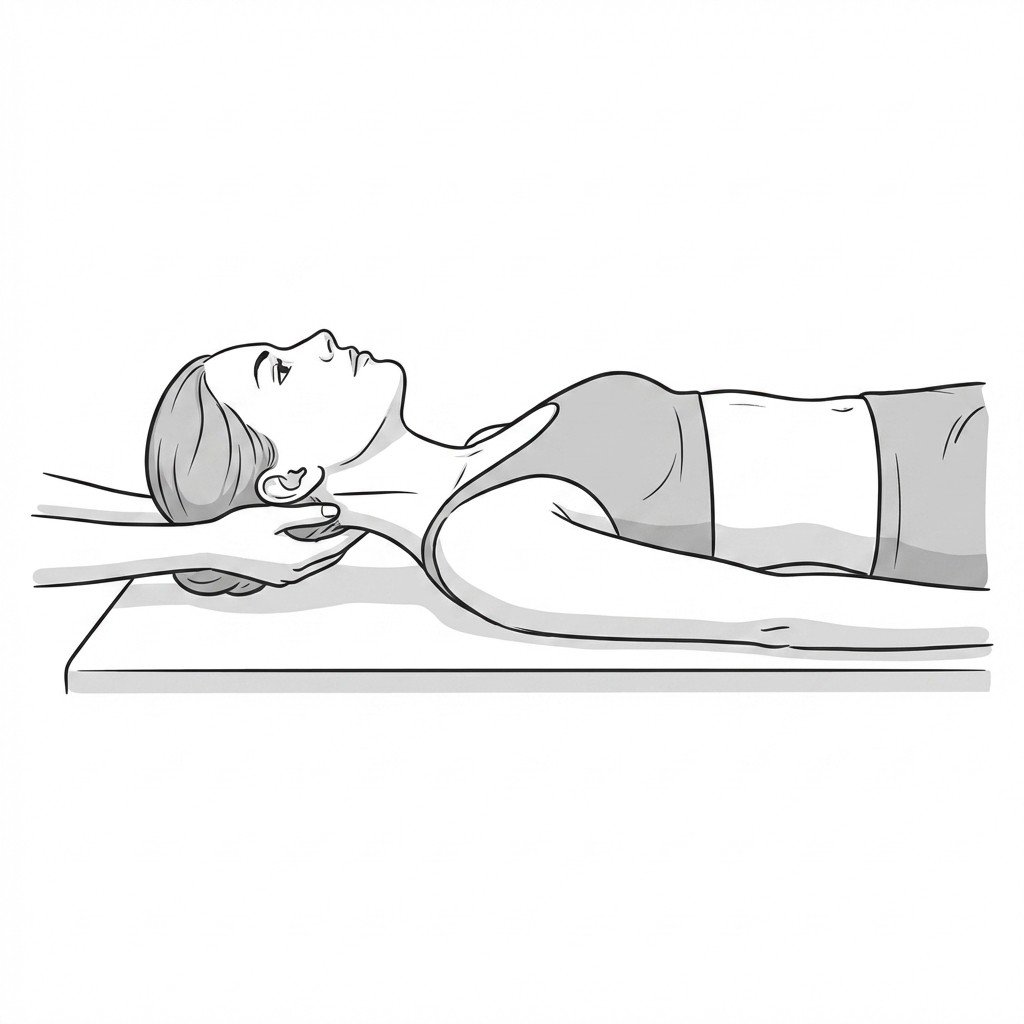
Cervical Distraction Test
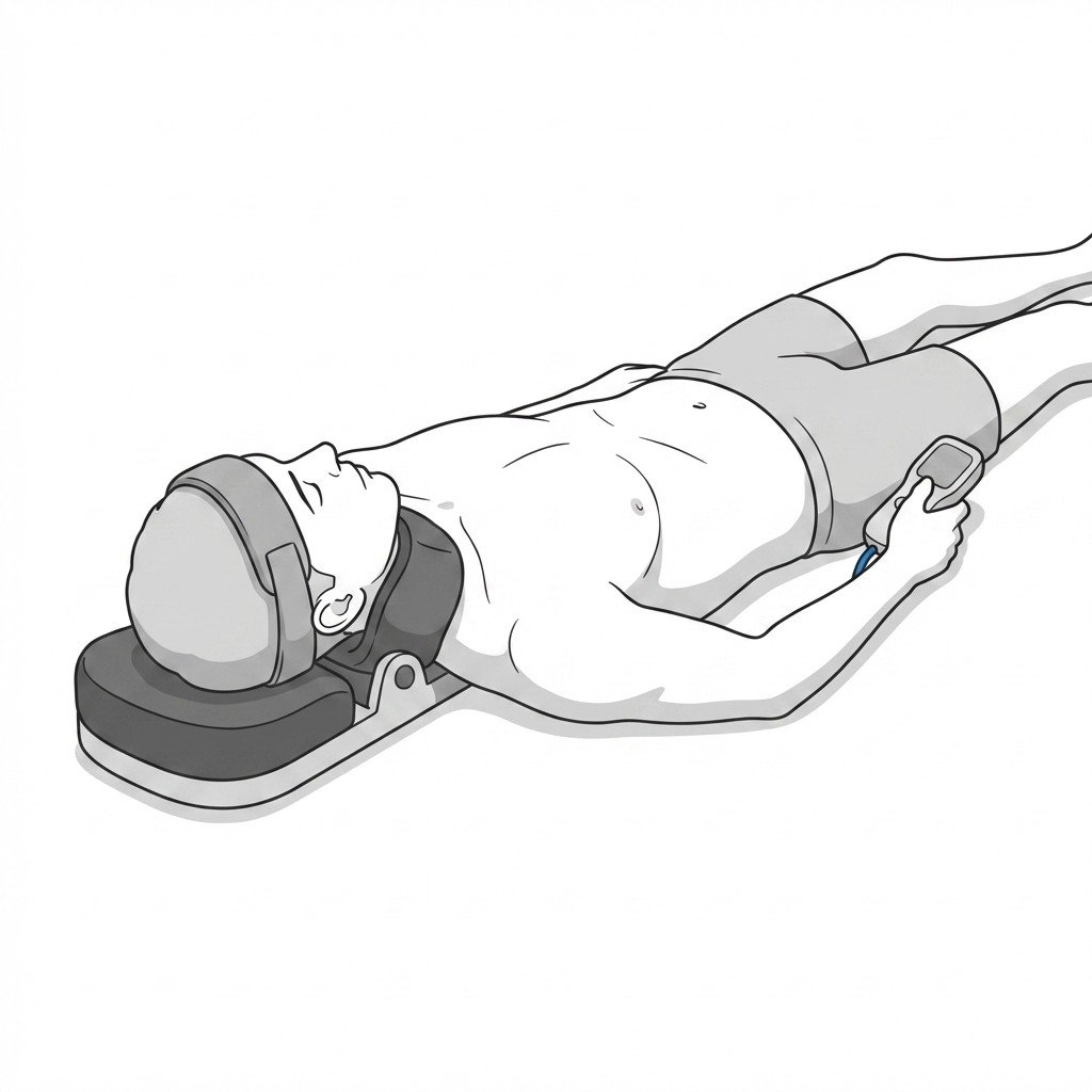
The patient lies supine. The clinician grasps under the patient's chin and occiput, applying a gradual, gentle superior traction force (approximately 10-15 lbs) to distract the cervical spine.
Positive: Reduction or complete relief of the radiating arm symptoms. This indicates that opening the neural foramen alleviates nerve root compression.
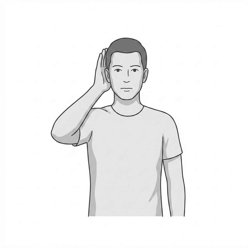
Scalene Relief Maneuver
The patient places the forearm of the affected arm across their forehead, elevating the elbow. This position slackens the lower roots of the brachial plexus and elevates the clavicle, potentially reducing tension on the scalene muscles and the neurovascular bundle.
Positive: Relief of arm pain or paresthesia. While traditionally associated with C5/C6 radiculopathy, it can also indicate relief of thoracic outlet syndrome or scalene-mediated myofascial pain.
Evidence-Based Treatment Pathways
Current guidelines generally recommend a multimodal approach to cervical radiculopathy and associated myofascial pain, emphasizing conservative care for 6 to 12 weeks before considering invasive options.
Self-Care
- Postural modification to avoid prolonged cervical flexion or extension
- Gentle cervical range of motion exercises within pain-free limits
- Application of heat to the cervical and periscapular musculature
- Ergonomic workstation adjustments to support the upper extremities
- Use of a supportive cervical pillow to maintain neutral alignment during sleep
Manual Therapy
- Ischemic compression of scalene and infraspinatus trigger points
- Cervical and thoracic spine mobilization to improve segmental mobility
- Manual cervical traction to decompress neural foramina
- Myofascial release of the upper trapezius and levator scapulae
- Nerve gliding/flossing techniques for the brachial plexus
Interventional
- Trigger point injections for refractory scalene or infraspinatus pain
- Cervical epidural steroid injections (under fluoroscopic guidance)
- Selective nerve root blocks for diagnostic and therapeutic purposes
- Oral pharmacotherapy (NSAIDs, membrane stabilizers like gabapentin)
- Acupuncture or dry needling targeting cervical and shoulder musculature
Rehabilitation
- Deep cervical flexor endurance training
- Scapular stabilization exercises (serratus anterior, lower trapezius)
- Mechanical cervical traction protocols
- Progressive upper extremity strengthening once neurological symptoms stabilize
- Proprioceptive retraining for the cervical spine
Red Flags — When to See a Doctor
While most neck and arm pain is benign and self-limiting, the following signs and symptoms require prompt medical investigation to rule out severe neurological compromise or systemic pathology.
Progressive Motor Weakness
Rapidly worsening weakness in a specific myotome (e.g., inability to extend the wrist or flex the elbow). This suggests acute, severe nerve root compromise requiring urgent evaluation.
UrgentSigns of Cervical Myelopathy
Presence of upper motor neuron signs such as hyperreflexia, Hoffmann's sign, Babinski reflex, clonus, or gait disturbances. This indicates spinal cord compression rather than isolated nerve root involvement.
EmergencyBowel or Bladder Dysfunction
New onset of urinary retention, incontinence, or bowel dysfunction. When combined with neurological signs, this is a hallmark of severe spinal cord compromise.
EmergencySevere, Unremitting Pain
Pain that is excruciating, constant, and completely unresponsive to changes in position or basic analgesics. This may warrant investigation for infection, malignancy, or severe acute disc herniation.
InvestigateHistory of Malignancy
New onset of cervical radicular pain in a patient with a history of cancer, particularly breast, lung, or prostate cancer, which have a propensity for spinal metastasis.
Urgent
Clinical Pearls
Cervical radiculopathy most commonly affects the C6 and C7 nerve roots, typically presenting as unilateral arm pain, numbness, and tingling.
Myofascial trigger points in the scalenes, infraspinatus, and subscapularis can perfectly mimic the pain distribution of cervical radiculopathy.
True radiculopathy is characterized by hard neurological signs (dermatomal sensory loss, myotomal weakness, diminished reflexes), which are absent in pure myofascial pain.
The "double-crush" phenomenon suggests that proximal muscle tension (e.g., scalenes) can compress nerves, making them more susceptible to distal irritation.
MRI findings must be correlated with clinical symptoms, as asymptomatic cervical disc bulges and foraminal stenosis are highly prevalent in the general population.
Current guidelines generally recommend a 6 to 12-week trial of multimodal conservative care before considering surgical intervention for non-progressive radiculopathy.