Understanding the Differential Diagnosis
Cervicogenic headache (CGH) is a secondary headache disorder typically arising from nociceptive sources in the upper cervical spine, most commonly the C2-C3 zygapophyseal (facet) joint. Epidemiological data suggest it accounts for approximately 15% to 20% of all chronic and recurrent headaches. Distinguishing CGH from primary headache disorders, such as migraine or tension-type headache, remains a diagnostic challenge due to overlapping phenotypic presentations. The International Headache Society (IHS) criteria emphasize clinical or imaging evidence of a cervical lesion or dysfunction, alongside headache exacerbation by neck movement or sustained postures.
The clinical picture is frequently complicated by the coexistence of myofascial pain syndrome (MPS). Suboccipital, sternocleidomastoid (SCM), and upper trapezius trigger points are among the most common mimickers of CGH. This overlap is largely explained by the neuroanatomical convergence of upper cervical afferents (C1-C3) and trigeminal nerve afferents at the trigeminocervical nucleus caudalis. Consequently, nociceptive input from cervical facet joints or myofascial trigger points can be perceived as pain in the frontal, orbital, or temporal regions. Furthermore, chronic joint dysfunction may perpetuate secondary myofascial sensitization, creating a complex, mixed pain presentation.
The neuroanatomical convergence at the trigeminocervical nucleus means that a C2-C3 joint irritation and a suboccipital trigger point can produce an identical frontal headache.
Differentiating primary joint pathology from primary myofascial pain requires a systematic clinical approach. While controlled diagnostic nerve blocks at the C2-C3 joint or third occipital nerve are generally considered the gold standard for confirming CGH, they are invasive and not universally accessible. In the clinical setting, the Cervical Flexion-Rotation Test (CFRT) has demonstrated high sensitivity and specificity in identifying upper cervical joint dysfunction. Conversely, if precise palpation of the suboccipital or SCM musculature reproduces the patient's familiar headache without significant articular restriction, a primary myofascial etiology should be strongly considered. Current guidelines generally recommend a trial of conservative, multimodal management before pursuing interventional diagnostics.
Anatomical Convergence
The upper cervical spine (C1-C3) is anatomically unique, lacking intervertebral discs between the occiput, atlas, and axis. This region relies heavily on the suboccipital musculature for stability, making it highly susceptible to both articular dysfunction and myofascial overload.
Suboccipital Group
A cluster of four small muscles located at the base of the skull (rectus capitis posterior major/minor, obliquus capitis inferior/superior). They are primarily responsible for fine proprioceptive control and micro-movements of the head. Trigger points in this region are among the most frequent mimickers of cervicogenic headache, often secondary to forward head posture or upper cervical joint dysfunction.
Deep, aching pain extending from the occiput to the temporal region, often concentrating behind the ipsilateral eye.Sternocleidomastoid (SCM)
A prominent two-headed muscle on the anterolateral neck. The SCM is frequently overloaded in whiplash-associated disorders and chronic postural faults. Its complex referral pattern is notorious for mimicking primary headaches, atypical facial pain, and autonomic symptoms like dizziness or tinnitus, complicating the differential diagnosis of cervicogenic headache.
Sternal head refers to the vertex, occiput, cheek, and over the eye. Clavicular head refers to the frontal region and deep into the ear.Upper Trapezius
The most superficial muscle of the posterior neck and upper back. It is highly susceptible to emotional stress and sustained postural loading. While it rarely causes isolated cervicogenic headache, its trigger points frequently coexist with upper cervical joint pathology, contributing to a mixed tension-type and cervicogenic presentation.
Unilateral pain extending up the posterolateral neck to the mastoid process, occasionally wrapping around the side of the head to the temple.Semispinalis Capitis
A thick, powerful extensor muscle lying deep to the trapezius and splenius capitis. It plays a significant role in maintaining upright head posture. Entrapment of the greater occipital nerve as it pierces this muscle is a proposed mechanism for some presentations of occipital neuralgia, which must be differentiated from cervicogenic headache.
Band-like pain across the side of the head, often peaking in the frontal region above the eye.Splenius Capitis
A broad, strap-like muscle that extends and rotates the cervical spine. Trigger points here are commonly activated by sudden trauma or prolonged rotation of the head. The referral pattern is highly specific and can mimic the vertex pain sometimes reported in complex cervicogenic or tension-type headaches.
A distinct, localized ache at the vertex (crown) of the head.Levator Scapulae
Connects the upper cervical transverse processes to the superior angle of the scapula. It is frequently implicated in "stiff neck" presentations. While its primary referral is to the neck and shoulder, the resulting biomechanical restriction often exacerbates upper cervical joint dysfunction, indirectly contributing to cervicogenic headache.
Angle of the neck and superior medial border of the scapula, occasionally extending to the posterior shoulder.
Trigeminocervical Convergence
Afferent signals from the upper three cervical nerves and the trigeminal nerve converge in the brainstem, causing cervical nociception to be perceived as head or facial pain.
Joint Sensitization
Inflammation or mechanical dysfunction at the C2-C3 or C1-C2 joints lowers the firing threshold of local nociceptors, leading to chronic afferent bombardment.
Myofascial Perpetuation
Protective muscle guarding secondary to joint dysfunction can lead to the development of active trigger points, which then become independent sources of referred pain.
Postural Overload
Sustained forward head posture increases the mechanical load on both the upper cervical facet joints and the suboccipital extensors, driving both articular and myofascial pathology.
Overlapping Referral Patterns
Trigger points in the cervical musculature frequently refer pain to the head and face. Recognizing these specific patterns is crucial for distinguishing myofascial pain from joint-referred cervicogenic headache or primary headache disorders.
- Suboccipital Group
- Pain typically originates at the base of the skull and radiates anteriorly over the side of the head, frequently settling deep within or behind the ipsilateral orbit. This pattern strongly mimics the C2-C3 facet joint referral.
- Sternocleidomastoid (Sternal Head)
- Refers pain to the occipital ridge, radiating to the vertex of the head, the cheek, and directly over the eye. It may also produce autonomic phenomena such as lacrimation or ptosis.
- Upper Trapezius
- Characterized by a "question mark" pattern: pain travels up the posterolateral neck, hooks behind the ear, and extends into the temporal region. It is a classic contributor to tension-type headache phenotypes.
- Semispinalis Capitis
- Produces a unilateral, band-like distribution of pain that wraps around the side of the head, often concentrating in the frontal region and mimicking a frontal tension headache.
Clinical Assessment
Clinical assessment aims to provoke or alleviate the familiar headache through mechanical testing. These assessments help differentiate between articular restrictions and myofascial hyperalgesia.
Cervical Flexion-Rotation Test (CFRT)
While lying supine, the neck is maximally flexed to lock out the lower cervical spine. The head is then rotated to the left and right. This test isolates movement at the C1-C2 (atlantoaxial) joint.
Positive: A restriction of 10 degrees or more compared to the asymptomatic side, or reproduction of the familiar headache, suggests upper cervical joint dysfunction.
Suboccipital Palpation
With the head supported in a neutral position, apply sustained, moderate pressure to the muscular tissue immediately inferior to the occipital ridge (base of the skull).
Positive: Reproduction of the patient's familiar frontal or orbital headache suggests a significant myofascial trigger point contribution, though it does not rule out underlying joint pathology.
Active Range of Motion (AROM) Assessment
The patient actively moves the head through full flexion, extension, lateral flexion, and rotation, noting any onset of headache symptoms or significant asymmetry in movement.
Positive: Headache provoked by specific, sustained cervical postures or movements is a key diagnostic criterion for cervicogenic headache according to the International Headache Society.
SCM Pincer Palpation
Gently grasp the sternocleidomastoid muscle between the thumb and index finger, rolling the muscle belly to identify taut bands and tender nodules along both the sternal and clavicular heads.
Positive: Eliciting referred pain to the forehead, ear, or vertex of the head indicates active SCM trigger points, which may mimic or complicate a cervicogenic presentation.
Evidence-Based Management
Management of cervicogenic headache and associated myofascial pain typically requires a multimodal approach. Systematic reviews suggest that combining manual therapy with specific therapeutic exercise yields the most favorable outcomes for upper cervical dysfunction.
Self-Care
- Ergonomic modification to reduce sustained forward head posture
- Gentle active range of motion exercises within pain-free limits
- Application of heat or ice to the suboccipital region based on patient preference
- Ischemic compression using a massage tool or tennis ball on upper trapezius
- Diaphragmatic breathing to reduce accessory respiratory muscle tone
Manual Therapy
- Upper cervical (C1-C3) joint mobilization or manipulation
- Sustained natural apophyseal glides (SNAGs) for cervical rotation
- Myofascial release targeting the suboccipital triangle and SCM
- Muscle energy techniques for the upper cervical extensors
- Soft tissue mobilization of the cervicothoracic junction
Interventional
- Diagnostic medial branch blocks (C2-C3, third occipital nerve)
- Fluoroscopically guided intra-articular facet joint injections
- Trigger point injections for refractory suboccipital or SCM myofascial pain
- Radiofrequency ablation of the medial branches for chronic, confirmed joint pain
- Greater occipital nerve blocks for overlapping occipital neuralgia
Rehabilitation
- Deep cervical flexor (longus colli/capitis) endurance training
- Scapulothoracic strengthening (lower/middle trapezius, serratus anterior)
- Sensorimotor and proprioceptive training (e.g., laser pointer tracking)
- Postural re-education targeting the cervicothoracic junction
- Graded exposure to provocative movements to reduce fear-avoidance
Red Flags & Differential Diagnosis
Headaches can occasionally be the presenting symptom of serious underlying pathology. The presence of any of the following "red flag" symptoms necessitates prompt medical investigation to rule out vascular, neoplastic, or infectious etiologies.
Thunderclap Headache
A sudden, explosive headache reaching maximum intensity within one minute. This requires immediate emergency evaluation to rule out subarachnoid hemorrhage or other acute vascular events.
EmergencyNew Onset in Older Adults
A new, severe headache presentation in a patient over 50 years of age. This warrants investigation to exclude temporal arteritis, mass lesions, or other secondary causes.
UrgentNeurological Deficits
Headache accompanied by focal neurological signs, altered mental status, unilateral weakness, or visual changes. Suggests central nervous system pathology.
EmergencySystemic Symptoms
Headache associated with fever, unexplained weight loss, night sweats, or a history of malignancy. May indicate infection (e.g., meningitis) or metastasis.
UrgentStrictly Positional Headache
Headache that significantly worsens upon standing and improves when lying flat (or vice versa). May suggest intracranial hypotension (CSF leak) or hypertension.
Investigate
Key Clinical Takeaways
Cervicogenic headache and suboccipital myofascial pain frequently overlap due to neuroanatomical convergence at the trigeminocervical nucleus.
The C2-C3 zygapophyseal joint is the most common articular source of cervicogenic headache, often referring pain to the occiput and frontal regions.
Trigger points in the suboccipital muscles and sternocleidomastoid can perfectly mimic the referral patterns of upper cervical joint pathology.
The Cervical Flexion-Rotation Test (CFRT) is a highly useful clinical tool for identifying upper cervical joint dysfunction, though it does not rule out concurrent myofascial pain.
Current guidelines generally recommend a trial of multimodal conservative care, including manual therapy and exercise, before pursuing invasive diagnostic blocks.
Diagnostic nerve blocks remain the gold standard for definitively confirming a joint-mediated cervicogenic headache in refractory cases.
Movement therapy
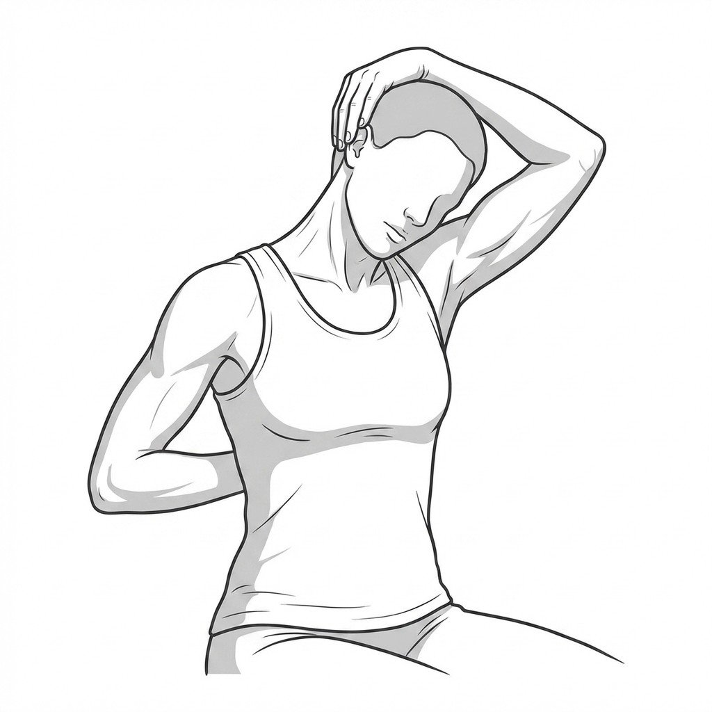
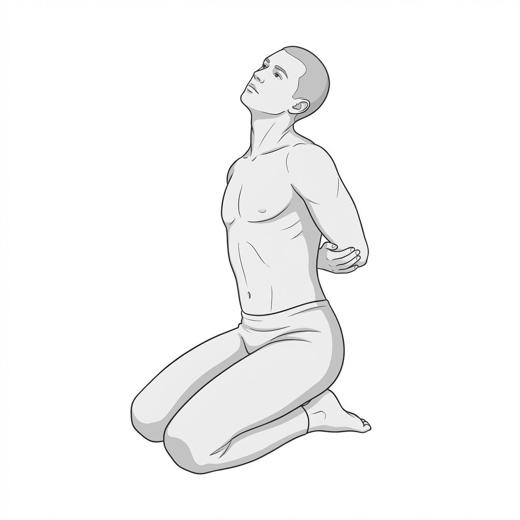
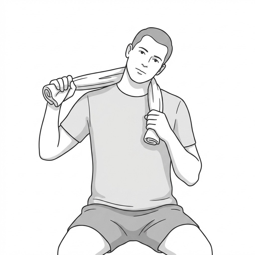
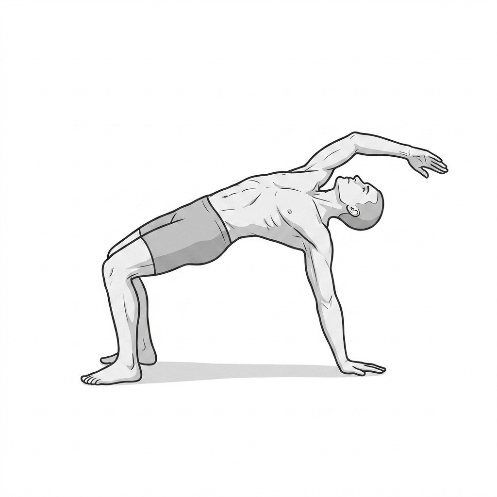
Exercises for Cervicogenic Headache
These exercises restore upper cervical mobility and retrain the deep flexors that are commonly inhibited in cervicogenic headache — thoracic extension helps offload the suboccipital joints that drive referred head pain. Start with 2–3 and progress as tolerated.