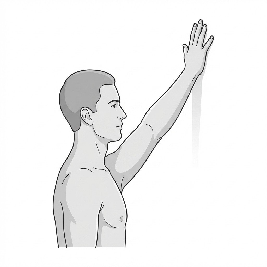
Understanding Adhesive Capsulitis
Adhesive capsulitis, commonly known as frozen shoulder, is an insidious condition characterized by progressive pain and global restriction of both active and passive shoulder movement. The pathophysiology involves fibroblastic proliferation and multilocular capsular thickening, leading to a contracted glenohumeral joint space. The condition typically progresses through three distinct phases: the painful "freezing" stage, the stiff "frozen" stage, and the recovery "thawing" stage. Epidemiological data indicates a strong association with systemic conditions, particularly diabetes mellitus and thyroid dysfunction, which often correlate with a more severe and treatment-resistant clinical course.
The clinical presentation of adhesive capsulitis frequently overlaps with severe myofascial pain syndrome (MPS) of the shoulder girdle. During the early freezing stage, intense pain prompts profound muscular guarding, particularly in the subscapularis, pectoralis minor, and infraspinatus. This sustained contraction not only exacerbates pain but can accelerate capsular contracture by chronically limiting joint excursion. Furthermore, trigger points in the subscapularis can restrict external rotation so severely that they can closely mimic the capsular pattern of frozen shoulder, complicating early diagnosis.
Differentiating the true capsular restriction of a frozen shoulder from the muscular guarding of myofascial pain is a cornerstone of effective shoulder rehabilitation.
Differentiating between primary capsular restriction and secondary myofascial restriction requires careful clinical assessment. The hallmark of true adhesive capsulitis is a proportional loss of passive range of motion—most notably external rotation—accompanied by a firm, leathery end-feel. In contrast, restriction driven primarily by myofascial trigger points often demonstrates a more yielding, muscular end-feel, and passive motion may improve significantly following targeted soft tissue release or trigger point injection. Current guidelines generally recommend a multimodal approach, addressing both the capsular inflammation and the inevitable secondary myofascial adaptations.
True Frozen Shoulder vs Myofascial Mimic
Passive external rotation
Pattern of motion loss
Course over time
First-line treatment
Myofascial Overlap & Anatomy
The glenohumeral joint relies on a delicate balance between capsular laxity for mobility and muscular control for stability. In adhesive capsulitis, the capsule thickens and adheres to the anatomical neck of the humerus, profoundly altering the mechanics of the surrounding musculature.
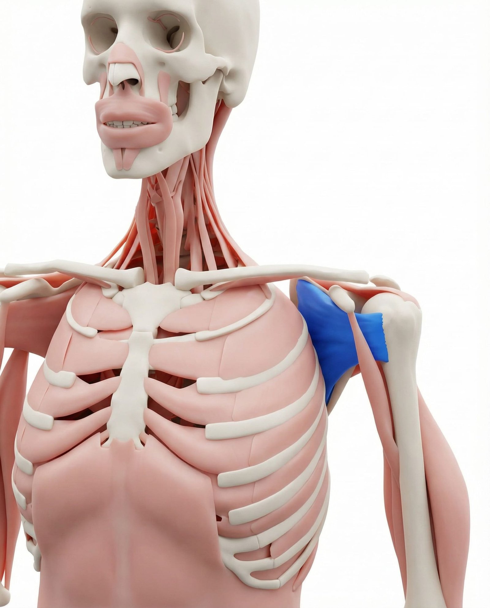
The subscapularis is a primary internal rotator of the shoulder. When affected by myofascial trigger points, it severely restricts external rotation, creating a clinical presentation that closely mimics the classic capsular pattern of adhesive capsulitis. Chronic guarding in this muscle is frequently observed during the freezing and frozen stages of true capsulitis.
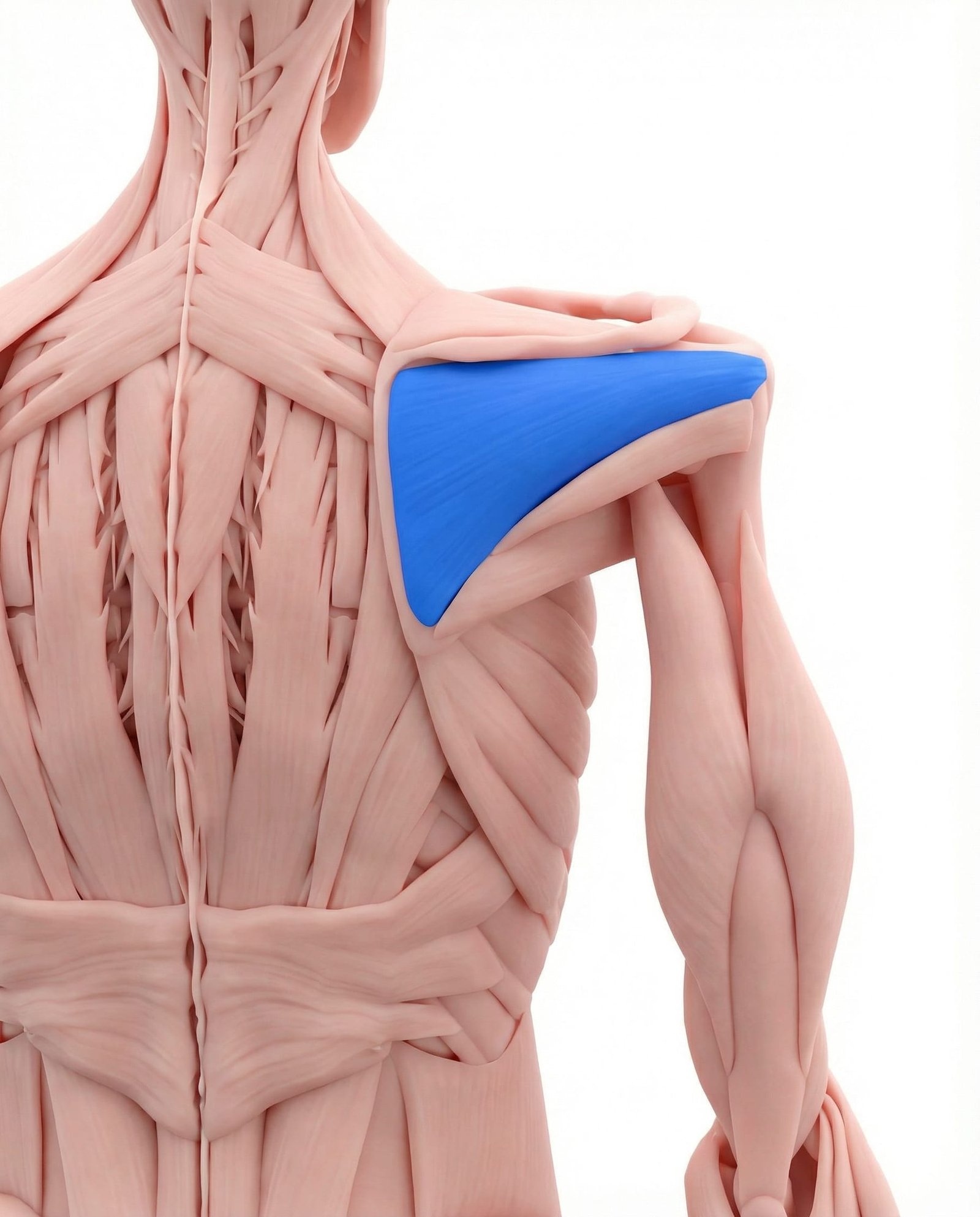
As a primary external rotator, the infraspinatus often develops secondary trigger points due to the altered biomechanics and chronic stretch placed upon it during adhesive capsulitis. Its referral pattern is frequently felt deep in the anterior shoulder joint, which patients often misinterpret as structural joint pain.
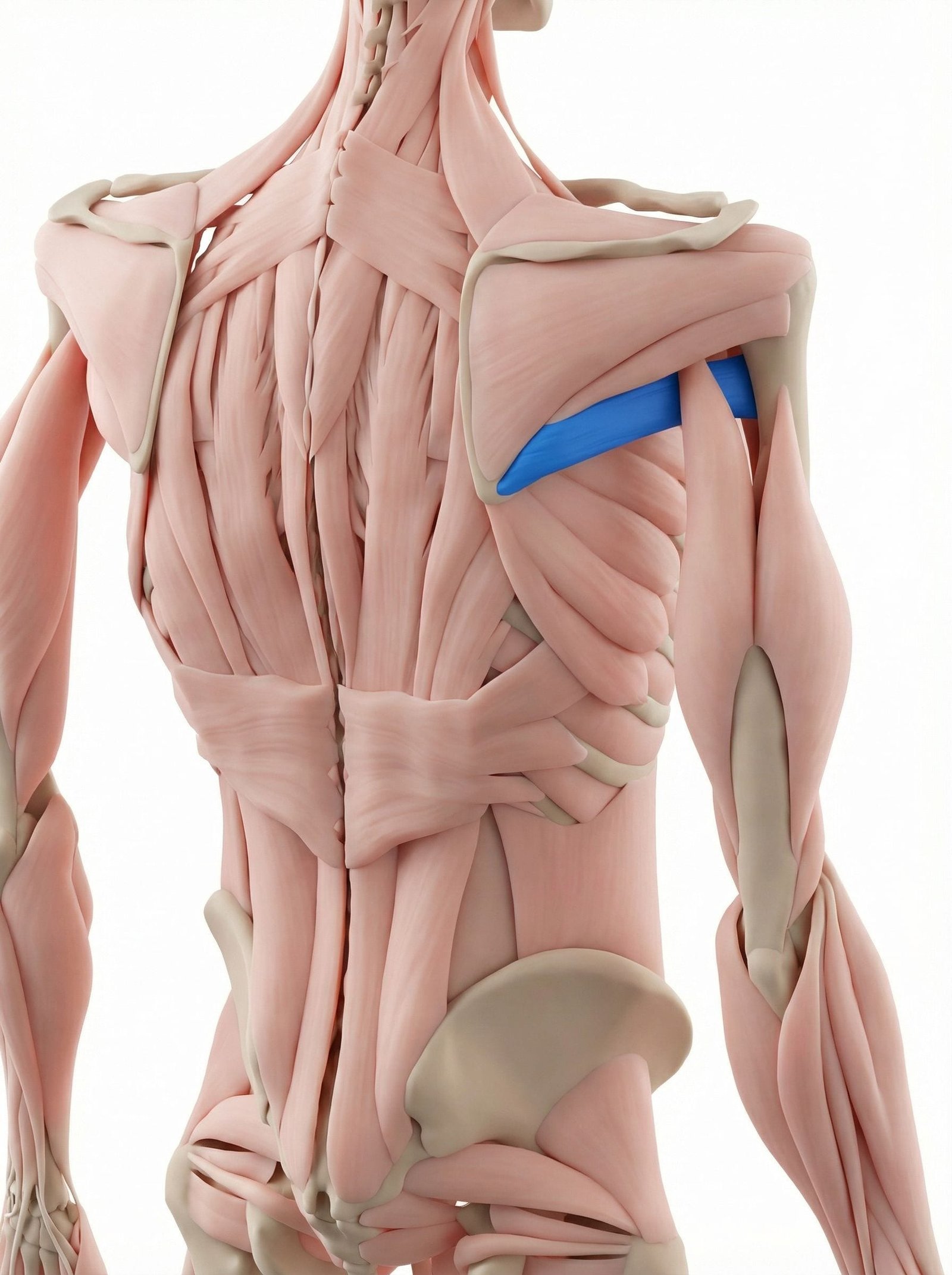
Working synergistically with the latissimus dorsi, the teres major assists in internal rotation and adduction. Myofascial restriction here limits overhead elevation and abduction, contributing to the global range-of-motion loss seen in frozen shoulder presentations.
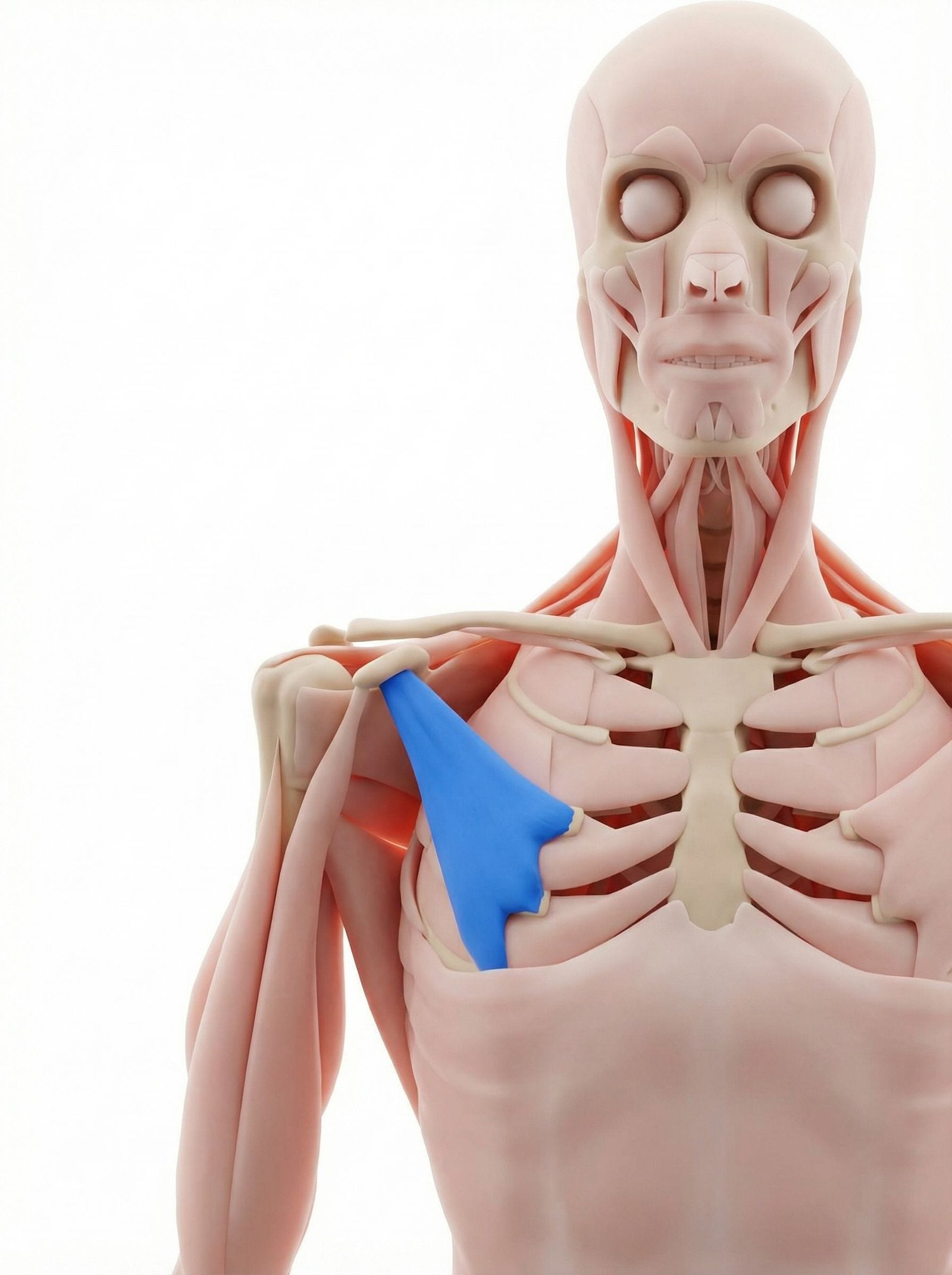
Adaptive shortening of the pectoralis minor is common in patients with shoulder pain who adopt a protective, protracted shoulder posture. This anterior tilt of the scapula further reduces the subacromial space and limits functional overhead reach, complicating the rehabilitation of adhesive capsulitis.
The deltoid is rarely the primary source of shoulder pathology but is a frequent site of referred pain from the rotator cuff and joint capsule. Secondary trigger points may develop here due to compensatory lifting mechanics when glenohumeral motion is restricted.
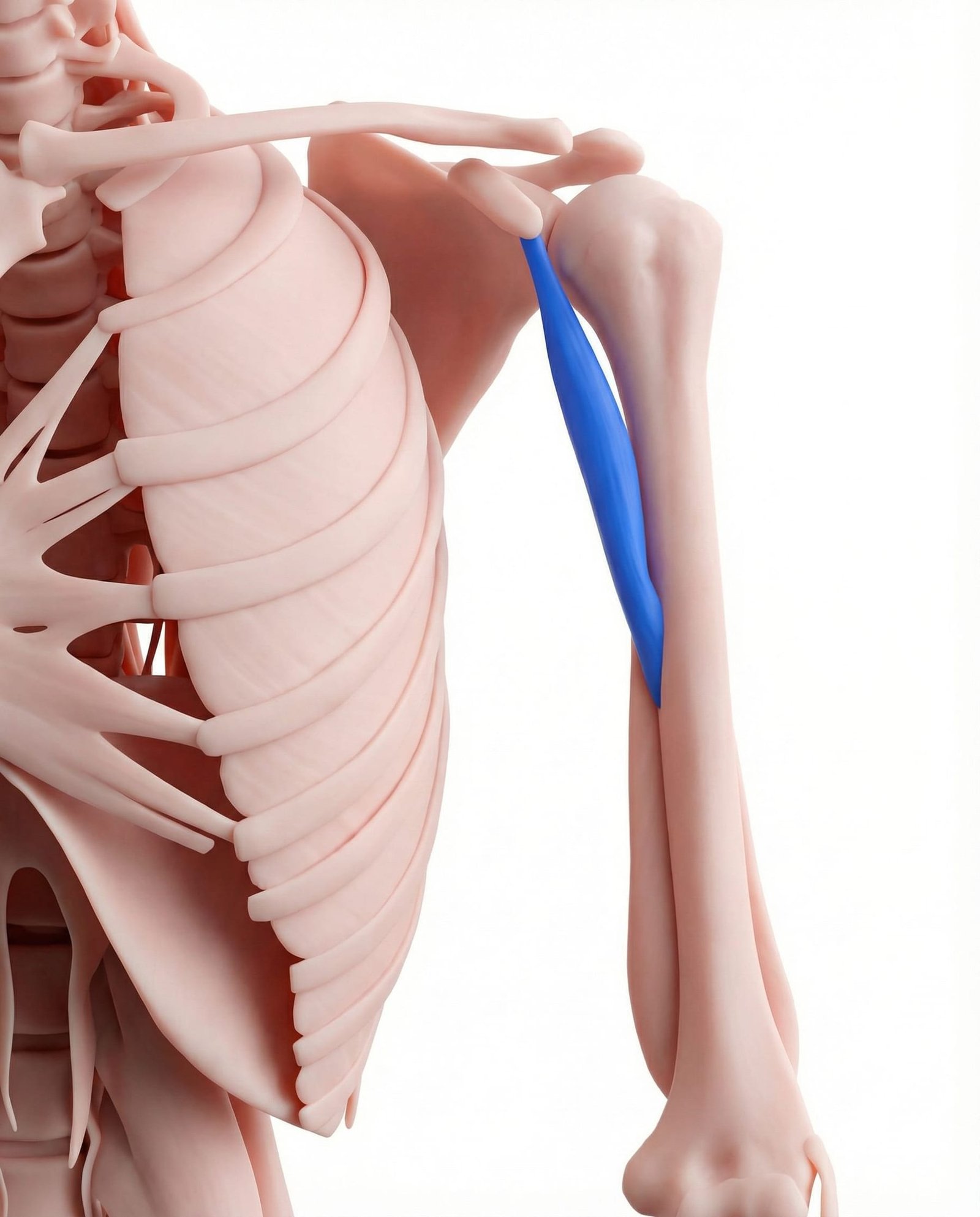
This muscle assists in shoulder flexion and adduction. When restricted, it limits end-range shoulder extension and abduction. Its involvement is often overlooked but can be a significant contributor to anterior shoulder pain that persists after capsular mobility begins to improve.
Stage 1: Freezing
Lasting 2-9 months. Characterized by severe, diffuse shoulder pain that is worse at night. Stiffness begins to develop, but pain is the primary limiting factor.
Stage 2: Frozen
Lasting 4-12 months. Pain gradually subsides, particularly at rest, but profound stiffness remains. Activities of daily living are significantly impaired.
Stage 3: Thawing
Lasting 5-24 months. Gradual return of range of motion. Pain is minimal and typically only occurs at the end ranges of available movement.
Myofascial Overlap
Throughout all stages, secondary trigger points in the rotator cuff and scapular stabilizers contribute to the pain experience and can perpetuate movement dysfunction.
Referred Pain Patterns
Pain in adhesive capsulitis is often diffuse, but the secondary development of myofascial trigger points creates specific, predictable patterns of referred pain that patients frequently mistake for joint or nerve damage.
Subscapularis
Infraspinatus
Pectoralis Minor
Clinical Assessment
Clinical evaluation focuses on distinguishing capsular stiffness from muscular guarding and ruling out structural rotator cuff pathology. These assessments help determine the primary driver of motion loss.
Passive External Rotation Test
Apley Scratch Test
Subscapularis Palpation
Painful Arc Observation
Evidence-Based Management
Management of adhesive capsulitis is highly stage-dependent. Interventions that are highly effective in the thawing stage may severely exacerbate symptoms if applied during the acute freezing stage.
Self-Care
- Frequent, gentle range-of-motion exercises within pain tolerance
- Use of moist heat prior to stretching to improve tissue extensibility
- Optimized sleep positioning using pillows to support the affected arm
- Activity modification to avoid forceful overhead or reaching movements
- Application of ice following activity to manage acute inflammatory flares
Manual Therapy
- Low-grade joint mobilizations during the freezing stage for pain modulation
- High-grade joint mobilizations during the thawing stage to restore mobility
- Ischemic compression of subscapularis and infraspinatus trigger points
- Scapulothoracic mobilization to optimize compensatory movement patterns
- Soft tissue release of the pectoralis minor to reduce anterior tilt
Interventional
- Intra-articular corticosteroid injections (most effective in early freezing stage)
- Glenohumeral joint hydrodilatation (distension) to stretch the contracted capsule
- Trigger point injections for refractory subscapularis or infraspinatus pain
- Suprascapular nerve blocks for severe, intractable night pain
- Manipulation under anesthesia (MUA) for recalcitrant frozen stage cases
Rehabilitation
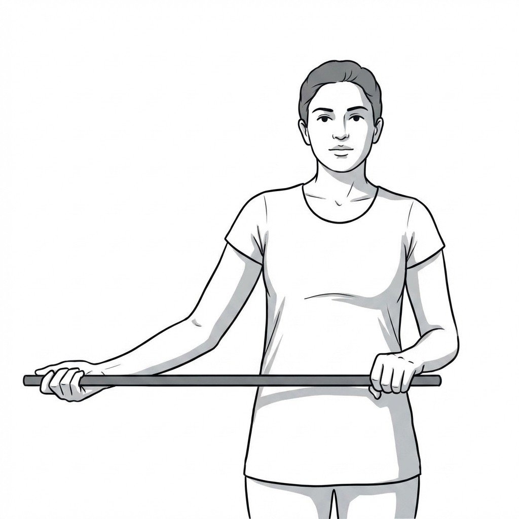
- Progressive passive and active-assisted stretching (e.g., wand exercises)
- Isometric strengthening of the rotator cuff in pain-free ranges
- Scapular stabilization exercises to prevent secondary impingement
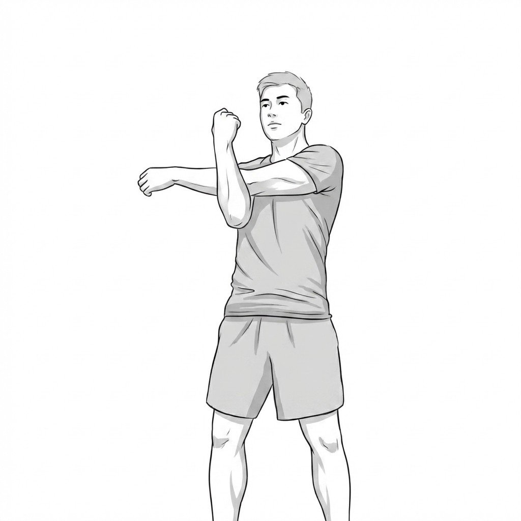
- Posterior capsule stretching (e.g., sleeper stretch) as tolerated
- Gradual return to functional overhead activities during the thawing stage
Red Flags & Differential Diagnosis
While adhesive capsulitis is a common cause of shoulder stiffness, several serious pathologies can present with similar symptoms. The following red flags require prompt medical investigation.
Adhesive capsulitis typically follows a three-stage course: freezing (pain predominant), frozen (stiffness predominant), and thawing (recovery).
The hallmark of true adhesive capsulitis is a significant restriction in passive external rotation with a firm, capsular end-feel.
Subscapularis trigger points can severely restrict external rotation, perfectly mimicking the clinical presentation of early frozen shoulder.
Patients with diabetes mellitus or thyroid disorders have a significantly higher risk of developing adhesive capsulitis, and often experience a more protracted course.
Current guidelines generally recommend intra-articular corticosteroid injections during the early freezing stage to reduce pain and shorten symptom duration.
Aggressive stretching during the highly inflammatory freezing stage is generally contraindicated and may exacerbate symptoms; gentle, pain-free motion is preferred.
Movement therapy
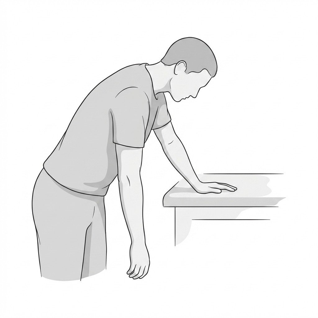
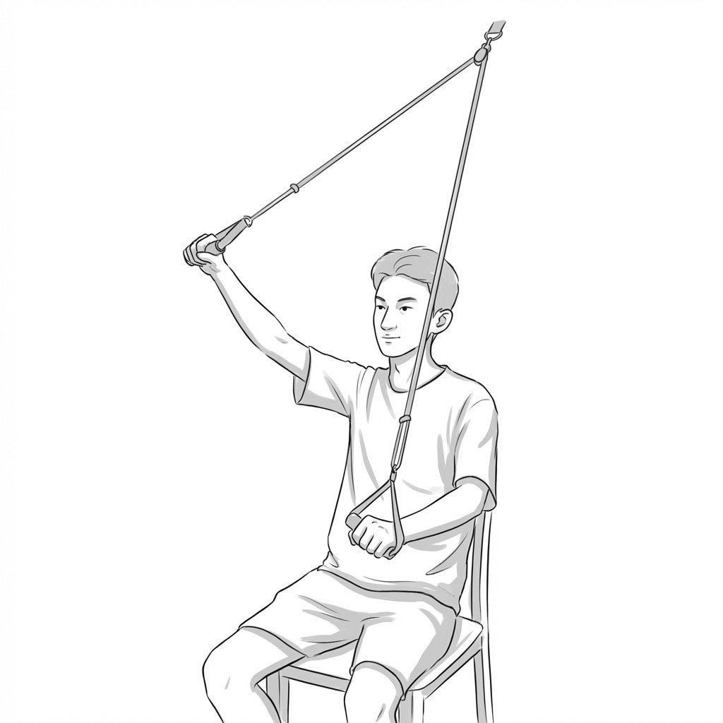
Exercises for Adhesive Capsulitis
Frozen shoulder requires gentle, pain-guided mobilization of the contracted glenohumeral capsule — forcing range prematurely prolongs the freezing stage. Work within a comfortable arc and start with 2–3, progressing as range returns.