Understanding Myofascial Low Back Pain
Low Back Pain
Anatomy and Pain Pattern OverviewLow back pain is one of the most common musculoskeletal complaints worldwide. Although many cases are labeled “nonspecific,” that label often means only that no single structural cause has been proven on imaging — not that muscles, movement, loading, and pain sensitization are irrelevant.
Myofascial trigger points in the QL, multifidus, psoas, gluteal muscles, and adjacent tissues can contribute meaningfully to pain patterns that patients experience as low back pain. These patterns may overlap with disc-related, SI-joint, facet, or hip pain presentations, which is why careful clinical examination matters.
The distinction from structural pathology is still important. Disc herniation with nerve root compression, spinal stenosis, fracture, inflammatory disease, infection, and visceral causes each have their own warning patterns and examination findings. A myofascial contributor should be added to the differential — not used to erase the rest of it.
Many low back pain cases are not “just the disc” or “just the joint.” Muscles, movement patterns, and sensitization often contribute more than patients are told.
Anatomy & Key Trigger Points
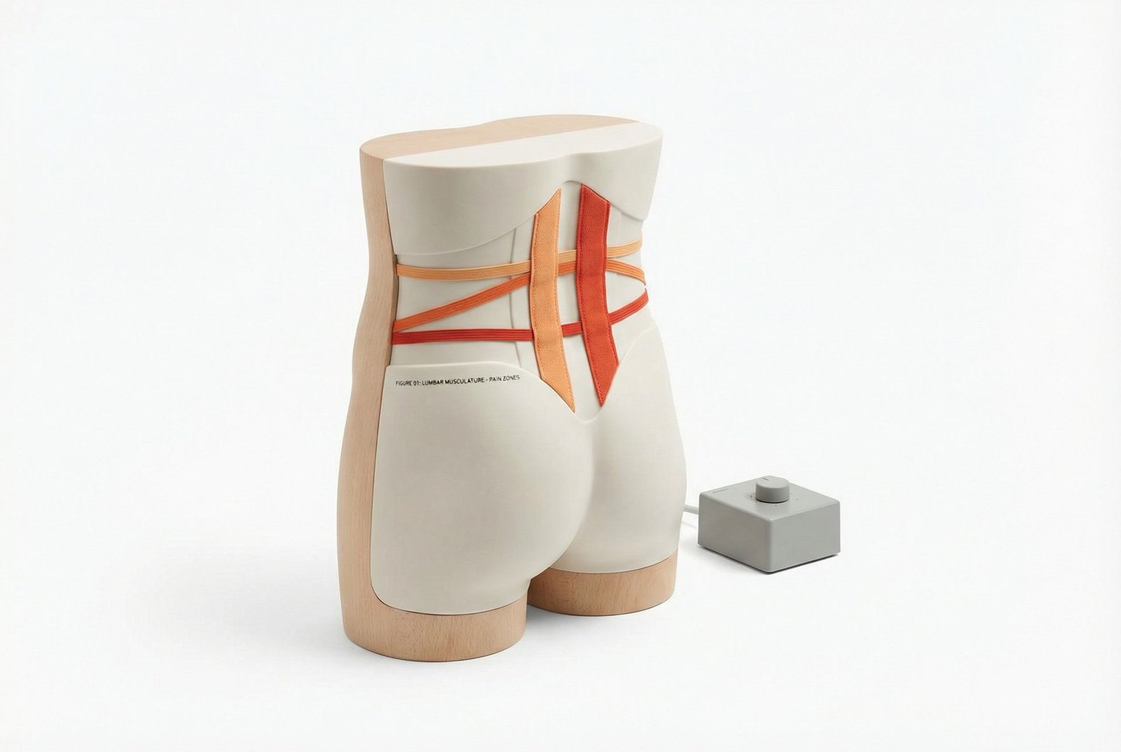
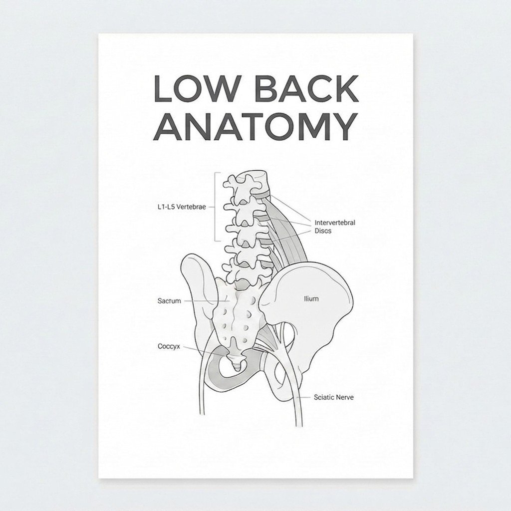
The lumbopelvic region contains multiple muscle layers that stabilize the spine, control the pelvis, and transfer load to the legs. Trigger points can develop in any of these muscles, but some are more commonly relevant in clinical low back pain patterns.
Low Back Pain
A frequent contributor to low back and flank-region pain. Trigger points in the QL may refer pain toward the sacroiliac region, iliac crest, greater trochanter, and lower abdomen. Patients often report pain with side-bending, rolling in bed, coughing, or prolonged standing. The deep flank-like quality is one reason QL pain is often mistaken for other sources.
The multifidus muscles are deep segmental stabilizers that span one to three vertebral levels. When irritated or inhibited, they may contribute to highly localized aching close to the spinous processes and may be part of recurrent low back pain patterns. Because they lie beneath more superficial muscles, they can be easy to miss without careful assessment.
The psoas is a deep hip flexor and lumbar stabilizing muscle. Irritability in this region may contribute to deep anterior low back or groin aching, difficulty standing fully upright after prolonged sitting, and discomfort with hip extension. Because of its depth and broad pain pattern, it is often part of a larger lumbopelvic problem rather than the only pain source.
The gluteus medius is a key hip abductor and pelvic stabilizer during single-leg stance. Trigger points or overload here may refer pain to the posterior iliac crest, sacrum, lateral hip, and upper lateral thigh. It is a common contributor when low back pain coexists with lateral hip pain or poor single-leg control.
The largest gluteal muscle often contributes to buttock, sacral, and coccygeal pain patterns when irritated. Patients may describe discomfort with prolonged sitting, climbing stairs, or rising from a chair. It commonly overlaps with other posterior-chain contributors rather than acting alone.
The piriformis is a deep external rotator of the hip that may contribute to deep buttock pain and posterior thigh referral. In some patients this overlaps with sciatic-type symptoms, though symptom location alone does not prove true nerve compression. Piriformis-related pain is best treated as one item in the differential rather than a stand-alone explanation for all posterior hip pain.
The most lateral column of the erector spinae group. Irritability here may contribute to broad paraspinal aching, flank discomfort, and lower rib-region referral. These symptoms often worsen with prolonged static postures, repetitive asymmetrical loading, or end-of-day fatigue.
A major extensor within the erector spinae group. It may contribute to central paraspinal low back pain that is often perceived as diffuse or “nonspecific.” In practice, longissimus irritation often coexists with multifidus, iliocostalis, or gluteal dysfunction rather than acting as an isolated diagnosis.
An under-recognized contributor in some low back pain presentations. Trigger points in the rectus abdominis may refer pain to the mid-back or low back and may also alter trunk mechanics through guarding and abdominal wall stiffness. It is especially worth examining when low back pain coexists with abdominal wall tenderness.
The TFL contributes to hip flexion, abduction, and internal rotation and blends into the iliotibial band. Trigger points or overactivity here may contribute to lateral hip pain and altered pelvic mechanics, which in turn can influence low back loading patterns.
Quadratus Lumborum
Deep lateral trunk muscle that may contribute to SI-region, flank, or iliac crest pain patterns.
Iliopsoas Complex
Deep hip flexor system that may contribute to anterior low back, groin, or standing-upright difficulty.
Gluteus Medius
Hip stabilizer often involved when low back pain overlaps with lateral hip pain or poor single-leg control.
Multifidus
Deep segmental stabilizer commonly implicated in recurrent or very localized paraspinal pain patterns.
Referral Patterns
Referral patterns help explain why patients often feel pain away from the muscle that is actually irritable. These patterns are guides, not rigid maps, and several muscles can overlap in the same patient.
Quadratus Lumborum
Multifidus
Psoas Major
Gluteus Medius
Piriformis
Self-Assessment
These tests and self-assessment maneuvers can help identify likely muscular contributors to low back pain. They are best used as part of a broader assessment, not as definitive standalone diagnostic tools.
QL Palpation at L3-L4
Thomas Test for Hip Flexor Contribution
FABER/Patrick Test
Prone Press-Up Test
Single-Leg Stand Test
Treatment Pathways
Effective treatment of low back pain usually requires matching the plan to the presentation. In many patients, muscular symptom relief, movement retraining, and progressive strengthening all need to work together.
Treatment Pathways
Mechanism DiagramSelf-Care
- Symptom-guided stretching of the QL, hip flexors, piriformis, hamstrings, or gluteal region when clearly relevant
- Foam rolling or ball release for gluteal and posterior hip muscles if tolerated
- Heat may help some patients with muscular guarding; ice may help if there is acute irritation or swelling
- Breaking up prolonged sitting or static standing with short movement breaks
- Reducing obvious aggravators such as asymmetric loading, repeated hip hiking, or poor workstation setup
Manual Therapy
- Sustained pressure release of clearly relevant trigger points
- Myofascial treatment of the thoracolumbar fascia and surrounding muscle groups when indicated
- Muscle energy techniques for the psoas, piriformis, or QL in selected cases
- Joint mobilization of the lumbar spine, SI joint, or hips when mobility restriction appears clinically relevant
- Manual care should ideally be paired with follow-up movement or exercise rather than used in isolation
Interventional
- Dry needling of clearly identified trigger point regions in selected patients
- Trigger point injection with local anesthetic or saline where clinically appropriate
- Ultrasound guidance may be useful for deeper or higher-risk targets such as psoas or deep paraspinals
- Botulinum toxin should be viewed as a selective specialist option, not routine care for ordinary low back pain
Rehabilitation
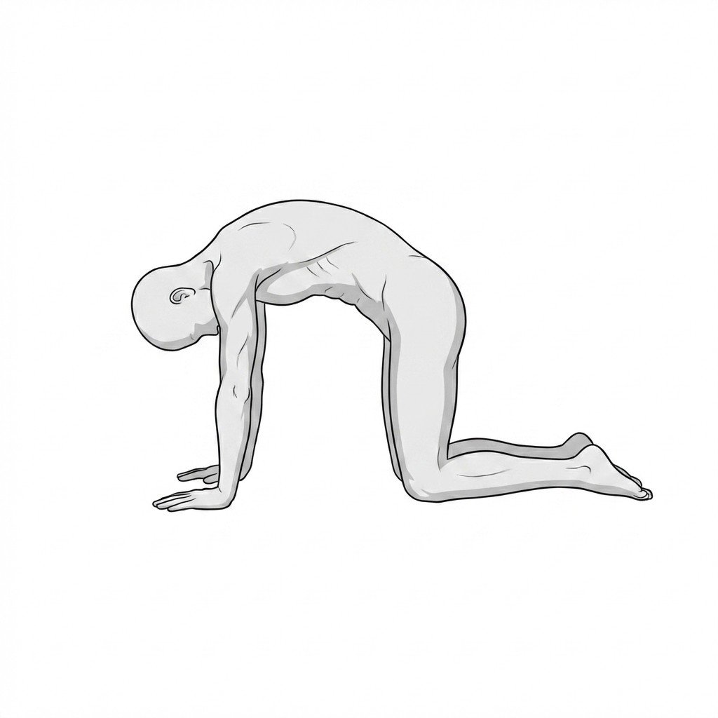
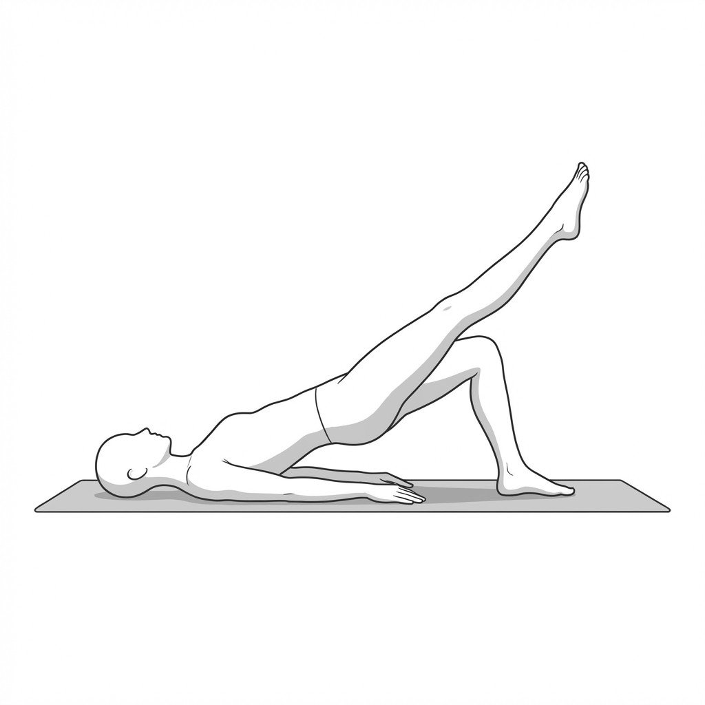
- Core endurance and control work such as bird dog, side bridge, and other trunk-stability exercises
- Progressive hip strengthening, especially gluteus medius and maximus
- Movement retraining with emphasis on lumbopelvic control and tolerance
- Graded return to bending, lifting, walking, stairs, or sport rather than avoidance
- Ergonomic and sleep-position review when symptoms are repeatedly aggravated by posture or environment
Red Flags — When to See a Doctor
Before attributing low back pain to a myofascial source, screen for the following red flags that suggest urgent or non-routine pathology.
Low back pain is often multifactorial, and myofascial trigger points can be an important part of the picture rather than a separate fringe diagnosis.
The quadratus lumborum, multifidus, psoas, and gluteal muscles are among the most common muscular contributors in persistent low back pain.
Simple tests and palpation can help generate hypotheses about muscular contributors, but they do not replace a full examination when structural or neurological causes are possible.
Treatment tends to work best when symptom relief, load modification, and rehabilitation are combined rather than when any one modality is used alone.
Red flags still matter. Not all low back pain is benign or muscular, and urgent symptoms must be screened before settling on a myofascial explanation.
Pseudo-sciatic and mixed low-back/hip pain patterns are common, which is why careful differential diagnosis is more useful than assuming every referral pattern is discogenic.
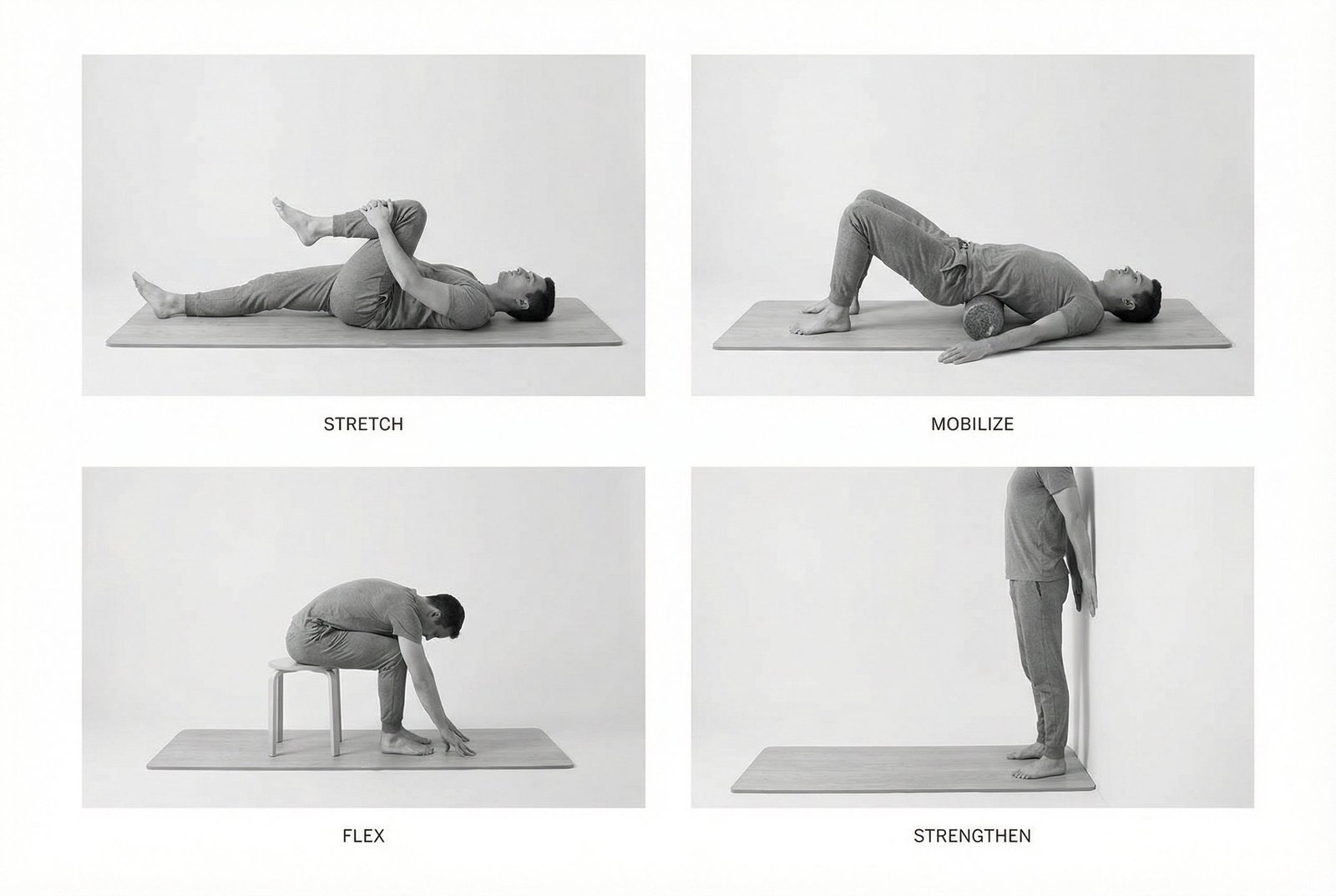
Movement therapy
Exercises for Low Back Pain
These exercises work the lumbar extensors and core stabilizers that bear the load in low back pain — building the endurance and segmental control that reduces recurrence. Start with 2–3 and progress as tolerated.