Understanding Scapular Dyskinesis & MPS
Scapular dyskinesis refers to altered scapular position or movement during shoulder motion. It is common in patients with shoulder pain, but it is also seen in people with no pain at all, which is why dyskinesis should be interpreted as a clinical sign rather than a diagnosis by itself.
There is a strong clinical overlap between scapular dyskinesis and periscapular myofascial pain. Trigger points and muscle inhibition can change scapular timing, while altered scapular timing can overload the very muscles that then develop trigger points.
Scapular dyskinesis is often the visible movement expression of a deeper problem — myofascial pain, shoulder pathology, cervical referral, or true neurological deficit.
A major assessment task is separating true neurological weakness from pain-mediated inhibition or motor-control failure. A winged or poorly controlled scapula does not automatically mean nerve palsy — but nerve palsy must stay on the differential when weakness is marked or persistent.
Key Periscapular Muscles
Normal scapular motion depends on coordinated force sharing among serratus anterior, trapezius, rhomboids, levator scapulae, the thoracic spine, and the glenohumeral joint.
Serratus Anterior
A major stabilizer and upward rotator of the scapula. Pain, inhibition, or poor recruitment here can contribute to winging-like patterns and poor scapulohumeral rhythm, especially during overhead elevation.
Pain may refer to the lateral chest wall, inferior angle of the scapula, and occasionally the medial arm.Lower Trapezius
Helps with scapular depression, upward rotation, and posterior control. It is often underperforming in dyskinesis patterns, especially when the upper trapezius is doing too much of the work.
May refer upward toward the lower cervical region, acromial area, or upper thoracic paraspinals.Upper Trapezius
Often becomes overactive as a compensatory elevator when lower trapezius and serratus anterior are not doing enough. This is one reason dyskinesis commonly coexists with neck pain and tension-type headache patterns.
Typical referral includes the posterolateral neck, behind the ear, and temporal region.Rhomboid Major & Minor
Act as retractors and downward rotators. They are frequently overloaded in protracted shoulder postures and may develop secondary trigger points when anterior chest wall tightness or scapular instability is not addressed.
Deep aching pain along the medial border of the scapula, often described as a knot or a burning ache.Levator Scapulae
Elevates and downwardly rotates the scapula. It often becomes overworked in the “elevated shoulder” posture and commonly overlaps with upper trapezius and cervical pain patterns.
Pain near the side of the neck and superior angle of the scapula, sometimes extending into the posterior shoulder.
Altered Kinematics
Changes in upward rotation, posterior tilt, or timing may affect shoulder comfort and load sharing.
Myofascial Inhibition
Trigger points and pain can inhibit key stabilizers, making a muscle appear weaker than it truly is structurally.
Compensatory Overload
The upper trapezius, levator scapulae, and rhomboids often overwork when other stabilizers underperform.
Neurological Mimicry
Severe pain-mediated inhibition can sometimes resemble nerve-related scapular weakness, which is why the differential matters.
Myofascial Referral Patterns
Periscapular trigger points often refer pain locally, but the broader pain pattern may overlap with shoulder, cervical, or thoracic problems.
- Serratus Anterior
- May refer pain into the lateral chest wall, axillary line, or inferior angle of the scapula. In some patients the pattern overlaps with chest-wall or lower brachial discomfort.
- Lower Trapezius
- May refer superiorly toward the neck, upper thoracic paraspinals, or acromial region and can overlap with shoulder impingement complaints.
- Levator Scapulae
- Typically produces neck stiffness and pain around the superior angle of the scapula, usually staying above the level of the scapular spine.
- Infraspinatus (Associated)
- Although not a classic periscapular stabilizer, it may coexist and refer pain deeply into the anterior shoulder, complicating the dyskinesis / shoulder-pain picture.
Clinical Assessment Tests
Clinical assessment relies on visual observation, symptom-modification tests, and the ability to tell whether changing scapular mechanics changes the patient’s pain or strength.
Visual Scapular Dyskinesis Test
Observe the scapulae from behind during repeated arm elevation, with or without light load depending on the patient. Look for asymmetry, early shrugging, medial border prominence, or loss of smooth rhythm.
Positive: Visible asymmetry, winging, or premature scapular elevation suggests altered scapular control, though it does not identify the exact structural cause by itself.
Scapular Assistance Test (SAT)
The clinician assists scapular upward rotation and posterior tilt during arm elevation to see whether the patient’s familiar pain or motion improves.
Positive: Reduced pain or improved elevation suggests that scapular mechanics are contributing meaningfully to the patient’s symptoms.
Scapular Retraction Test (SRT)
The clinician manually stabilizes the scapula in a more retracted position while the patient performs a resisted shoulder test.
Positive: Improved strength or reduced pain suggests that scapular control is affecting apparent rotator cuff performance.
Wall Push-Up Test
The patient performs a wall push-up while the clinician observes scapular border control, especially during the eccentric phase.
Positive: Clear winging or loss of control suggests serratus anterior weakness, pain-related inhibition, or — less commonly — a true nerve palsy pattern.
Evidence-Based Management
Management usually follows a phased logic: calm the painful tissues, reduce the overload pattern, and then rebuild scapular motor control and strength.
Self-Care
- Postural awareness and reduction of prolonged shoulder protraction
- Gentle stretching of the pectoralis minor and overactive upper-quarter tissues
- Heat for reactive upper cervical or upper-trapezius tension if it helps
- Temporary reduction of repetitive overhead loading during flare phases
- Basic scapular setting and low-level control exercises
Manual Therapy
- Trigger point treatment of painful periscapular muscles
- Myofascial work to pectoralis minor, levator scapulae, and related tissues
- Thoracic mobility work to improve extension and ribcage motion
- Glenohumeral or thoracic-joint mobilization when indicated
- Soft-tissue treatment to serratus anterior or lateral chest wall when relevant
Interventional
- Trigger point injections when myofascial pain is clearly refractory
- Dry needling of selected upper-quarter muscles
- Neurological testing when true palsy is suspected
- Image-guided subacromial treatment only when a true concurrent shoulder pathology is present
- More invasive approaches only after the movement diagnosis is clearer
Rehabilitation
- Motor control training for scapular resting and movement position
- Progressive serratus anterior activation
- Lower and middle trapezius strengthening
- Integration into closed-chain and task-specific movement
- Gradual return to overhead work, sport, or training
Red Flags & Differential Diagnosis
Periscapular pain and altered scapular motion are often benign musculoskeletal findings, but some presentations suggest neurological or structural pathology that needs more urgent assessment.
True Scapular Winging at Rest
Pronounced winging that is present even at rest raises concern for a real neurological deficit such as long thoracic nerve palsy and warrants further evaluation.
InvestigateLateral Winging with Shoulder Droop
This may suggest spinal accessory nerve involvement, especially with trauma or prior neck surgery/history.
UrgentSevere Radiating Pain Below the Elbow
Pain, numbness, or tingling extending clearly into the forearm or hand raises concern for cervical radiculopathy or brachial plexus pathology rather than isolated periscapular MPS.
InvestigateTraumatic Onset of Weakness
Sudden inability to elevate the arm or marked weakness after trauma requires structural and neurological assessment.
UrgentUnrelenting Night Pain
Deep pain that is not mechanically influenced and repeatedly wakes the patient should prompt evaluation for non-myofascial causes.
Investigate
Clinical Pearls
Scapular dyskinesis is best understood as a clinical sign or movement impairment, not automatically a stand-alone diagnosis.
Periscapular myofascial pain can drive dyskinesis through pain, inhibition, compensation, or poor motor control.
Distinguishing pain-mediated inhibition from a true nerve palsy is a critical step in assessment.
Upper trapezius and levator scapulae often become overactive secondarily, creating additional neck and scapular pain.
Current rehabilitation usually works best when myofascial pain is addressed first and scapular control is then rebuilt progressively.
The Scapular Assistance Test can be useful for showing whether improving scapular motion temporarily changes the patient’s shoulder pain.
Movement therapy
Exercises for Scapular Dyskinesis
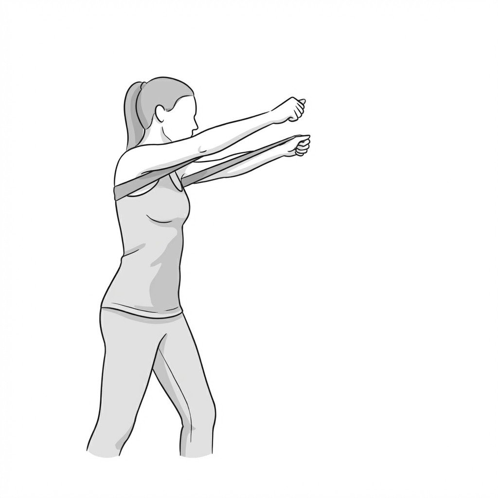
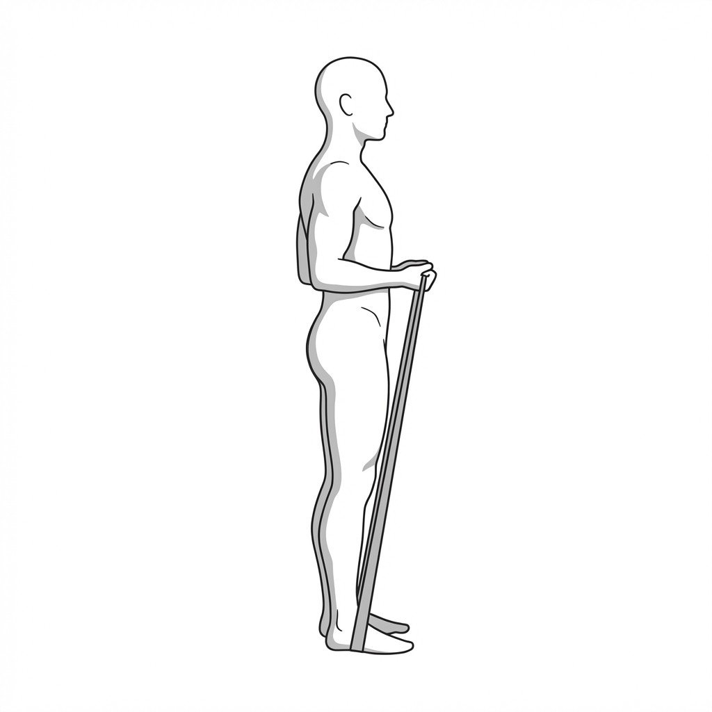
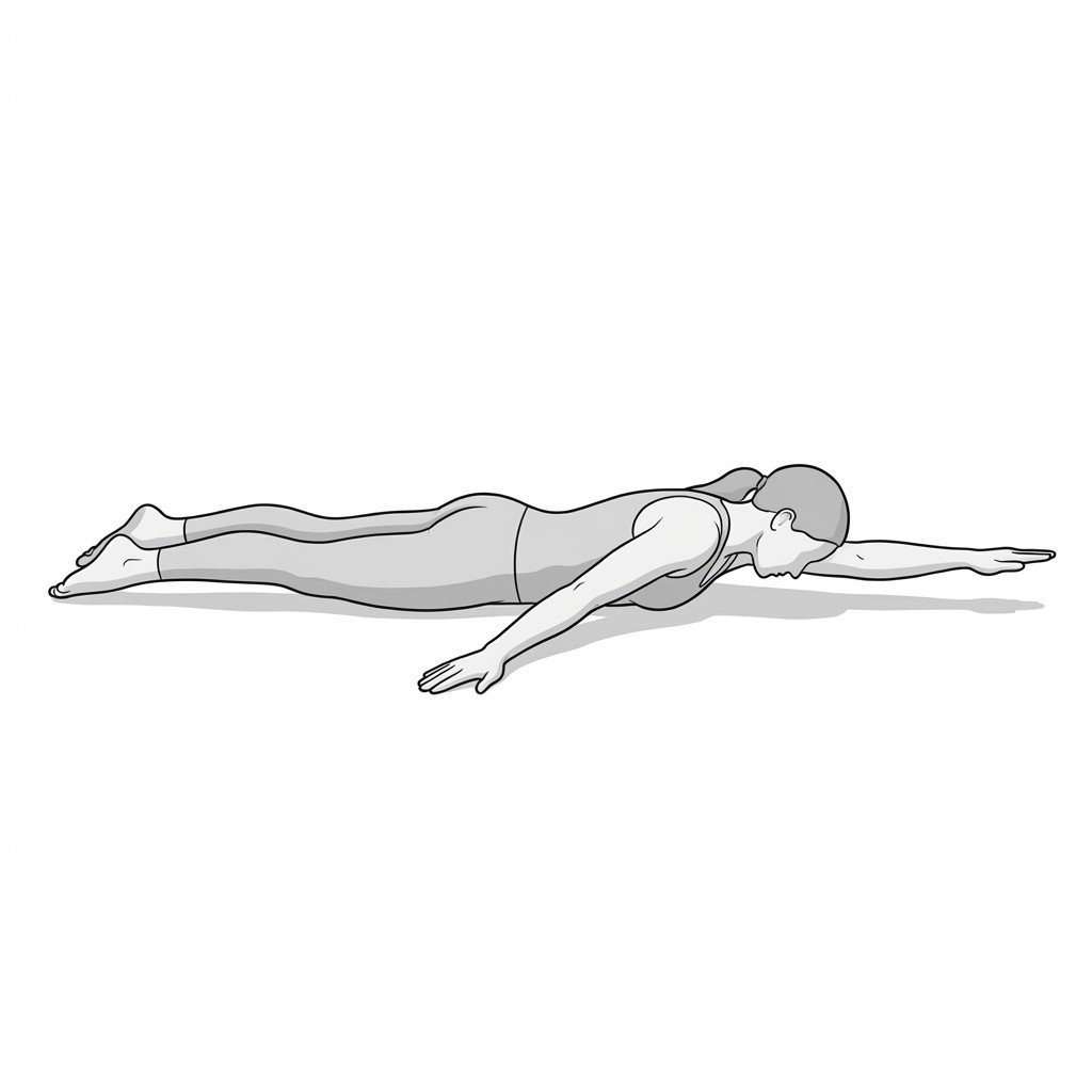
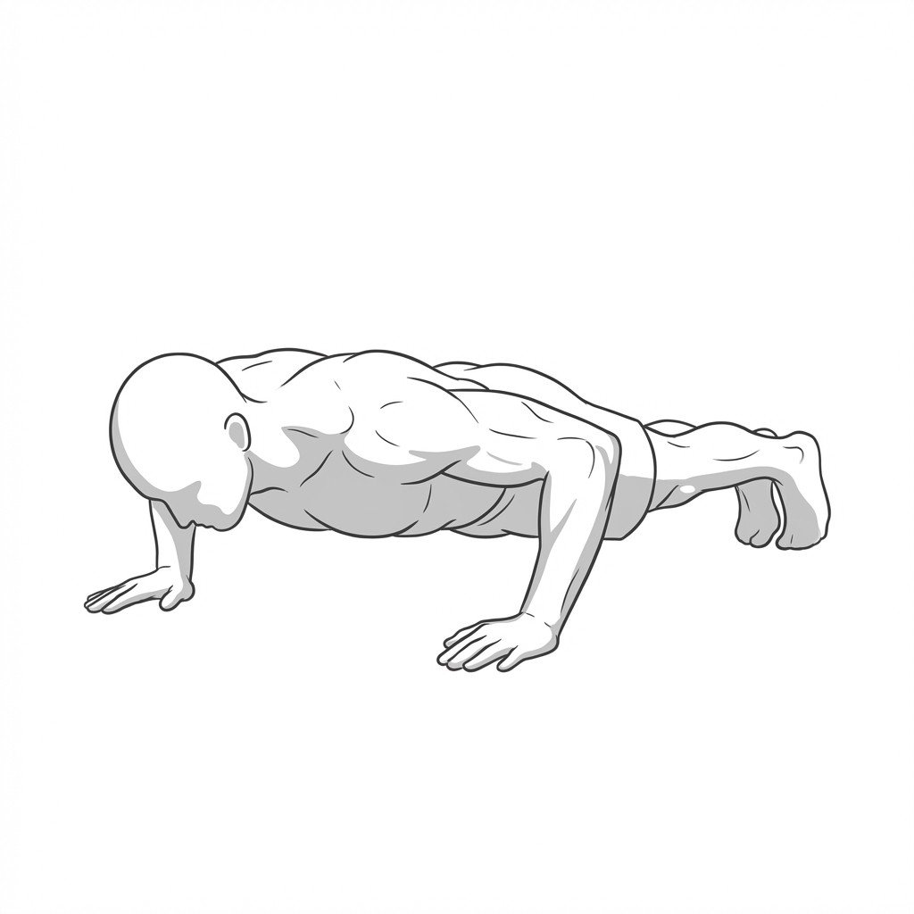
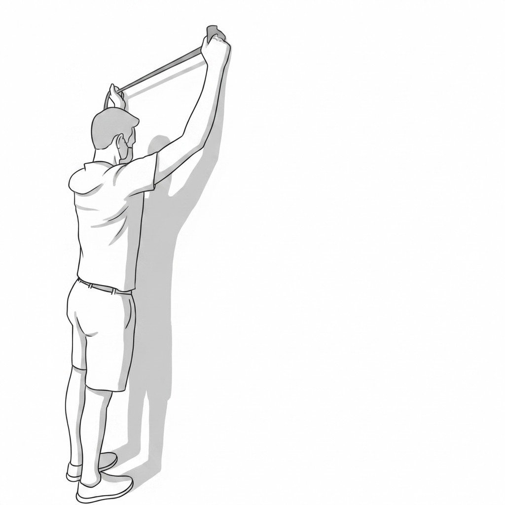
These exercises retrain the lower trapezius and serratus anterior that are typically inhibited in scapular dyskinesis — Y-raises, wall slides, and push-up plus work progressively to restore posterior tilt and upward rotation of the scapula under load. Start with 2–3 and progress as tolerated.