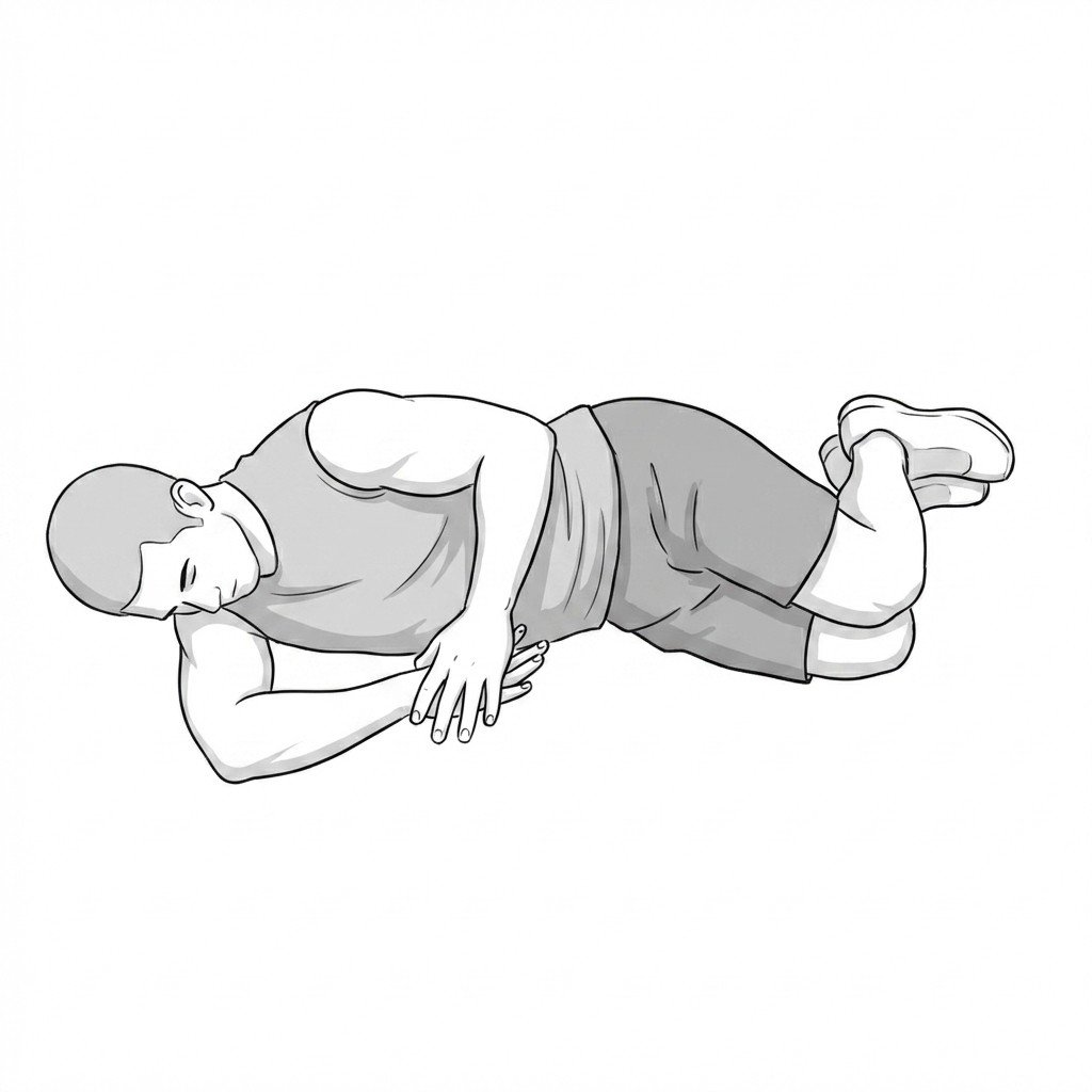
Understanding SAPS & Myofascial Overlap
Subacromial pain syndrome (SAPS) is now often used as a broader clinical term for non-traumatic lateral or anterolateral shoulder pain thought to involve the subacromial tissues. It usually includes bursitis, rotator cuff tendinopathy, and related overload states rather than one single discrete diagnosis.
Modern thinking places less emphasis on a simple “bone pinching tendon” model and more emphasis on tendon reactivity, load mismatch, altered scapular mechanics, and movement-system contributors. That said, mechanical narrowing of space may still matter in some patients, especially when scapular control is poor.
In many shoulder patients, subacromial pain is not purely structural or purely myofascial — it is a layered problem.
Myofascial pain frequently overlaps with SAPS. Trigger points in the infraspinatus, supraspinatus, upper trapezius, pectoralis minor, and related muscles can mimic or amplify shoulder pain that otherwise looks structural.
The practical implication is that good shoulder care often means treating both the tissue tolerance problem and the motor-control / myofascial problem, rather than trying to force the case into one category only.
Subacromial Pain vs Rotator Cuff Tear
Onset pattern
Active vs passive motion
Strength testing
Imaging implication
Anatomical & Kinematic Considerations
The subacromial region is dynamic. During arm elevation, the rotator cuff, scapular stabilizers, thoracic spine, and humeral head all influence how much stress is placed on the subacromial tissues.
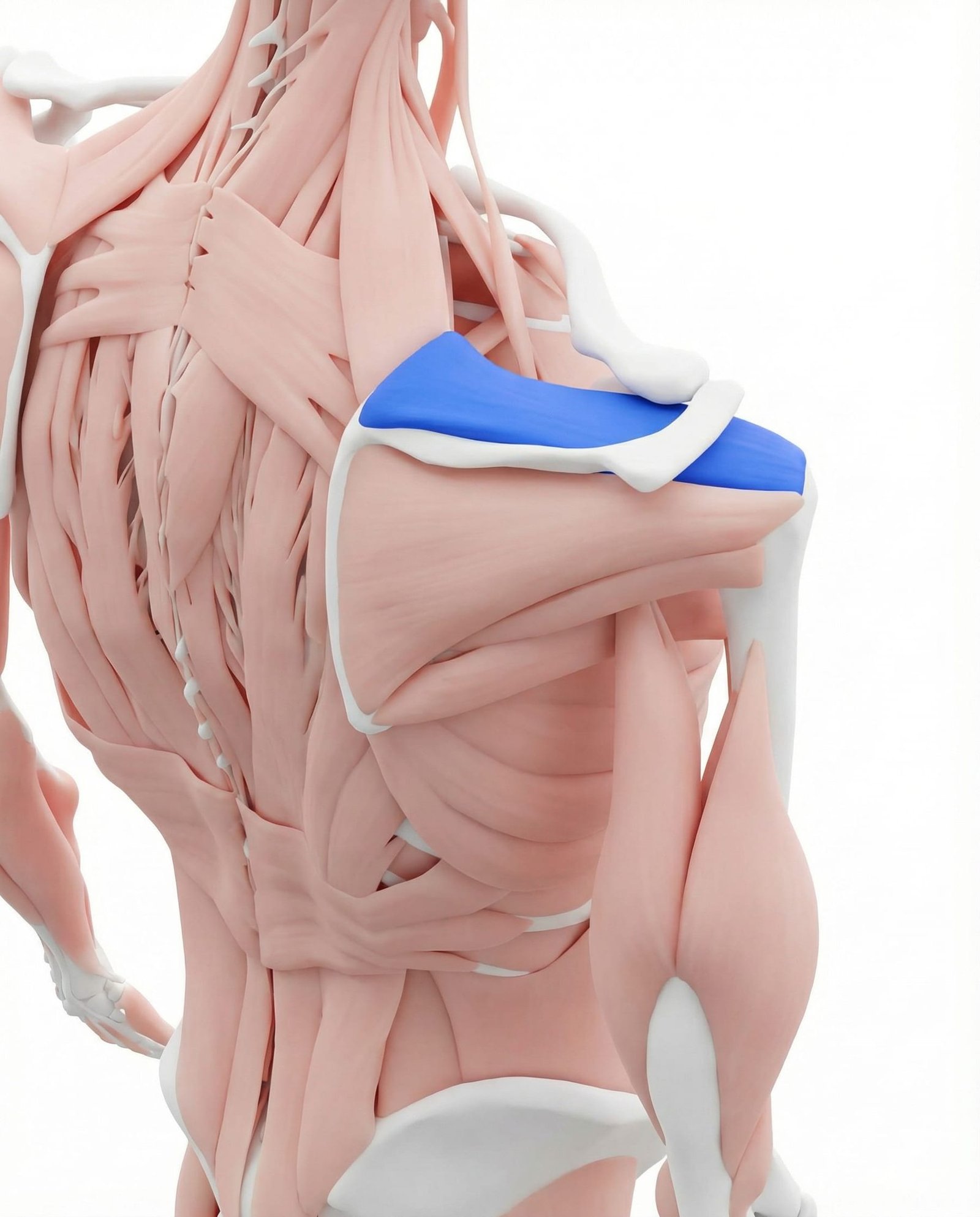
The supraspinatus is commonly involved in subacromial pain presentations, both as a tendon and as a myofascial pain source. It helps initiate abduction and contributes to glenohumeral stability. Trigger points here may coexist with tendon overload and can blur the line between “tendon pain” and referred muscular pain.
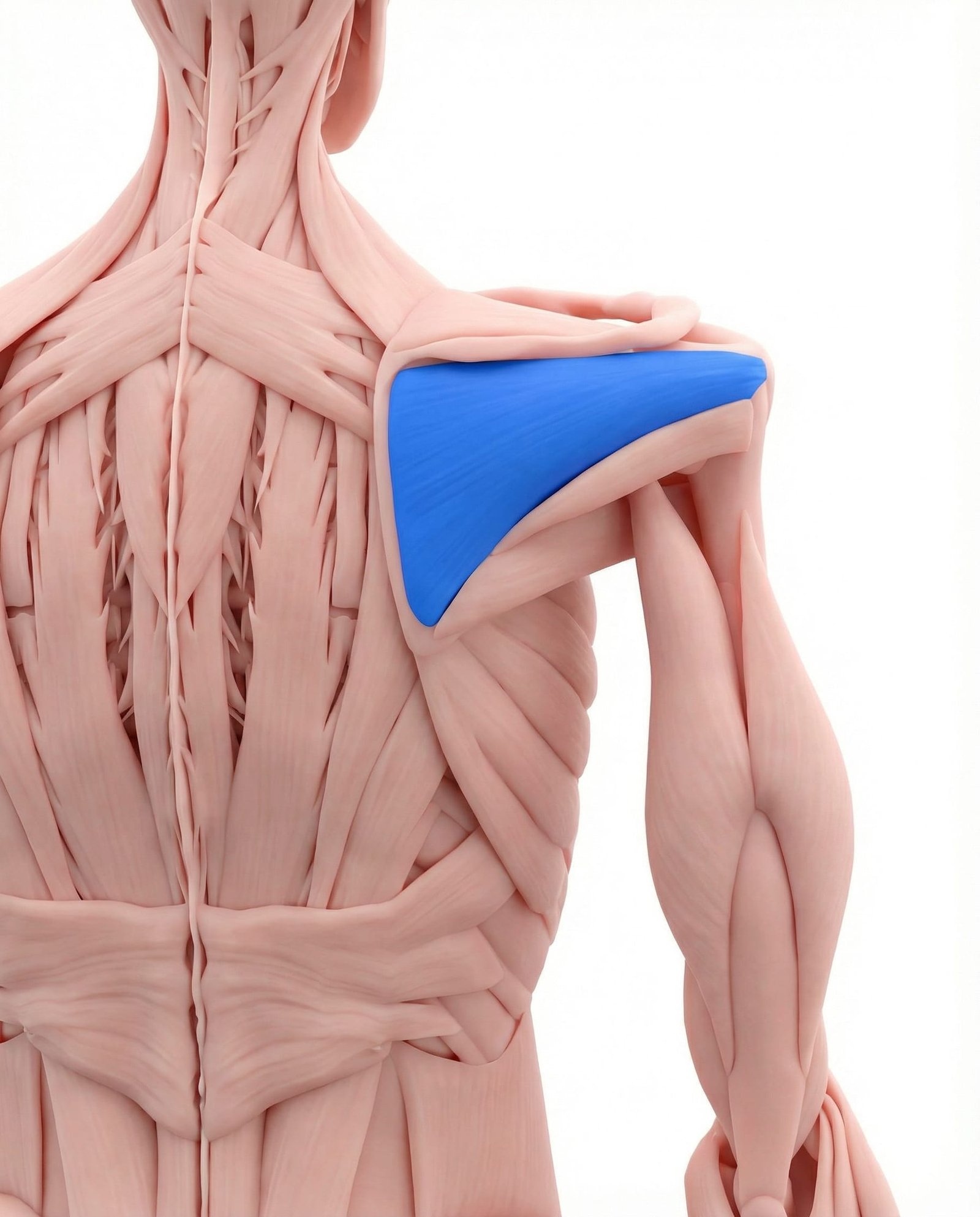
A key external rotator and posterior stabilizer of the shoulder. Infraspinatus trigger points are very common and often mistaken for primary anterior-shoulder pathology because the referral pattern may be felt more in the front of the shoulder than over the muscle itself.
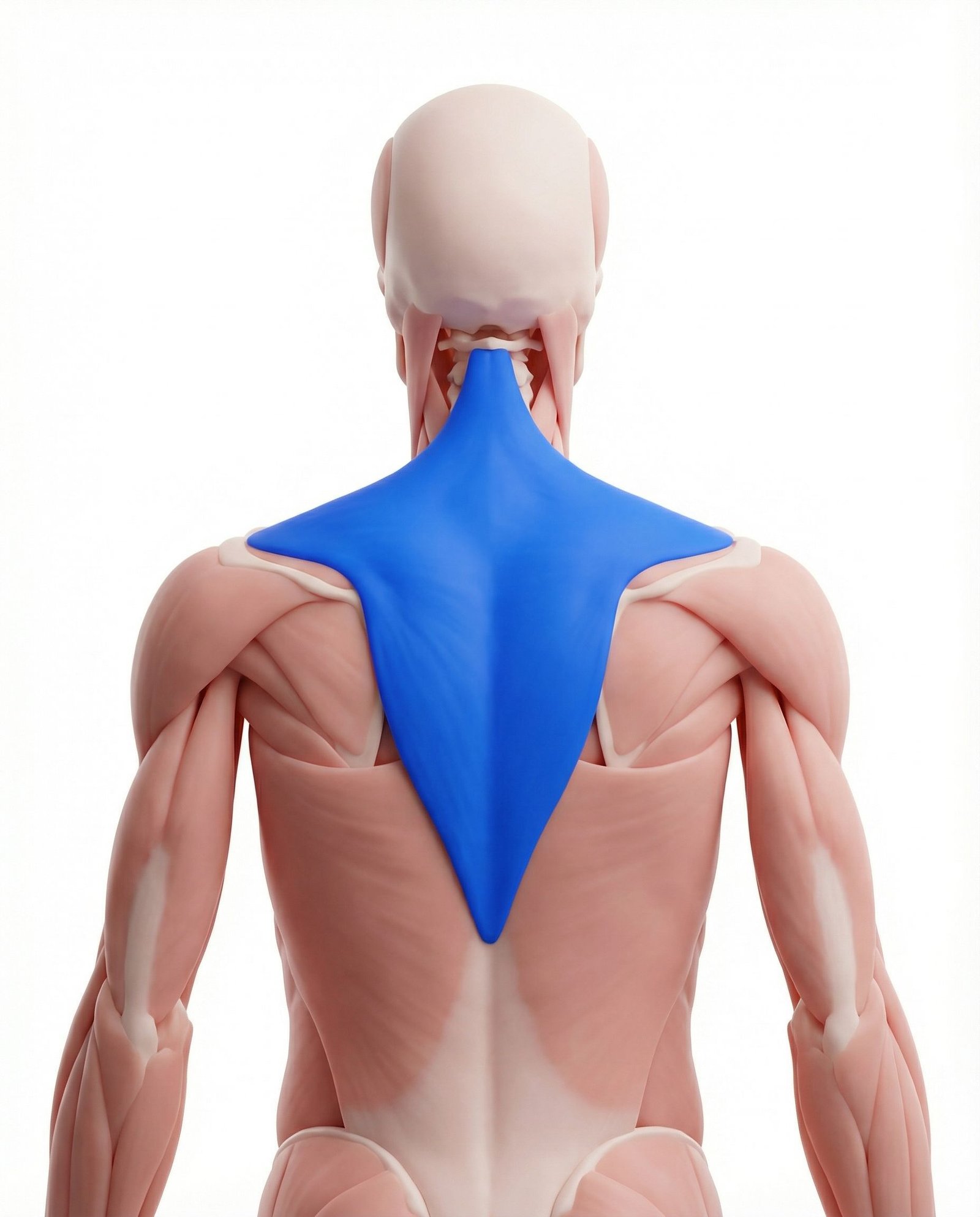
The upper trapezius helps elevate and upwardly rotate the scapula. In many patients with shoulder pain it becomes overactive as a compensation for poor scapular control, especially when serratus anterior and lower trapezius are underperforming. Trigger points here often reflect that compensation rather than the primary pathology.
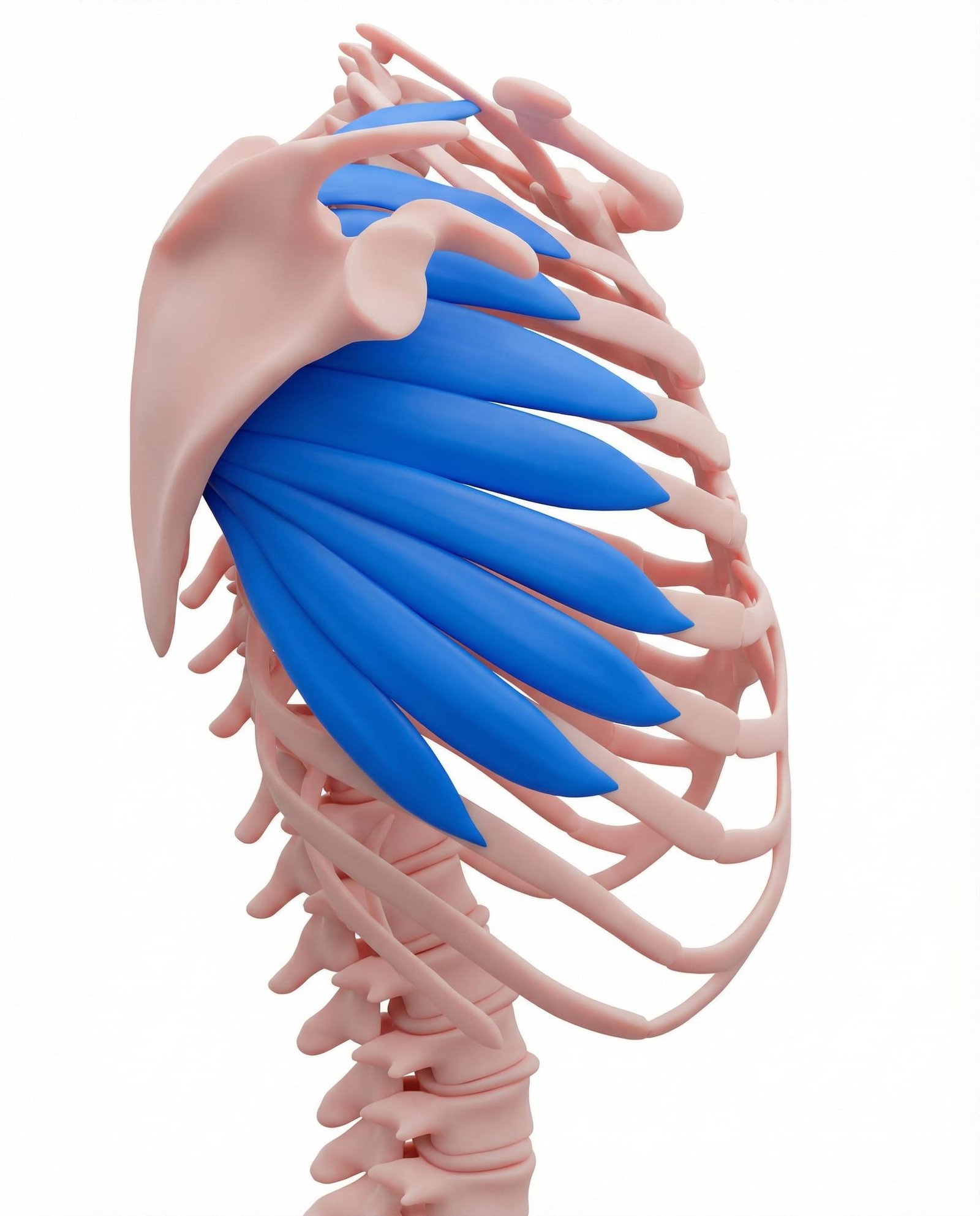
The serratus anterior is crucial for scapular upward rotation and posterior tilt during arm elevation. Weakness, inhibition, or poor timing may contribute to scapular dyskinesis, which in turn can aggravate subacromial symptoms. Trigger points here may further reduce efficient scapular control.
The deltoid is the primary mover for abduction and flexion, but when rotator cuff or scapular support is inadequate it may dominate the movement too early. Trigger points in the anterior or middle deltoid can add local pain that overlaps with bursal or tendon pain.
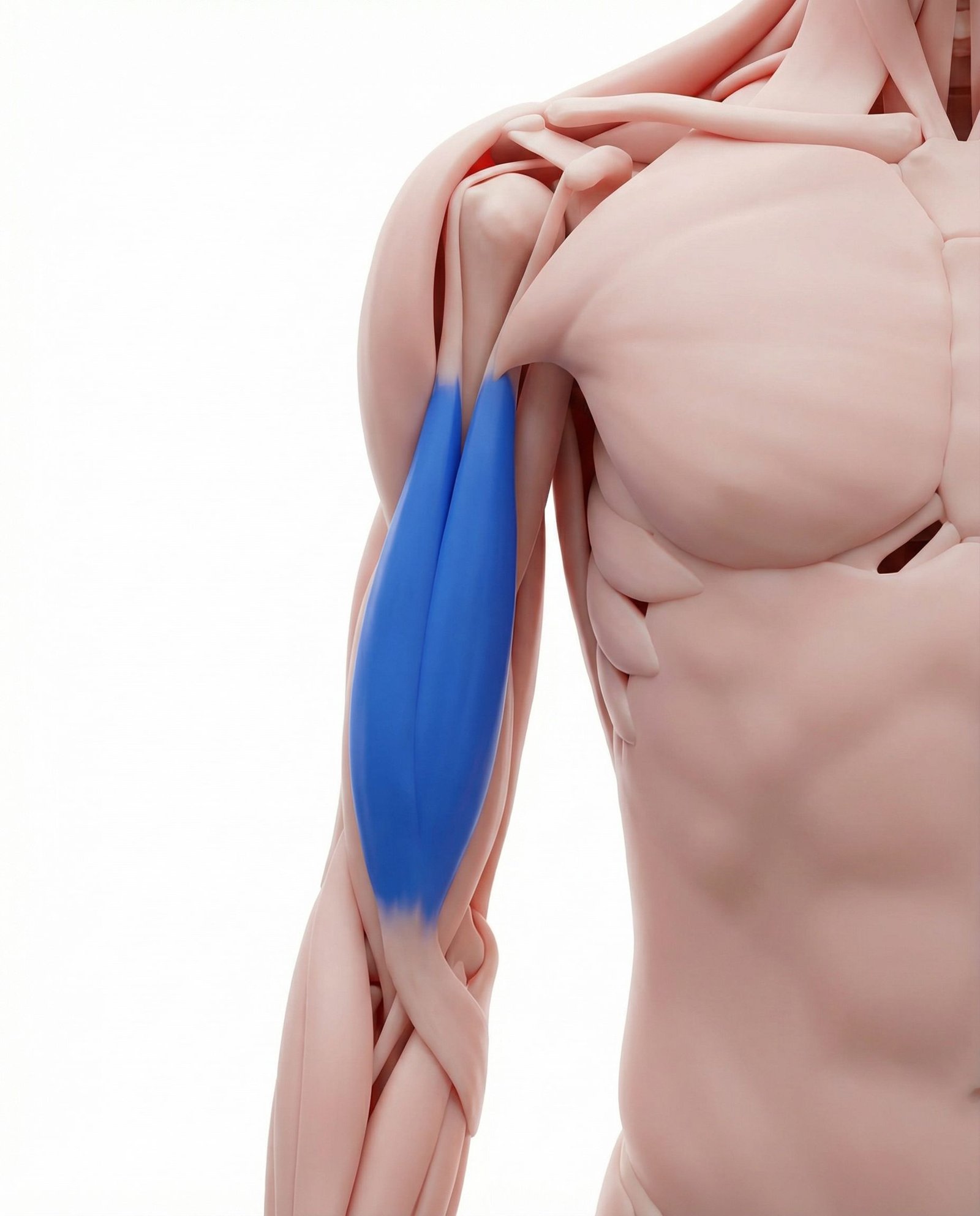
The long head of the biceps tendon is frequently involved in anterior shoulder pain presentations and often coexists with rotator cuff or subacromial symptoms. Trigger points in the muscle belly can add a secondary anterior-arm pain component.
Reactive Tendinopathy
Rotator cuff tendon tissue may become painful and reactive when load exceeds capacity.
Altered Kinematics
Poor scapular timing or humeral-head control can increase stress on the subacromial tissues.
Myofascial Referral
Trigger points in cuff and scapular muscles may refer pain into the same region as SAPS.
Capsular / Mobility Factors
Posterior shoulder tightness or thoracic stiffness may change how the shoulder moves under load.
Trigger Point Referral Patterns
Myofascial trigger points around the shoulder girdle can create pain that looks almost identical to primary subacromial pathology. That is why referral-pattern knowledge matters in shoulder diagnosis.
Infraspinatus
Supraspinatus
Upper Trapezius
Subscapularis
Clinical Assessment & Differentiation
Shoulder assessment is most useful when orthopedic tests, movement observation, palpation, and symptom-modification procedures are combined rather than treated as isolated proofs.
Painful Arc Test
Hawkins-Kennedy Impingement Test
Empty Can (Jobe) Test
Scapular Assistance Test
Evidence-Based Treatment Pathways
Current best practice usually starts with a multimodal conservative plan built around load management, progressive rehabilitation, and treatment of clearly relevant myofascial or mobility contributors.
Self-Care
- Modify repetitive overhead activity temporarily when it clearly provokes symptoms
- Use ice for reactive flare-ups if the patient finds it helpful
- Use pendulum exercises or gentle unloaded motion to maintain confidence in movement
- Apply self-release to upper trapezius, posterior shoulder, or pectoral tissues when these are clearly involved
- Reduce prolonged rounded-shoulder positions when posture is a major perpetuating factor
Manual Therapy
- Trigger point treatment for infraspinatus, supraspinatus, upper trapezius, and related tissues
- Pectoralis minor or anterior shoulder soft-tissue treatment when scapular posterior tilt is limited
- Selected glenohumeral or thoracic mobility techniques when indicated by the exam
- Posterior shoulder and thoracic mobility work
- Reassessment of pain and movement after treatment rather than relying on technique alone
Rehabilitation
- Serratus anterior activation and lower-trapezius strengthening
- Rotator cuff strengthening in symptom-tolerable ranges
- Progressive load exposure below or around the painful arc as tolerated
- Thoracic and scapular control work to improve upward rotation and posterior tilt
- Gradual return to overhead function rather than reflexive avoidance
Interventional
- Subacromial injection when short-term pain reduction is needed to support rehabilitation
- Trigger point injections when myofascial pain is clearly a major contributor
- Diagnostic injection when differentiating joint / bursal vs. referred pain is clinically important
- Shockwave therapy in selected calcific or chronic tendon-related cases
- Surgical consultation only when conservative care has been appropriate, well executed, and unsuccessful
Red Flags & Differential Diagnosis
Most subacromial-type shoulder pain is mechanical and non-emergent, but severe weakness, trauma, infection, neurologic change, and systemic symptoms should always be screened for carefully.
Subacromial pain syndrome is a broad clinical label rather than one single lesion, and it commonly includes tendon, bursal, scapular, and myofascial contributors.
Exercise-based rehabilitation is usually the cornerstone of treatment and often performs as well as more invasive strategies in many non-traumatic cases.
Trigger points in the infraspinatus, supraspinatus, and periscapular system can closely mimic structural subacromial pain patterns.
Scapular dyskinesis often matters, especially when tight pectoralis minor, overactive upper trapezius, or weak serratus anterior are part of the picture.
Diagnostic injections can be useful in selected cases, but they should support — not replace — careful clinical reasoning.
Cervical referral and neural causes remain part of the differential whenever the presentation is not behaving like a straightforward shoulder problem.
Movement therapy
Exercises for Subacromial Pain Syndrome
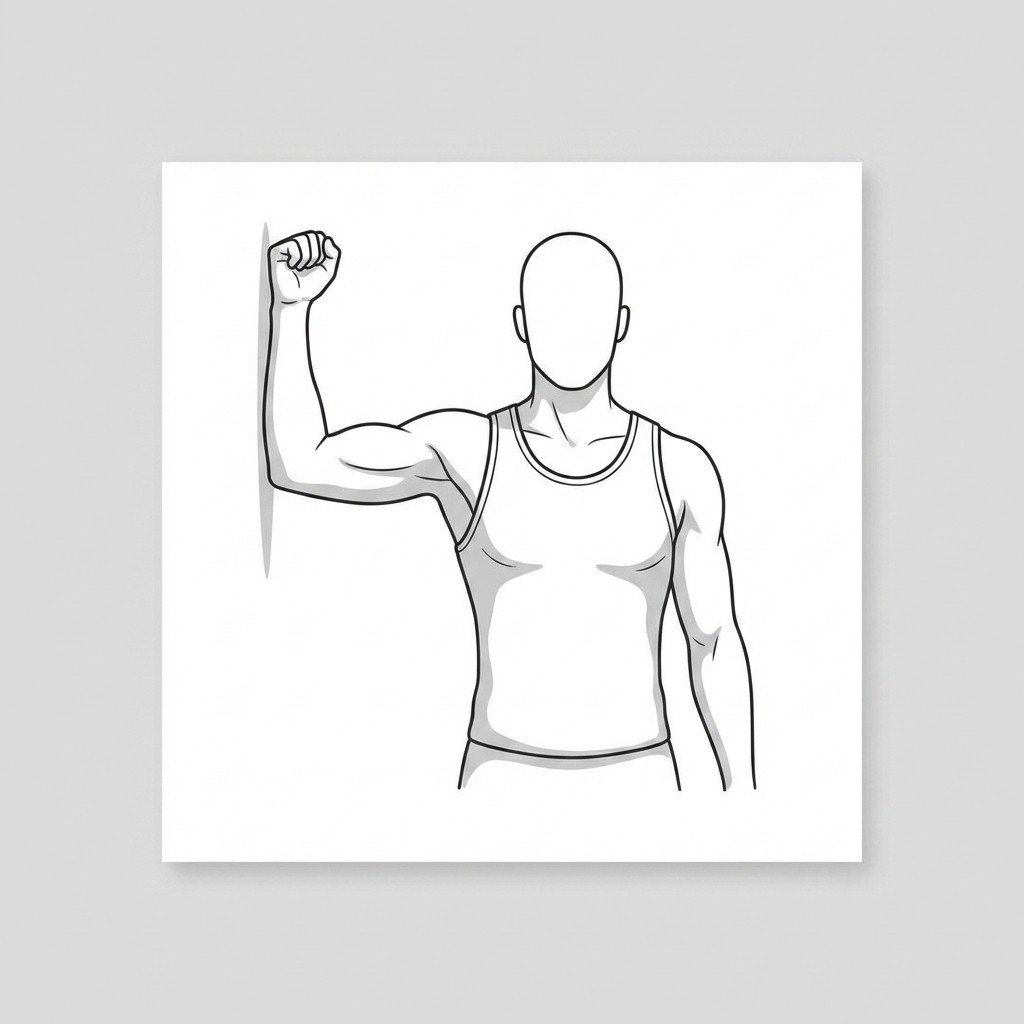
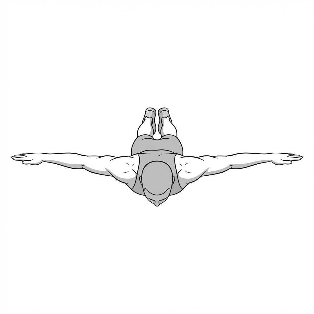
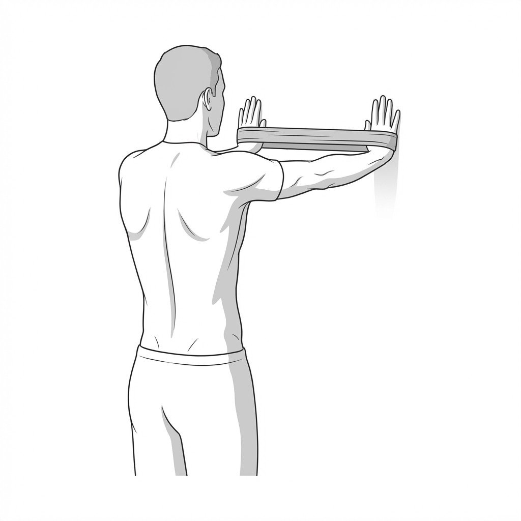
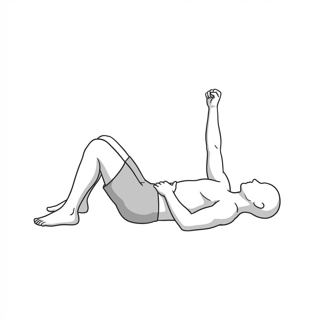
These exercises target the muscles and movement patterns most often involved in this condition. Start with 2-3 and progress based on symptom response.