Understanding the Clinical Picture
Cubital tunnel syndrome (CuTS) represents the second most common peripheral nerve entrapment in the upper extremity, characterized by compression or traction of the ulnar nerve at the medial elbow. Epidemiological data suggest a higher prevalence among manual laborers, overhead athletes, and individuals whose occupations require prolonged elbow flexion. The clinical presentation typically involves paresthesia in the ring and little fingers, medial elbow pain, and, in advanced cases, intrinsic hand muscle weakness. Differentiating CuTS from myofascial pain syndrome (MPS) is critical, as medial elbow pain is frequently multifactorial, and misdiagnosis can lead to delayed appropriate care or unnecessary surgical interventions.
The anatomical relationship between the ulnar nerve and surrounding musculature creates significant potential for overlap between CuTS and MPS. The ulnar nerve passes posterior to the medial epicondyle and enters the forearm between the two heads of the flexor carpi ulnaris (FCU), beneath Osborne's ligament. Myofascial trigger points in the medial head of the triceps or the FCU can refer pain directly to the medial elbow and ulnar forearm, closely mimicking the aching discomfort of early CuTS. Furthermore, increased tension or fascial restrictions in these muscles may mechanically load the cubital tunnel, exacerbating true nerve compression. This interplay often results in a mixed clinical picture where neuropathic and somatic pain generators coexist.
Medial elbow pain is frequently multifactorial; distinguishing true ulnar neuropathy from myofascial referral is essential to avoid unnecessary surgical intervention.
A rigorous clinical approach is required to differentiate isolated ulnar neuropathy from myofascial referral or to identify when both conditions are present. Current guidelines generally recommend a combination of provocative testing, such as the elbow flexion test and Tinel's sign, alongside a detailed neurological examination assessing two-point discrimination and intrinsic motor function. Electrodiagnostic studies (EMG/NCS) remain the gold standard for confirming axonal loss or demyelination, though they may be negative in early or dynamic compression. Conversely, identifying reproducible pain patterns through systematic palpation of the triceps, FCU, and flexor-pronator mass can isolate a myofascial component, guiding targeted conservative therapy before considering surgical decompression.
Anatomical Considerations
The medial elbow is a complex anatomical junction where the ulnar nerve is vulnerable to both dynamic traction and static compression from surrounding myofascial structures.
Triceps Brachii (Medial Head)
The medial head of the triceps brachii lies deep and medial, attaching to the olecranon process. Trigger points in this region frequently refer pain directly to the medial epicondyle, closely mimicking the somatic pain of medial epicondylalgia or early cubital tunnel syndrome. Because the ulnar nerve courses adjacent to the medial head before entering the cubital tunnel, increased muscle tension may mechanically irritate the nerve. Clinical evaluation often requires differentiating this local myofascial pain from true neuropathic pain.
Refers deep, aching pain to the medial epicondyle and occasionally down the ulnar aspect of the forearm.Flexor Carpi Ulnaris (FCU)
The FCU forms the distal roof of the cubital tunnel via Osborne's fascia, creating a critical anatomical chokepoint for the ulnar nerve. Hypertrophy, fascial thickening, or myofascial trigger points within the FCU can directly compress the nerve during elbow flexion. Referral patterns from the FCU typically project to the ulnar aspect of the wrist and hand, which can be easily confused with ulnar neuropathy. However, isolated myofascial referral generally lacks the true paresthesia or sensory deficits seen in nerve entrapment.
Refers pain along the ulnar volar forearm, extending into the ulnar side of the wrist and hand.Flexor Digitorum Superficialis (FDS)
The FDS originates from the medial epicondyle and coronoid process, contributing significantly to the common flexor tendon. While primarily associated with median nerve dynamics, its proximity to the medial elbow means trigger points can contribute to the overall medial elbow pain presentation. Pain is often described as a deep ache in the volar forearm extending to the digits. It is frequently involved in generalized flexor-pronator mass tendinopathy.
Refers pain down the anterior forearm, often concentrating in the middle and distal digits.Pronator Teres
Although the pronator teres is primarily implicated in median nerve entrapment (pronator syndrome), its origin at the medial epicondyle makes it a key structure in medial elbow pain syndromes. Trigger points here cause deep aching in the volar forearm and wrist. Careful assessment is necessary to rule out concurrent median nerve involvement or to identify a broader myofascial pain syndrome affecting the entire flexor compartment.
Refers pain deep into the anterior forearm and anterior wrist, occasionally mimicking carpal tunnel symptoms.Anconeus & Epitrochleoanconeus
The anconeus is a small muscle at the posterolateral elbow, but anatomical variants like the epitrochleoanconeus can cross the medial elbow and directly compress the ulnar nerve. Even the standard anconeus can develop trigger points that refer pain to the lateral and posterior elbow, complicating the regional pain picture. Assessment of elbow mechanics should include this often-overlooked stabilizer, particularly when dynamic snapping of the ulnar nerve is suspected.
Refers pain locally to the lateral and posterior aspects of the elbow joint.
Direct Nerve Compression
Mechanical narrowing of the cubital tunnel via Osborne's fascia, osteophytes, or prolonged elbow flexion leading to ulnar nerve ischemia.
Myofascial Referral
Trigger points in the medial triceps or FCU projecting somatic pain to the medial epicondyle and ulnar forearm without true nerve involvement.
Double-Crush Syndrome
Proximal compromise of the C8-T1 nerve roots or lower trunk of the brachial plexus rendering the ulnar nerve more susceptible to distal compression at the elbow.
Medial Epicondylalgia
Degenerative tendinopathy of the common flexor origin, often coexisting with or predisposing the area to secondary ulnar nerve irritation.
Referral Patterns & Mimics
Trigger points in the medial triceps and forearm flexors can project pain into the medial elbow and ulnar hand, creating a sensory map that closely mimics ulnar nerve entrapment.
- Triceps Brachii (Medial Head)
- Produces a strong, localized ache at the medial epicondyle. This pattern is frequently misdiagnosed as medial epicondylitis (golfer's elbow) or early ulnar nerve irritation.
- Flexor Carpi Ulnaris
- Generates a diffuse ache along the ulnar side of the forearm, wrist, and hand. It mimics the spatial distribution of ulnar neuropathy but typically does not cause numbness or tingling.
- Flexor Digitorum Superficialis
- Projects pain into the anterior forearm and fingers. Patients may report a sensation of hand fatigue or cramping that overlaps with the weakness seen in advanced nerve compression.
Clinical Assessment
Clinical evaluation relies on provocative maneuvers to stress the ulnar nerve, alongside careful palpation to identify concurrent myofascial pain generators.
Tinel's Sign at the Cubital Tunnel
With the elbow slightly flexed, gently tap the ulnar nerve in the groove between the medial epicondyle and the olecranon. Perform this 4-6 times.
Positive: Reproduction of tingling or "electric shocks" radiating into the ring and little fingers suggests ulnar nerve irritability.
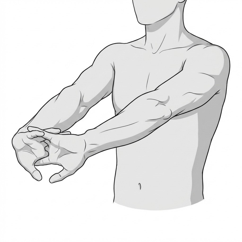
Elbow Flexion Test
Fully flex both elbows and extend the wrists, holding the position for up to 60 seconds. This maximally decreases the volume of the cubital tunnel while placing traction on the nerve.
Positive: Onset or exacerbation of numbness and tingling in the ulnar nerve distribution (ring and small fingers).
Froment's Sign
Attempt to hold a piece of paper tightly between the thumb and index finger while an examiner tries to pull it away. This tests the strength of the adductor pollicis (ulnar nerve innervated).
Positive: Compensatory flexion of the thumb interphalangeal joint (using the median nerve-innervated FPL) indicates ulnar nerve motor weakness.
Myofascial Palpation (Medial Triceps & FCU)
Apply sustained, moderate pressure to the muscle bellies of the medial triceps (just above the elbow) and the proximal FCU (just below the elbow).
Positive: Reproduction of the patient's familiar medial elbow or forearm ache without the induction of paresthesia or numbness, suggesting a myofascial component.
Treatment Pathways
Management typically begins with conservative measures focused on reducing nerve compression and addressing myofascial tension, progressing to interventional or surgical options only when conservative care fails or severe deficits are present.
Self-Care
- Nocturnal elbow splinting (keeping the elbow at 30-45 degrees of flexion)
- Activity modification to avoid prolonged or repetitive elbow flexion
- Use of elbow pads to prevent direct pressure on the medial epicondyle
- Ergonomic adjustments to computer workstations and phone usage
- Gentle self-massage of the triceps and forearm flexors
Manual Therapy
- Ulnar nerve gliding and tensioning exercises (prescribed carefully)
- Ischemic compression of trigger points in the medial triceps and FCU
- Myofascial release of Osborne's fascia and the flexor-pronator mass
- Joint mobilization of the elbow and proximal radioulnar joint
- Cervical and thoracic spine assessment to rule out double-crush phenomena
Interventional
- Ultrasound-guided corticosteroid injection (evidence is mixed; generally used cautiously)
- Nerve hydrodissection to mechanically free the ulnar nerve from adhesions
- Electrodiagnostic testing (EMG/NCS) to quantify nerve damage
- Surgical decompression (in situ release) for refractory cases
- Anterior transposition of the ulnar nerve (subcutaneous or submuscular) if instability is present
Rehabilitation
- Isometric strengthening of the flexor-pronator mass
- Eccentric loading protocols for concurrent medial epicondylalgia
- Postural re-education to reduce cervical and thoracic contributions
- Progressive intrinsic hand muscle strengthening
- Gradual return-to-play or return-to-work protocols
Red Flags — When to See a Doctor
While many cases of medial elbow pain are benign and self-limiting, certain neurological signs indicate severe nerve compromise that requires prompt specialist evaluation.
Intrinsic Muscle Wasting
Visible atrophy of the hypothenar eminence or the first dorsal interosseous muscle. This suggests severe, chronic axonal loss.
UrgentClawing of the Hand
A resting posture where the ring and small fingers are hyperextended at the MCP joints and flexed at the IP joints (ulnar claw). Indicates advanced motor neuropathy.
UrgentConstant Numbness
Paresthesia or anesthesia that does not resolve with changes in elbow position. Suggests persistent compression rather than dynamic irritation.
InvestigateSudden Onset Following Trauma
Acute medial elbow pain and ulnar nerve symptoms following a fall or direct blow. May indicate a fracture, dislocation, or acute nerve contusion.
UrgentSevere Night Pain
Unremitting pain that awakens the patient from sleep and is not relieved by splinting or positional changes. Warrants evaluation for other pathologies.
Investigate
Key Takeaways
Medial elbow pain requires careful differentiation between ulnar nerve entrapment, medial epicondylalgia, and myofascial trigger points.
Paresthesia in the ring and small fingers is a hallmark of ulnar neuropathy, whereas myofascial referral typically produces an aching sensation without true numbness.
The medial head of the triceps and the flexor carpi ulnaris are the primary muscular structures that can either mimic or mechanically contribute to cubital tunnel syndrome.
Conservative management, including nocturnal elbow extension splinting, is generally recommended as the first-line treatment for mild to moderate cases.
Electrodiagnostic testing (EMG/NCS) is valuable for confirming nerve compression severity, particularly when surgical intervention is being considered.
A "double-crush" syndrome should be considered if symptoms overlap with cervical radiculopathy or thoracic outlet syndrome.
Movement therapy
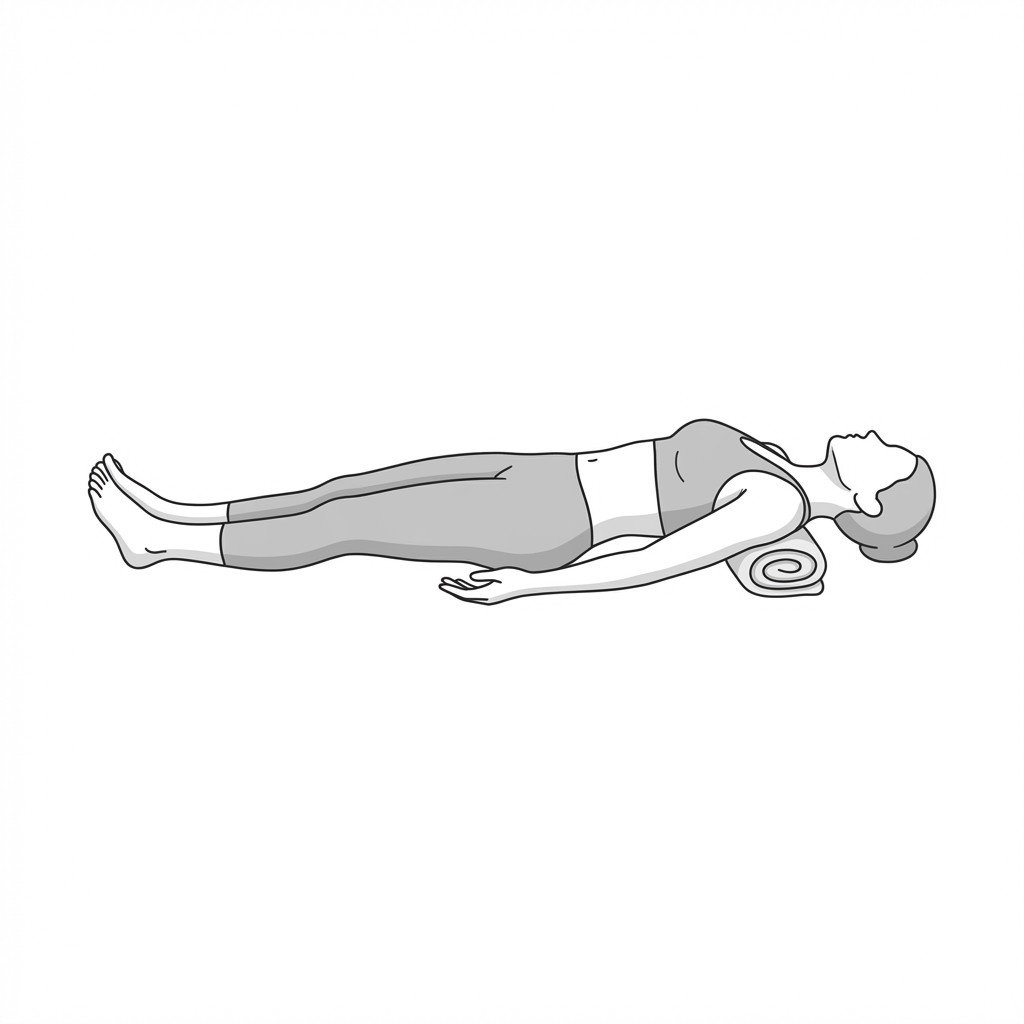
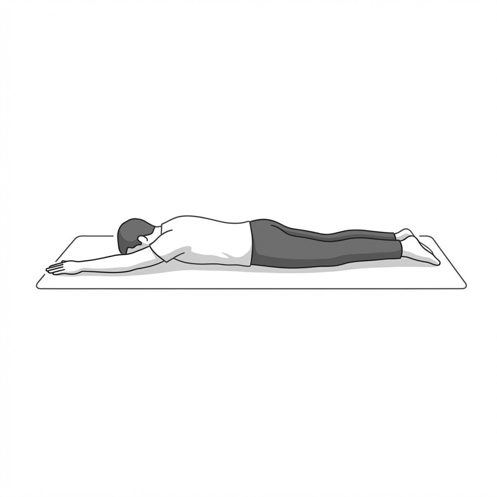
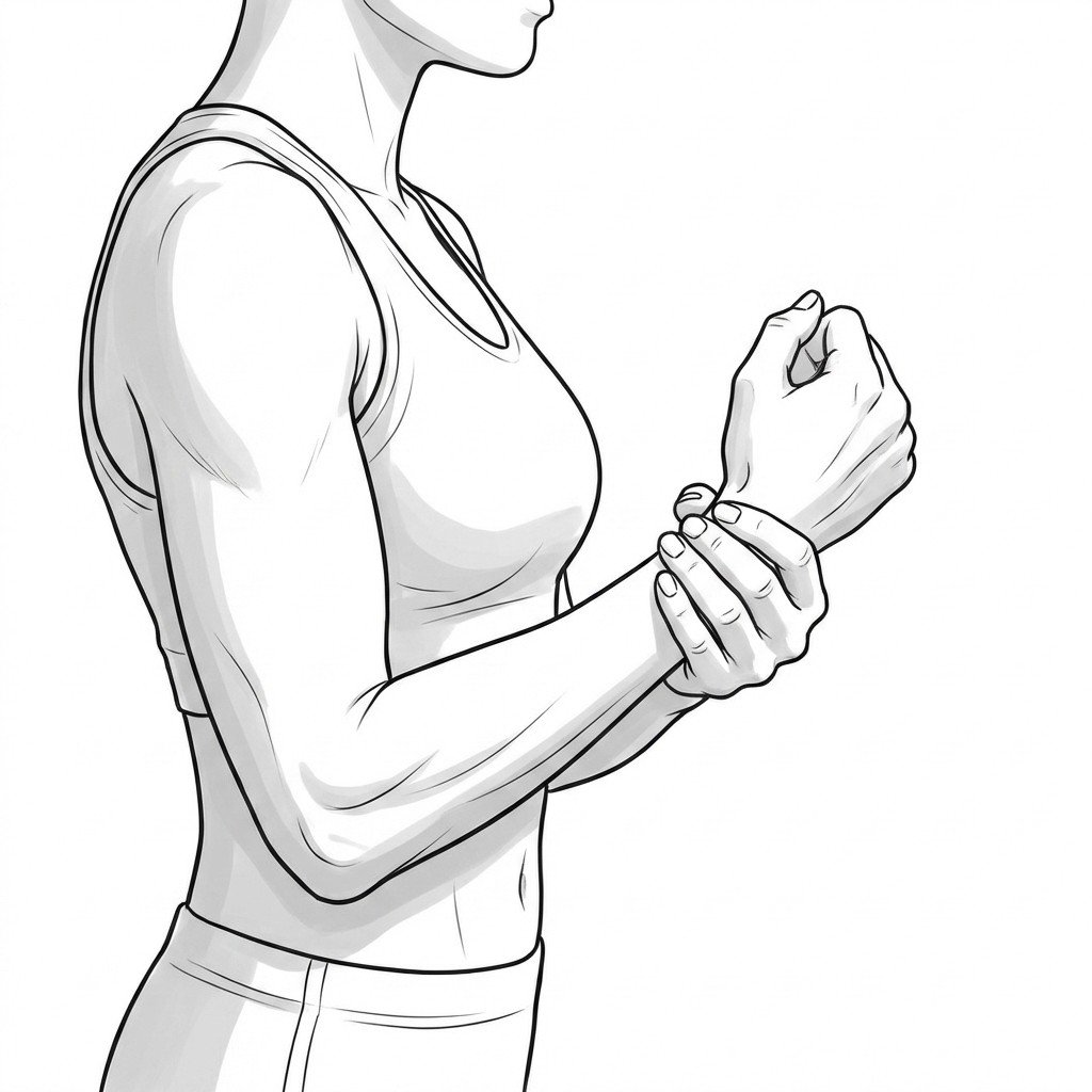
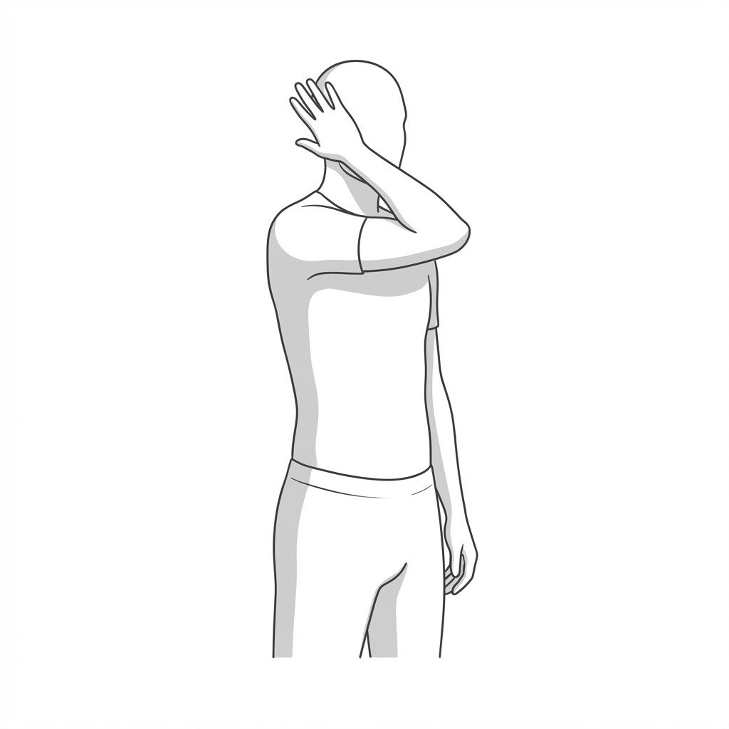
Exercises for Cubital Tunnel Syndrome
These exercises mobilise the ulnar nerve along its full course, reduce compressive tension at the cubital tunnel through elbow extension and forearm pronation, and stabilise the shoulder girdle to decrease neural tension from above. Start with 2–3 and progress as tolerated.