Understanding the Clinical Picture
De Quervain's tenosynovitis is classically described as a stenosing tenosynovitis of the first dorsal compartment of the wrist, affecting the abductor pollicis longus (APL) and extensor pollicis brevis (EPB) tendons. The condition is characterized by myxoid degeneration and thickening of the extensor retinaculum rather than acute inflammation, making "tendinosis" or "tendinopathy" more accurate histopathological descriptors. Epidemiological data indicate a higher prevalence among individuals engaging in repetitive thumb abduction and ulnar deviation, with a notably high incidence in postpartum populations due to the biomechanics of lifting infants. Accurate diagnosis is essential, as the radial wrist is a complex anatomical crossroads susceptible to various overlapping pathologies.
The clinical presentation of De Quervain's is frequently complicated by, or entirely mimicked by, myofascial pain syndrome (MPS). The brachioradialis muscle, in particular, harbors trigger points that refer pain directly to the radial styloid and the dorsal web space of the thumb. Furthermore, mechanical overloading of the forearm musculature—often seen in repetitive strain scenarios—can lead to the simultaneous development of true compartment stenosis and proximal myofascial trigger points. Scalene trigger points can also refer pain distally along the C6 dermatomal distribution, creating a complex pain pattern that may be misdiagnosed as isolated distal tendinopathy or peripheral nerve entrapment.
Differentiating true stenosing tenosynovitis from brachioradialis referred pain is critical, as the former often responds to localized corticosteroid injection, while the latter requires proximal myofascial intervention.
Differentiating true stenosing tenosynovitis from myofascial referred pain requires a meticulous physical examination. While traditional provocative maneuvers like Finkelstein's and Eichhoff's tests place tension on the APL and EPB, they also stretch the overlying superficial radial nerve and can provoke pain in a sensitized brachioradialis. Current clinical guidelines generally recommend combining these distal provocative tests with proximal palpation of the forearm and cervical musculature. High-resolution diagnostic ultrasound has emerged as a critical tool in this differential diagnosis, allowing clinicians to visualize retinacular thickening, peritendinous fluid, and the presence of an EPB sub-compartment, which is a known risk factor for conservative treatment failure.
Anatomical & Myofascial Context
The radial aspect of the wrist is a densely packed anatomical region where tendons, nerves, and myofascial referral zones intersect. Understanding the relationship between the first dorsal compartment and proximal musculature is vital for accurate diagnosis.
Brachioradialis
A superficial forearm muscle that flexes the elbow, especially in a semi-pronated position. Trigger points in the brachioradialis frequently refer pain directly to the radial styloid and the dorsal web space of the thumb. This referral pattern is a primary myofascial mimic of De Quervain's tenosynovitis. Clinical differentiation requires palpation of the proximal muscle belly rather than the distal tendon insertions.
Strong referral to the radial styloid, base of the thumb, and the first dorsal web space.Abductor Pollicis Longus (APL)
One of the two tendons passing through the first dorsal compartment of the wrist. The APL frequently features multiple tendon slips, which anatomical studies suggest may contribute to compartment crowding and stenosing tenosynovitis. Myofascial trigger points in the APL belly can cause localized pain that exacerbates tension on the distal tendon, complicating the clinical picture.
Pain localized to the radial aspect of the wrist, occasionally extending distally to the base of the first metacarpal.Extensor Pollicis Brevis (EPB)
The second tendon of the first dorsal compartment. The EPB is often housed in its own distinct sub-compartment within the first dorsal compartment, a variation noted in surgical and ultrasound studies that may explain cases refractory to single-compartment corticosteroid injections. Tension from the EPB muscle belly directly loads this potentially stenosed sheath.
Pain primarily at the radial styloid, worsening with thumb extension and abduction.Scalene Group
The anterior, middle, and posterior scalenes are lateral neck muscles involved in respiration and cervical movement. Trigger points in the anterior and middle scalenes can refer pain down the radial aspect of the arm, forearm, and into the thumb and index finger. This C6-like distribution can mimic or coexist with distal peripheral nerve entrapments or tenosynovitis.
Broad referral pattern extending down the anterolateral arm, radial forearm, and into the thumb and index finger.Pronator Teres
A forearm pronator and weak elbow flexor. While its primary referral is to the volar forearm and wrist, altered biomechanics secondary to pronator teres tightness can change the functional axis of the wrist. This compensatory shift often places increased mechanical demand on the APL and EPB during gripping and lifting tasks.
Deep aching pain in the volar forearm, occasionally extending to the palmar aspect of the radial wrist.
Stenosing Tenosynovitis
Thickening of the extensor retinaculum causing mechanical friction and impaired glide of the APL and EPB tendons.
Myofascial Referral
Trigger points in the brachioradialis or scalenes projecting nociceptive signals to the radial styloid and thumb.
Intersection Syndrome
Friction at the crossing point of the first dorsal compartment (APL/EPB) and second dorsal compartment (ECRL/ECRB) tendons, typically 4-6 cm proximal to the wrist.
Nerve Entrapment
Irritation of the superficial branch of the radial nerve (Wartenberg's syndrome), often exacerbated by tight wrist wear or handcuffs.
Referral Patterns & Mimics
Pain at the radial styloid is not pathognomonic for De Quervain's tenosynovitis. Several proximal muscles can project pain into this exact region, confounding the clinical picture.
- Brachioradialis
- Referred pain concentrates intensely over the radial styloid and the base of the thumb, often indistinguishable from the localized pain of first dorsal compartment tenosynovitis based on patient history alone.
- Scalene Group
- Pain and aching radiate from the lateral neck down the radial aspect of the upper extremity, terminating in the thumb and index finger, frequently mimicking a C6 radiculopathy or contributing to double-crush phenomena.
- Extensor Pollicis Longus (EPL)
- Pain refers to the dorsum of the thumb and occasionally the radial aspect of the wrist. The EPL passes around Lister's tubercle (third dorsal compartment), making its pain pattern slightly more dorsal than the APL/EPB.
Clinical Assessment Techniques
Clinical assessment must differentiate between local tendon pathology, nerve irritation, and myofascial referred pain. The following tests help isolate the involved structures.
Finkelstein's Test
The examiner grasps the patient's thumb and passively deviates the hand in an ulnar direction. This places isolated stretch on the APL and EPB tendons. It is generally considered more specific than Eichhoff's test.
Positive: Sharp pain over the radial styloid (first dorsal compartment), suggesting tenosynovitis. Note that severe brachioradialis trigger points may also be provoked by this stretch.
Eichhoff's Test
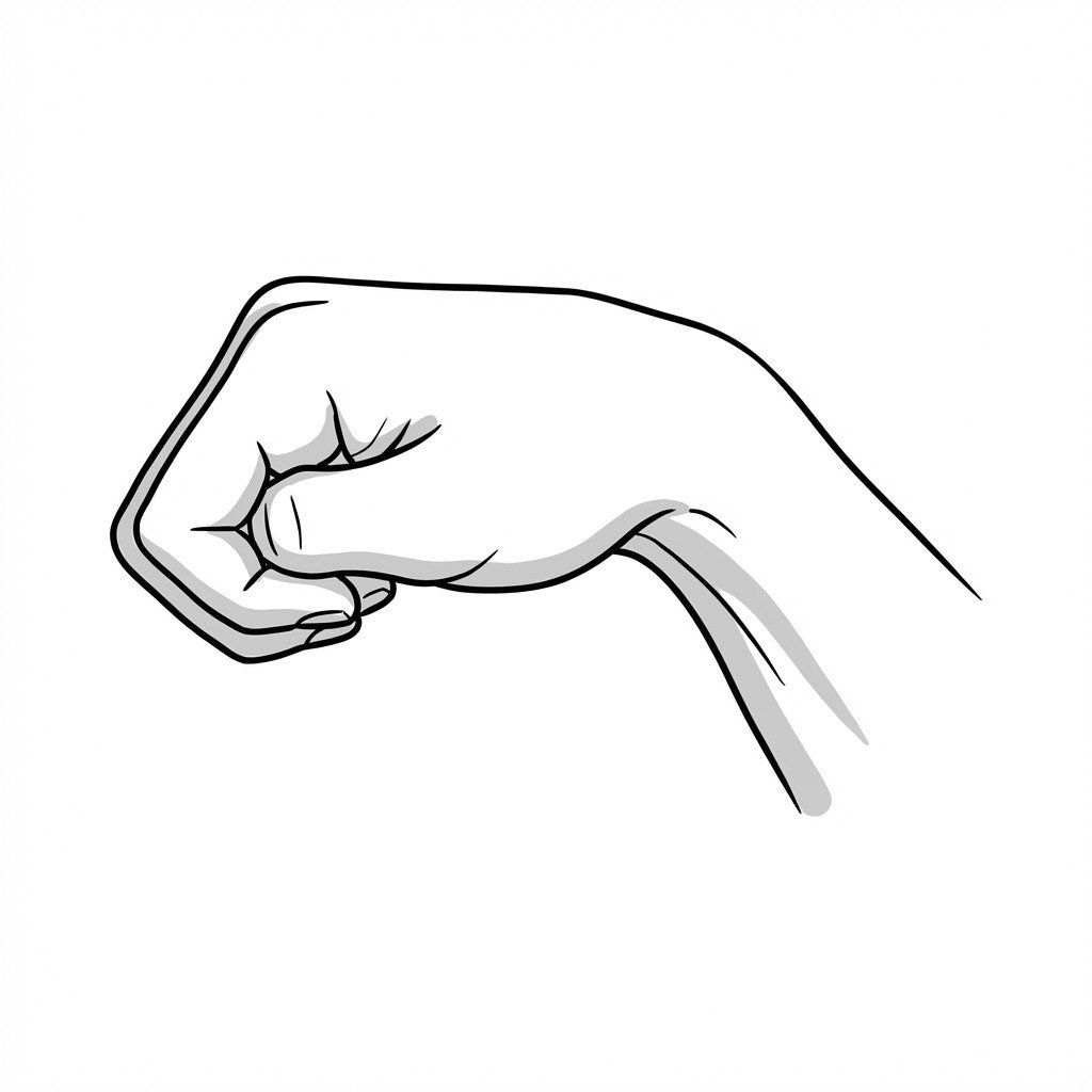
The patient actively places their thumb within their palm, closes their fingers over the thumb to make a fist, and actively ulnar deviates the wrist. This test is highly provocative.
Positive: Pain at the radial styloid. Literature suggests this test produces a higher rate of false positives compared to Finkelstein's test due to the broad tension placed on multiple structures.
WHAT (Wrist Hyperflexion and Abduction of the Thumb) Test
The patient hyperflexes the wrist and actively abducts the thumb against the examiner's resistance. This actively loads the APL and EPB rather than passively stretching them.
Positive: Pain at the first dorsal compartment. Some studies suggest the WHAT test may have higher sensitivity and specificity for De Quervain's than traditional passive stretch tests.
Brachioradialis Pincer Palpation
The examiner uses a pincer grasp to palpate the proximal third of the brachioradialis muscle belly, searching for taut bands and applying sustained pressure to suspected trigger points.
Positive: Reproduction of the patient's familiar radial wrist and thumb pain, indicating a myofascial referral component rather than, or in addition to, local tendon pathology.
Evidence-Based Management
Management strategies should be tailored to the specific etiology identified during assessment. Current evidence supports a multimodal approach, particularly when myofascial and tendinopathic conditions coexist.
Self-Care
- Thumb spica splinting (typically leaving the interphalangeal joint free)
- Activity modification to avoid repetitive ulnar deviation with thumb flexion
- Ergonomic assessment of lifting mechanics (e.g., avoiding "L-shaped" lifting of infants)
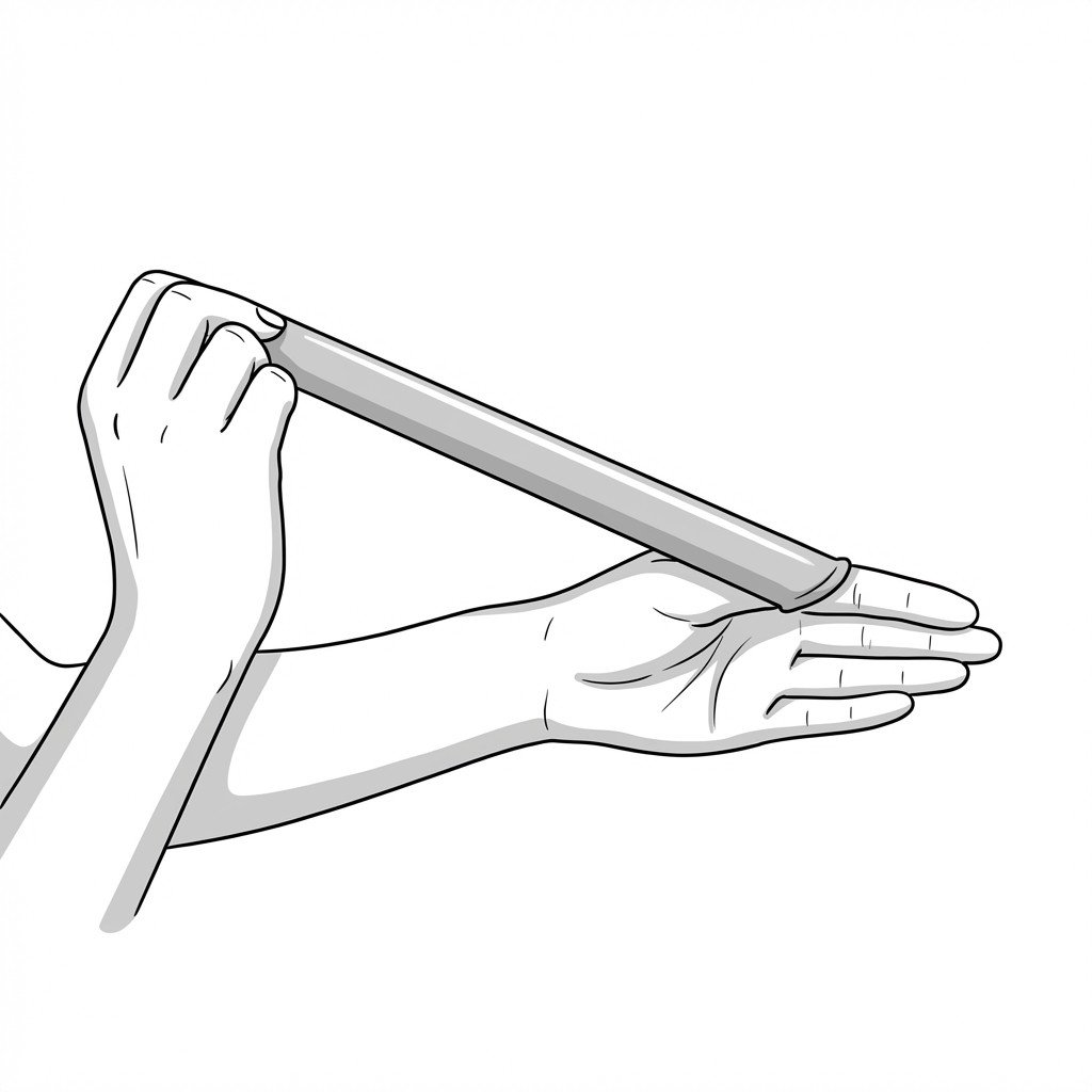
- Gentle, pain-free range of motion exercises to maintain glide
- Application of ice to the radial styloid during acute exacerbations
Manual Therapy
- Ischemic compression of brachioradialis and scalene trigger points
- Soft tissue mobilization of the proximal APL and EPB muscle bellies
- Cross-friction massage proximal to the extensor retinaculum (avoiding the inflamed sheath)
- Joint mobilization of the radiocarpal and trapeziometacarpal joints
- Nerve gliding exercises for the superficial radial nerve
Interventional
- Ultrasound-guided corticosteroid injection into the first dorsal compartment
- Verification of EPB sub-compartmentalization during ultrasound assessment
- Trigger point injections into the brachioradialis for refractory referred pain
- Diagnostic local anesthetic block to differentiate local vs. referred pain
- Surgical release of the first dorsal compartment (for cases failing conservative management)
Rehabilitation
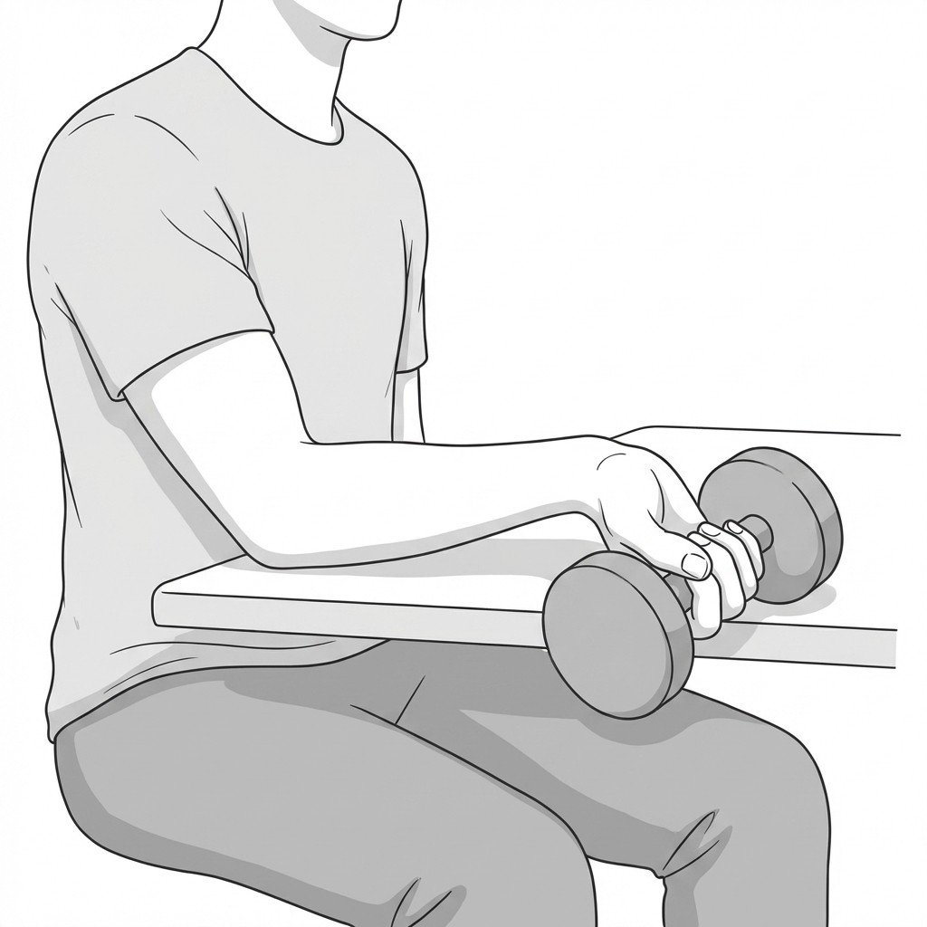
- Progressive eccentric loading of the thumb extensors and abductors
- Isometric strengthening of the wrist and hand in neutral positions
- Forearm supination and pronation strengthening
- Scapular and proximal shoulder stabilization exercises
- Gradual return-to-work or return-to-sport activity simulation
Red Flags & Differential Diagnosis
While radial wrist pain is typically benign and mechanically driven, certain signs and symptoms warrant immediate medical imaging or specialist referral to rule out severe pathology.
Traumatic Tendon Rupture
Sudden loss of ability to actively extend the thumb, often following a distal radius fracture or rheumatoid arthritis flare. Requires urgent orthopedic evaluation.
UrgentSeptic Tenosynovitis
Erythema, significant swelling, severe pain with minimal movement, and systemic signs such as fever. This is a medical emergency requiring immediate surgical consultation and antibiotics.
EmergencyScaphoid Fracture
Tenderness in the anatomical snuffbox following a fall on an outstretched hand (FOOSH). High risk of avascular necrosis if missed; requires radiographic imaging.
UrgentCervical Radiculopathy (C6)
Radial wrist pain accompanied by progressive motor weakness in wrist extension or elbow flexion, diminished brachioradialis reflex, and dermatomal sensory changes.
InvestigateBasal Joint Osteoarthritis
Deep, aching pain at the base of the thumb, positive grind test, and radiographic evidence of trapeziometacarpal joint degeneration. Often coexists with De Quervain's.
Investigate
Key Clinical Takeaways
De Quervain's tenosynovitis involves stenosing of the APL and EPB tendons within the first dorsal compartment, but its symptoms are frequently mimicked by myofascial trigger points.
The brachioradialis muscle is a primary myofascial mimic, capable of referring intense pain directly to the radial styloid and thumb base.
Clinical literature suggests Eichhoff's test produces more false positives than Finkelstein's test; both should be interpreted with caution if proximal trigger points are present.
Ultrasound imaging is highly valuable for confirming tenosynovial thickening, identifying EPB sub-compartments, and guiding corticosteroid injections.
Corticosteroid injections show meaningful short-term benefit for true De Quervain's in pooled analyses, but failure to respond may indicate an unrecognized EPB sub-compartment or a primary myofascial etiology.
Treatment should address both the local tendon pathology and proximal biomechanical contributors, including forearm and cervical myofascial restrictions.
Movement therapy
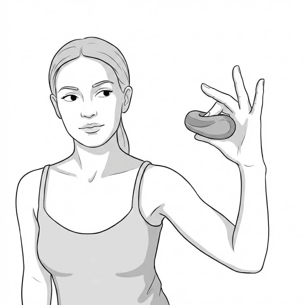
Exercises for De Quervains Tenosynovitis
De Quervain's rehabilitation moves from tissue unloading to controlled eccentric loading of the APL and EPB tendons — restoring thumb mechanics without aggravating the narrowed first dorsal compartment. Start with 2–3 and progress as tolerated.