Understanding the Erector Spinae
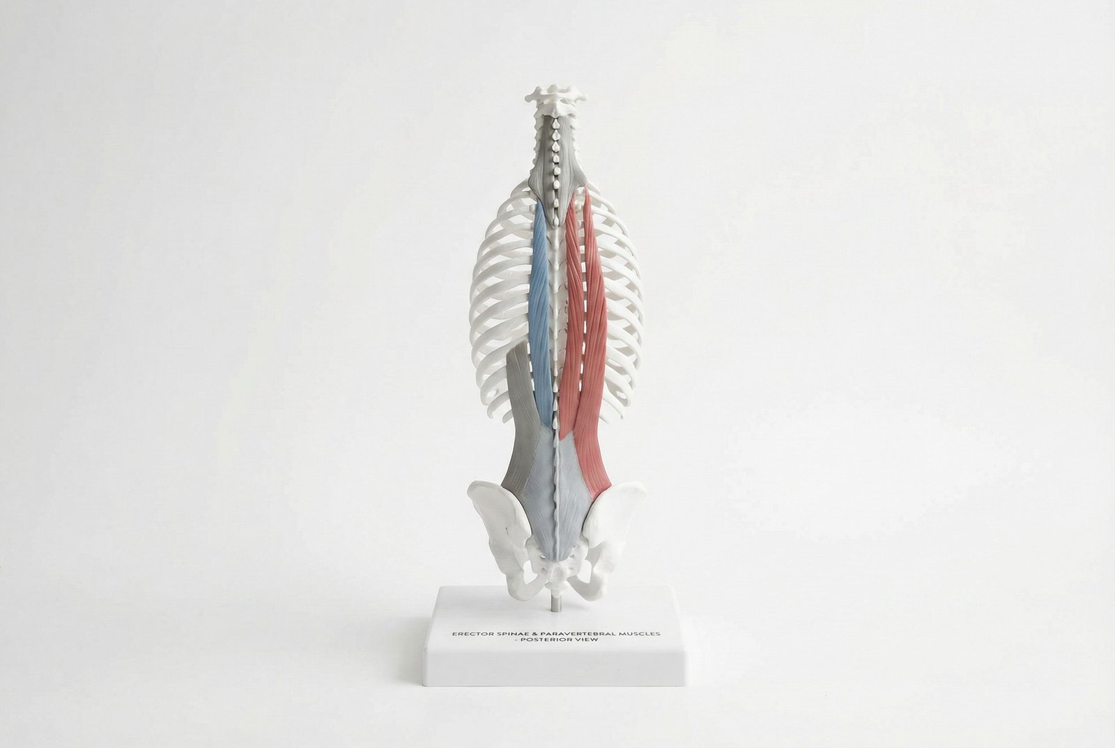
Erector Spinae and Paravertebral Muscles Anatomy Posterior View
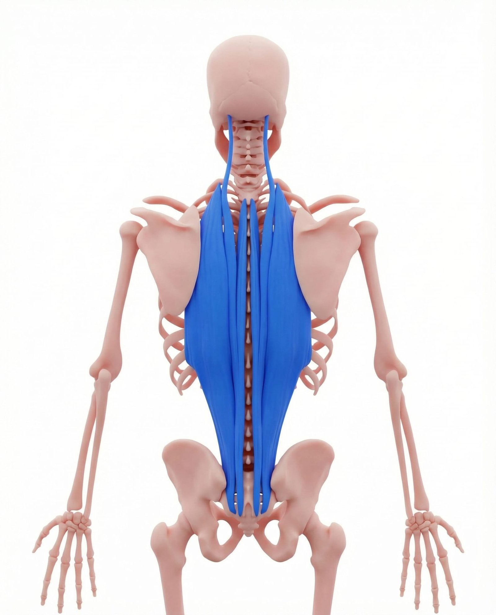
The erector spinae is not one muscle but a group of three parallel columns — the iliocostalis (lateral), longissimus (intermediate), and spinalis (medial) — running the length of the spine from the sacrum and iliac crest toward the cervical region and skull. They share broad lower attachments through the sacrum, posterior iliac crest, and thoracolumbar fascia, then branch upward to the ribs and vertebral structures at multiple levels.
Their primary function is spinal extension and postural support against gravity, but the erectors also contribute to controlled forward bending, trunk rotation, and load transfer during lifting. Because they are active during so much upright activity, they are especially sensitive to sustained postural demand and repeated eccentric loading.
Trigger points may develop in the erectors through several routes: acute overload injury, sustained postural loading such as desk work or prolonged standing, and secondary activation related to other pain generators including visceral or pelvic pain. This bidirectional relationship helps explain why treating only the muscles without considering broader drivers sometimes leads to incomplete results.
The deep multifidus, lying in the groove between the spinous and transverse processes, plays a more segmental stabilizing role. Rather than generating gross motion, it helps prepare the spine for movement and contributes to intersegmental stiffness. After acute pain, multifidus function may be reduced early, which may leave the spine feeling less stable and more vulnerable to recurrence if control is not rebuilt.
Anatomy & Trigger Points
Erector spinae trigger points can develop at virtually any spinal level, but they commonly cluster in the lower lumbar region and the mid-thoracic region. Lumbar clusters more often contribute to buttock and posterior-thigh referral, while thoracic clusters more often produce rib-wrapping, chest wall, or parascapular discomfort. Multifidus trigger points are palpated more medially, in the groove immediately lateral to the spinous processes.
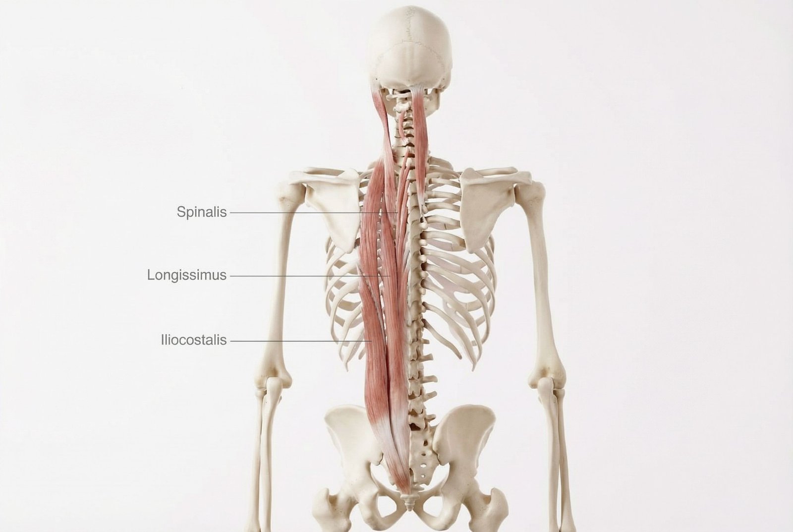
Erector Spinae Trigger Point Locations and Referral Zones
Lateral trunk and flank pain, buttock pain, and in the thoracic region a wrapping rib pain that may be mistaken for pleuritic or costochondral pain. Iliocostalis trigger points at the lumbar level may refer to the iliac crest and upper buttock; thoracic-level trigger points may refer anteriorly around the ribcage and occasionally toward the abdomen, creating a pain pattern that can resemble visceral or chest wall pain.
Deep, local paravertebral aching directly lateral to the spinous processes — the classic "pain in the spine itself" description many patients use. Longissimus trigger points at different spinal levels often generate focal segmental pain and may also refer caudally. Lumbar longissimus trigger points commonly contribute to buttock and upper posterior thigh aching.
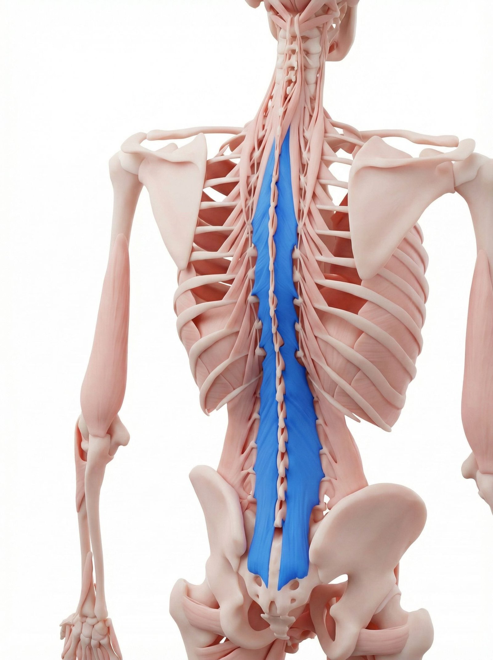
Deeply local spinal pain and a sense of segmental instability or poor support. Multifidus trigger points generate pain immediately lateral to the spinous processes and may contribute to the "my back does not feel supported" sensation many chronic back pain patients describe. Multifidus inhibition and atrophy can occur early after an acute low back pain episode, and recovery may be incomplete without specific rehabilitation.
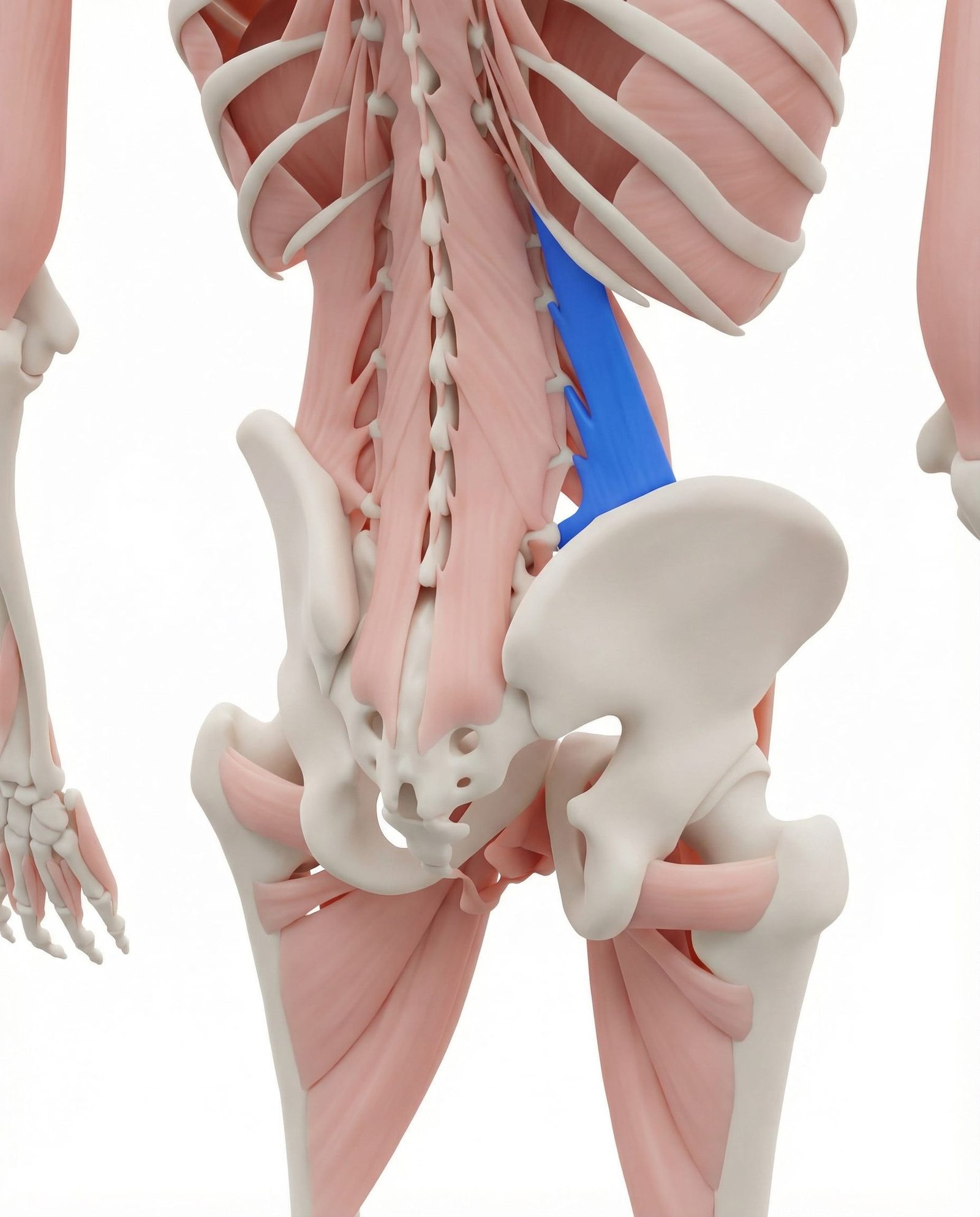
Iliac crest, lateral low back, and greater trochanteric pain — the QL is one of the most important co-activating muscles in lumbar pain patterns. QL and erector trigger points commonly coexist and may perpetuate each other. Treating one while ignoring the other is a frequent reason lumbar pain improves only partially.
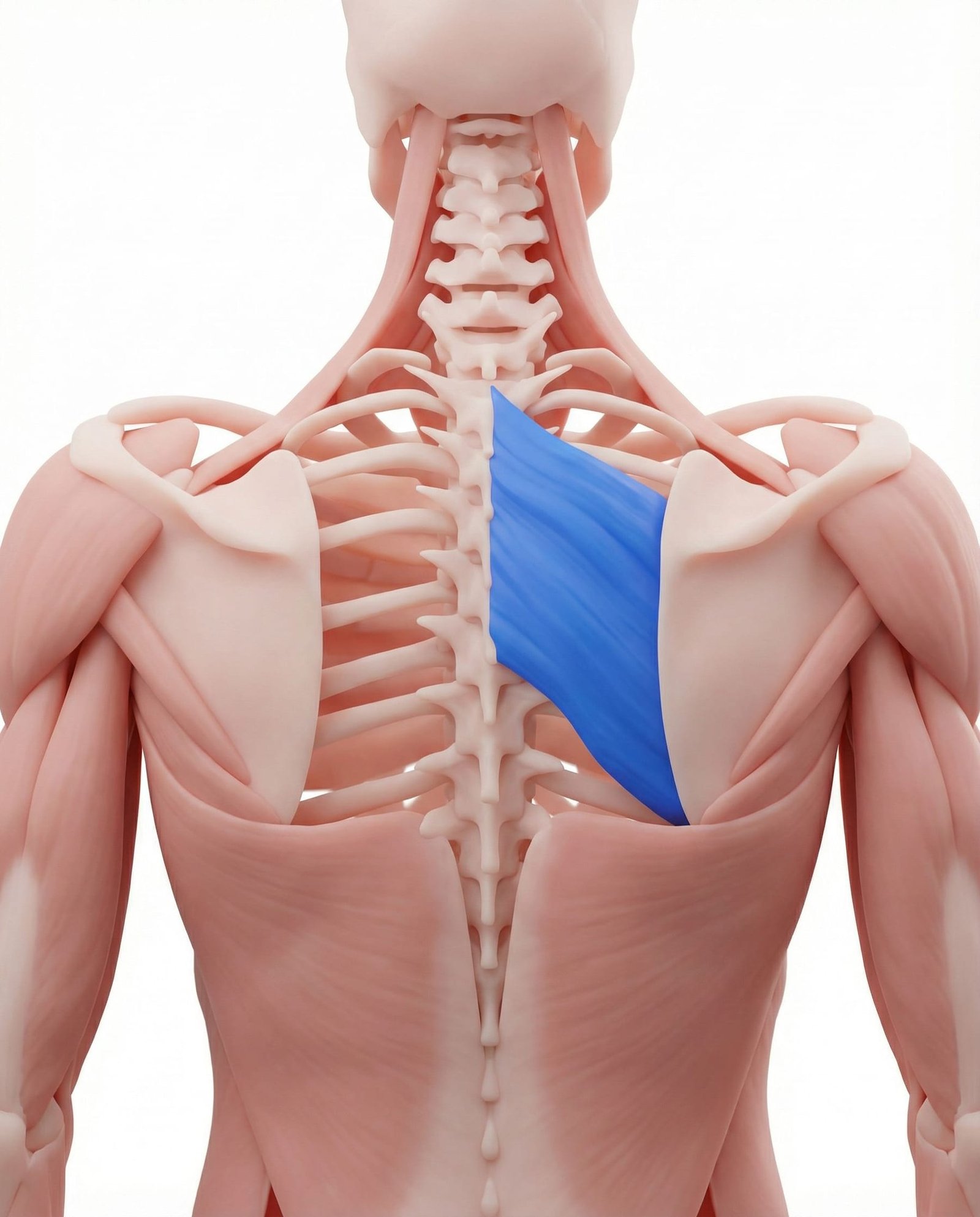
Deep inter-scapular aching medial to the scapula, often described as burning or a knife-like discomfort between the shoulder blades. Rhomboid trigger points may develop secondarily to chronic middle trapezius and thoracic erector dysfunction, contributing to the familiar "mid-back burning" pattern that builds through the workday.
Referral Patterns: The Back Pain Signature
Lumbar erector spinae trigger points often produce a diffuse deep aching that patients localize "into the spine" — a useful distinction from the more clearly lateral QL referral pattern. Pain is commonly worsened by prolonged sitting, prolonged standing, and forward bending, and may improve with unloading or lying down. These trigger points can also refer caudally to the buttock and upper posterior thigh, producing a "sciatica-like" ache that usually does not extend below the knee.
Thoracic erector trigger points generate a different pattern: unilateral or bilateral thoracic aching that may wrap around the ribcage. Patients may describe "band-like" chest or rib tightness that worsens with deep inspiration, rotation, or sustained forward bending. This can resemble pleuritic, rib, or chest wall pain closely enough that thoracic paraspinals are often overlooked unless they are examined directly.
The back is not a static structure that occasionally fails — it is a dynamic system that becomes more resilient when trained and more vulnerable when repeatedly overloaded without recovery.
Buttock
Rib Pain
Instability
Self-Assessment Tests
These tests assess erector spinae and multifidus trigger point activity, lumbar mobility, and the differential between muscular, discogenic, and neural pain sources. A particularly useful pattern to track is whether exercises centralize your pain — move it from the leg toward the back — because that often has prognostic value.
Forward Flexion Screen
Standing Lumbar Flexion Range Assessment
Paraspinal Furrow Palpation
Bilateral Paravertebral Trigger Point Assessment
Sphinx Position Test
Prone Extension Tolerance & Erector Length
Seated Slump Test
Neural Tension Differentiation from Muscular Pain
Treatment Pathways
Effective erector spinae treatment often combines three complementary approaches: mechanical decompression or mobility restoration, direct trigger point treatment, and rebuilding of deep trunk stability. Addressing only one component may leave important vulnerabilities in place.
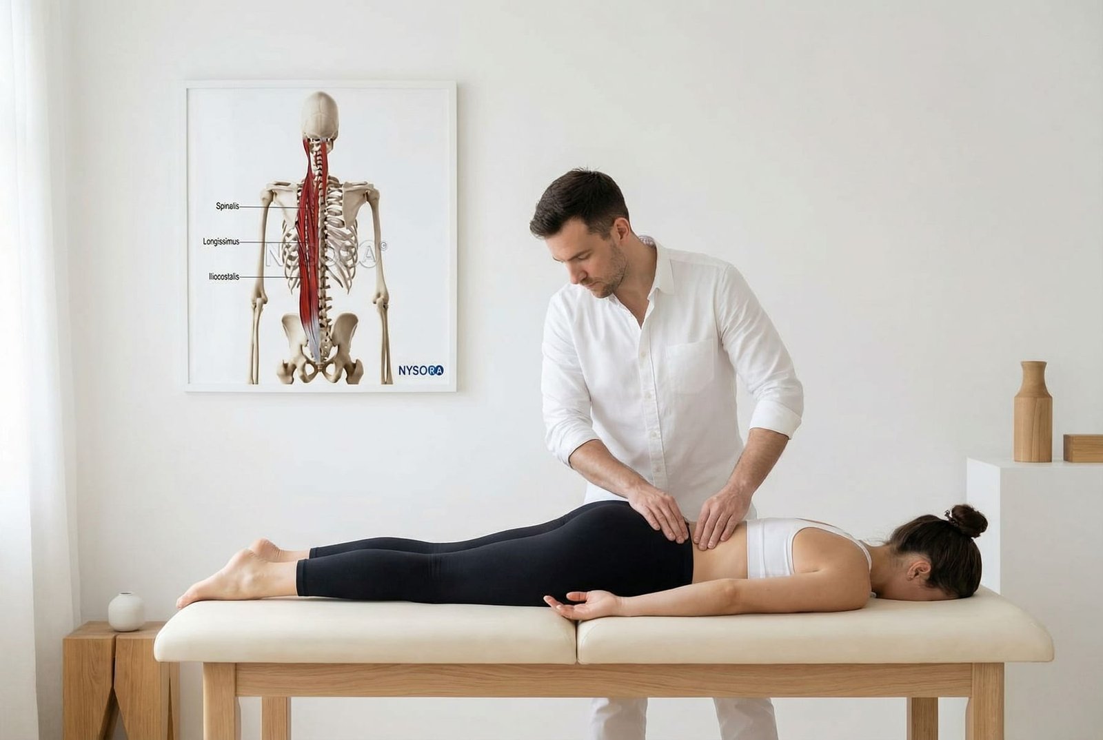
McKenzie Extension Protocol and Paraspinal Self-Release Techniques
Extension-Based Stretching (McKenzie)
Prone extension exercises from the McKenzie family are a commonly used first-line strategy in many lumbar back pain presentations, especially when symptoms centralize with extension. In paraspinal trigger point patterns they may help restore extension tolerance and counter the protective flexion posture seen in acute back pain.
- Prone press-up (cobra) — arms under shoulders, press chest up while hips remain on the floor
- Sphinx position — passive lumbar extension held briefly and repeated as tolerated
- Standing extension — hands on lower back, gentle repeated backward bending
- Progression can move from small repeated motions toward longer holds as irritability decreases
- Centralization monitoring can be useful: shifting pain from leg toward back is generally a favorable sign
Paraspinal Self-Release
Direct mechanical decompression of paraspinal trigger points using foam rolling, double-ball tools, or percussive therapy. The paravertebral muscles are relatively accessible to self-applied sustained pressure, making self-release a practical adjunct for many patients when done carefully.
- Foam roller thoracic extension — roll from T1-T12, pausing at tighter segments when tolerated
- Double lacrosse ball "peanut" along the paraspinal groove — targets erector trigger points segmentally
- Percussive therapy (massage gun) — lower or moderate settings along the paraspinal gutter rather than directly on the spine
- Supine knees-to-chest flexion stretch — sustained hold for lumbar erector unloading when flexion is tolerated
- Seated twist — gentle rotation that may compress paraspinals against deeper support structures
Dry Needling: Segmental Approach
Paravertebral dry needling may target erector spinae and multifidus at specific symptomatic levels. A segmental approach can be useful in multi-level trigger point patterns, though anatomy and depth control matter greatly — especially in the thoracic region.
- Lumbar paravertebral needling — often several needles per side at clinically identified levels
- Thoracic paravertebral needling — requires careful depth control near pleural structures
- Multifidus needling — a more medial and deeper approach targeting spinal stabilizers
- Electroacupuncture across paravertebral needles may be used by appropriately trained clinicians
- Chronic multi-level presentations may require a short treatment series rather than a single session
Core Stabilization: McGill Protocol
The McGill "Big 3" stabilization exercises are widely used to rebuild spinal control and tolerance. In patients with recurrent erector spinae pain, they may help reduce the mechanical demand placed on the paraspinals by improving deep trunk endurance and movement control.
- McGill curl-up — partial trunk lift with lumbar spine kept relatively neutral
- Side plank — elbow under shoulder, body in straight line, modified to knees if needed
- Bird-dog — quadruped alternating arm/leg extension with brief holds
- Progression toward dynamic or longer-duration versions as pain settles and control improves
- Programs often require several weeks of consistent practice rather than expecting immediate structural change
Lumbar Erectors (L3-L5)
Deep spinal aching with caudal referral to buttock and upper posterior thigh — may mimic sciatica but typically stays above the knee.
Thoracic Erectors (T4-T8)
Band-like rib pain wrapping around the chest wall — may be mistaken for cardiac, pleuritic, or costochondral pain.
Multifidus (Segmental)
Focal deep aching at the affected vertebral level with a sense of poor support or recurrent giving-way during bending.
Upper Lumbar (L1-L3)
Anterior referral to the abdomen or groin that can resemble visceral or pelvic pathology when workup is otherwise unrevealing.
Red Flags — When to See a Doctor
Most back pain is benign and musculoskeletal in origin, but a small and important group of presentations signal serious pathology requiring urgent evaluation. These red flags should be screened at every consultation because they change management immediately.
Key Takeaways
The erector spinae are not one muscle but a group of longitudinal columns running along the spine. Their trigger points are common contributors to axial back pain, acute "back attack" patterns, and segmental thoracic or lumbar discomfort.
The multifidus, lying beneath the erectors, is a key deep stabilizer. Early inhibition and atrophy have been documented after acute low back pain episodes, and recovery may be incomplete without targeted rehabilitation.
Thoracic erector spinae trigger points can refer pain around the ribcage in a pattern that sometimes resembles rib, pleural, or chest wall pain, which is one reason thoracic paraspinal examination matters in "non-cardiac" chest or rib complaints.
The "stuck in flexion" posture after an acute back episode is often driven by pain, guarding, and protective splinting rather than by structural damage alone. Understanding that can support more active, graded treatment strategies.
Lumbar erector trigger points can sometimes refer toward the buttock, posterior thigh, abdomen, or groin, which is why normal abdominal or pelvic investigations should not end the evaluation if posterior spinal muscles have not been assessed.
Prolonged static sitting is one important perpetuating factor for lumbar erector trigger points. For many desk workers, regular movement breaks are a practical part of treatment, not just general wellness advice.