Understanding Diastasis Recti & Myofascial Overlap
Diastasis recti abdominis (DRA) is characterized by an abnormal widening of the linea alba, the central connective tissue that joins the two bellies of the rectus abdominis muscle. An inter-recti distance (IRD) of greater than 2cm is generally considered diagnostic, though clinical consensus increasingly emphasizes functional impairment over absolute measurement. It is highly prevalent in postpartum populations: most studies report near-universal occurrence in late pregnancy with persistence in roughly 30% to 60% of women at six months postpartum, and 30% or more remaining at one year. While often viewed purely as a cosmetic or structural issue, the widening of the linea alba fundamentally alters the biomechanics of the anterior abdominal wall, compromising load transfer and intra-abdominal pressure regulation.
The relationship between diastasis recti and Myofascial Pain Syndrome (MPS) is clinically significant but frequently overlooked. When the linea alba loses its structural integrity, the rectus abdominis, obliques, and transversus abdominis must alter their activation patterns to maintain lumbo-pelvic stability. This compensatory mechanical strain frequently leads to the development of myofascial trigger points within the abdominal musculature. These trigger points can produce localized tenderness, but more importantly, they often refer pain to the mid-lumbar spine, lower back, and pelvic region. Consequently, patients presenting with persistent low back or abdominal pain postpartum may be suffering from secondary myofascial pain driven by the underlying biomechanical deficits of DRA.
Rehabilitation should focus on the ability to generate tension across the linea alba during functional tasks, rather than merely attempting to close the inter-recti gap.
Differentiating primary myofascial pain from pain directly caused by diastasis recti—or ruling out visceral pathology—requires a nuanced clinical approach. Abdominal wall trigger points are notorious for mimicking visceral disease, presenting with pseudo-visceral symptoms such as nausea, dysmenorrhea-like cramping, or heartburn. A positive Carnett's sign (increased pain upon abdominal muscle contraction) can help localize the pain to the abdominal wall rather than internal organs. Management requires addressing both the myofascial pain through targeted release techniques and the underlying biomechanical deficit through graded functional loading, ensuring the patient can generate appropriate tension across the midline during exertion.
Anatomical & Biomechanical Context
The anterior abdominal wall is a complex myofascial cylinder. The linea alba acts as the central anchor point for the aponeuroses of the transversus abdominis and oblique muscles, making its integrity vital for core mechanics.
Rectus Abdominis
The paired, vertically oriented anterior abdominal muscles separated by the linea alba. In the presence of diastasis recti, altered tension across these muscle bellies frequently leads to compensatory overload. This mechanical strain may precipitate the development of myofascial trigger points, which can refer pain horizontally across the abdomen or into the mid-lumbar spine.
Bilateral mid-back pain, horizontal abdominal banding pain, and pseudo-visceral symptoms such as heartburn or dysmenorrhea mimics.Linea Alba (Connective Tissue)
While not a muscle, this central tendinous raphe is the primary structure involved in diastasis recti. It serves as the critical insertion point for the transverse abdominis and oblique muscles. Pathological widening (inter-recti distance >2cm) compromises the ability to transfer force across the midline, fundamentally altering abdominal wall mechanics and intra-abdominal pressure regulation.
Does not refer pain directly, but its laxity contributes to mechanical instability that provokes trigger points in surrounding musculature.Transversus Abdominis
The deepest abdominal muscle layer, functioning as a primary stabilizer of the trunk and regulator of intra-abdominal pressure. Clinical observations suggest that delayed or insufficient activation of this muscle often coexists with diastasis recti. Restoring its function is generally considered foundational for generating appropriate tension across the linea alba during rehabilitation.
Anterior abdominal pain, occasionally referring to the anterior costal margins or groin.External Oblique
The outermost of the lateral abdominal muscles. These muscles must generate balanced tension to support the abdominal contents and stabilize the pelvis. When the linea alba is compromised, the obliques may become hypertonic as a compensatory stabilization strategy, frequently developing trigger points that refer pain to the lower quadrant.
Lower abdominal quadrant pain, groin pain, and occasionally referral to the ipsilateral testicle or labia.Internal Oblique
Situated deep to the external oblique, this muscle plays a crucial role in forced respiration and core stability. Myofascial pain in the internal oblique often mimics pelvic floor dysfunction or lower gastrointestinal pathology. Its fascial connections to the linea alba mean it is directly affected by diastasis-related mechanical changes.
Deep pelvic pain, groin pain, and lower abdominal aching that may be mistaken for visceral pathology.Iliopsoas
The primary hip flexor complex. When the anterior abdominal wall fails to provide adequate lumbo-pelvic stability due to diastasis recti or abdominal myofascial pain, the iliopsoas frequently becomes overactive to compensate. This chronic shortening and overload commonly result in robust trigger point formation and secondary low back pain.
Vertical pain along the ipsilateral lumbar spine, extending down the anterior thigh.
Loss of Intra-Abdominal Pressure
Laxity in the linea alba compromises the abdominal cylinder, reducing the ability to generate and maintain the intra-abdominal pressure necessary for spinal stabilization.
Myofascial Trigger Point Activation
Altered tension and compensatory overuse in the rectus abdominis and obliques lead to the formation of trigger points, causing referred pain to the back and pelvis.
Pelvic Floor Overload
Inability of the anterior abdominal wall to manage pressure often shifts the mechanical burden downward, contributing to pelvic floor hypertonicity or dysfunction.
Compensatory Lumbar Hyperlordosis
Weakness in the anterior core frequently results in an anterior pelvic tilt and overactivity of the iliopsoas and lumbar paraspinals, driving secondary low back pain.
Trigger Point Referral Patterns
Trigger points in the abdominal musculature, often provoked by the mechanical instability of diastasis recti, produce distinct referral patterns that can easily be mistaken for spinal or visceral pathology.
- Rectus Abdominis
- Trigger points in the upper rectus abdominis often refer pain bilaterally to the mid-back (thoracolumbar junction) and can mimic symptoms of heartburn. Lower rectus trigger points typically refer pain horizontally across the lower abdomen and may mimic gynecological or urological pain.
- Oblique Complex
- Pain is frequently referred diagonally across the lower abdomen into the groin, pubic symphysis, and occasionally the genitalia. This pattern is often exacerbated by twisting movements or increases in intra-abdominal pressure.
- Iliopsoas (Compensatory)
- Presents as a strong, vertical band of pain adjacent to the lumbar spine, often accompanied by a diffuse ache in the anterior thigh. Patients typically report increased pain when standing fully erect from a seated position.
Clinical & Self-Assessment
Clinical evaluation of diastasis recti involves assessing both the structural gap and the functional competence of the abdominal wall under load. The following assessments help differentiate structural laxity from myofascial pain.
Inter-Recti Distance (IRD) Palpation
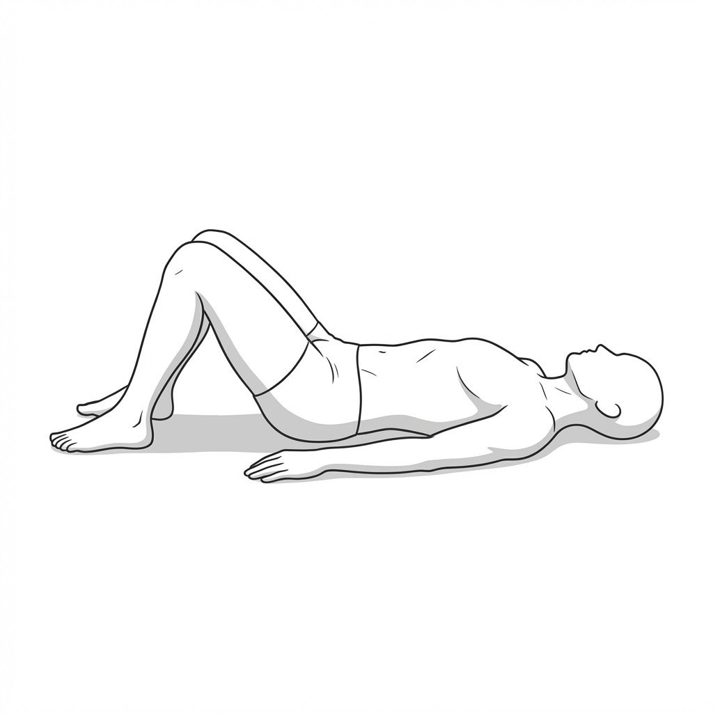
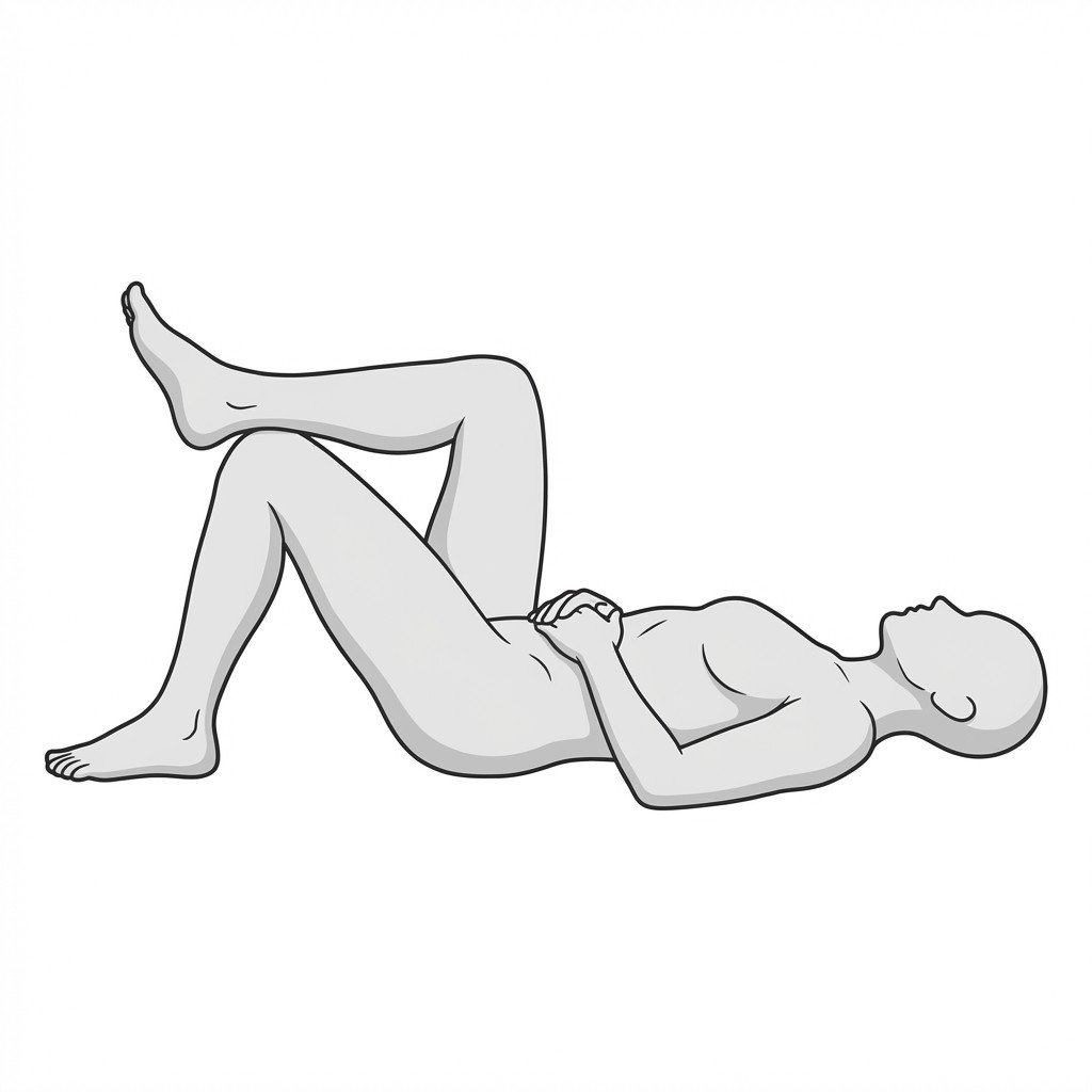
Lie supine with knees bent and feet flat. Place two fingers horizontally across the midline of the abdomen, just above the umbilicus. Slowly lift your head and shoulders off the floor (a partial crunch) while feeling for the borders of the rectus muscles closing in on your fingers.
Positive: A gap of more than two finger-widths (approximately 2cm) or the sensation of the fingers sinking deeply into the abdomen without firm fascial resistance.
Abdominal Doming Assessment
While in the same supine, knees-bent position, perform a partial curl-up or lift both feet slightly off the floor. Observe the midline of the abdomen for any bulging, tenting, or "doming" along the linea alba.
Positive: Visible protrusion along the midline, suggesting an inability to manage intra-abdominal pressure effectively during load.
Carnett's Sign Variant (Trigger Point Provocation)
Identify a tender spot on the abdominal wall while lying relaxed. Keep your finger on the tender spot and apply moderate pressure. Then, lift your head and shoulders to contract the abdominal muscles, noting if the pain changes.
Positive: Pain that increases or remains the same during muscle contraction suggests a myofascial or abdominal wall source (like a trigger point), whereas pain that decreases may suggest a deeper visceral source.
Active Straight Leg Raise (ASLR) Load Transfer Test
Lie supine with legs straight. Actively lift one leg about 20cm off the table. Note the effort required and any pelvic rotation. Then, have a clinician (or use your own hands) apply inward compressive force across the pelvis and repeat the lift.
Positive: The leg lift feels significantly easier or less painful when external pelvic compression is applied, indicating a deficit in intrinsic lumbo-pelvic load transfer, often associated with abdominal wall dysfunction.
Evidence-Based Management
Management of diastasis recti and associated myofascial pain requires a multimodal approach. Current guidelines generally recommend conservative rehabilitation focusing on intra-abdominal pressure management and graded loading before considering surgical intervention.
Self-Care
- Activity modification to avoid breath-holding during exertion
- Postural awareness to prevent chronic rib flaring or excessive anterior pelvic tilt
- Diaphragmatic breathing exercises to regulate intra-abdominal pressure
- Gentle self-massage of the lateral abdominal musculature
- Log-rolling to get out of bed to minimize sudden midline strain
Manual Therapy
- Myofascial release targeting the rectus abdominis and obliques
- Trigger point deactivation in compensatory muscles (e.g., iliopsoas, quadratus lumborum)
- Visceral mobilization techniques to address fascial restrictions
- Soft tissue mobilization of the thoracolumbar fascia
- Manual cueing for transversus abdominis activation
Interventional
- Trigger point injections for refractory abdominal wall pain
- Ultrasound-guided diagnostic blocks (e.g., rectus sheath block) to differentiate pain sources
- Dry needling of the oblique and rectus abdominis muscles
- Surgical consultation for severe, functionally limiting diastasis recti (rarely the first-line approach)
Rehabilitation
- Graded abdominal loading focusing on linea alba tension rather than gap closure
- Pelvic floor physical therapy to address co-occurring dysfunction
- Integration of transversus abdominis activation with functional movements
- Progressive core stabilization exercises tailored to load tolerance
- Neuromuscular re-education for optimal intra-abdominal pressure management
Red Flags & Differential Diagnosis
While diastasis recti and abdominal myofascial pain are generally benign, abdominal pain can be a symptom of serious visceral or systemic pathology. The following red flags require prompt medical investigation.
Incarcerated or Strangulated Hernia
A painful, firm bulge in the abdomen or groin that cannot be pushed back in, especially if accompanied by nausea, vomiting, or skin color changes. This requires immediate emergency evaluation.
EmergencyPulsatile Abdominal Mass
A rhythmic, pulsing sensation or visible mass in the midline of the abdomen, which may indicate an abdominal aortic aneurysm.
EmergencyUnexplained Visceral Symptoms
Abdominal pain accompanied by unexplained weight loss, blood in stool, persistent vomiting, or jaundice, suggesting internal organ pathology rather than musculoskeletal pain.
InvestigateSevere Pelvic Floor Dysfunction
New onset of severe urinary or fecal incontinence, or complete inability to void, which may indicate significant neurological or structural compromise.
UrgentPain Disproportionate to Findings
Severe, unrelenting abdominal pain that is not altered by movement, posture, or abdominal contraction (negative Carnett's sign), warranting investigation for visceral disease.
Investigate
Clinical Pearls
Diastasis recti is defined by a widening of the linea alba (>2cm), but functional impairment is often more clinically relevant than the exact measurement of the gap.
Myofascial trigger points in the rectus abdominis frequently coexist with diastasis recti, developing secondary to altered biomechanics and mechanical strain.
Abdominal wall trigger points can refer pain to the mid-back, lower back, and groin, often mimicking visceral or gynecological pathology.
Current rehabilitation guidelines emphasize generating appropriate tension across the linea alba during movement, rather than strictly attempting to "close the gap."
Evidence suggests that graded abdominal loading is generally safe and beneficial, challenging historical advice to permanently avoid all flexion-based exercises.
Co-occurring pelvic floor dysfunction is reported in some studies (with older work suggesting >60%) but recent prospective studies have found no association with diastasis itself; intra-abdominal pressure regulation should still be addressed holistically across the diaphragm, abdominal wall, and pelvic floor.
Movement therapy
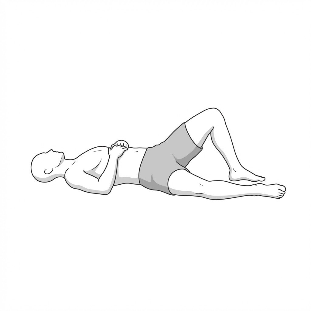
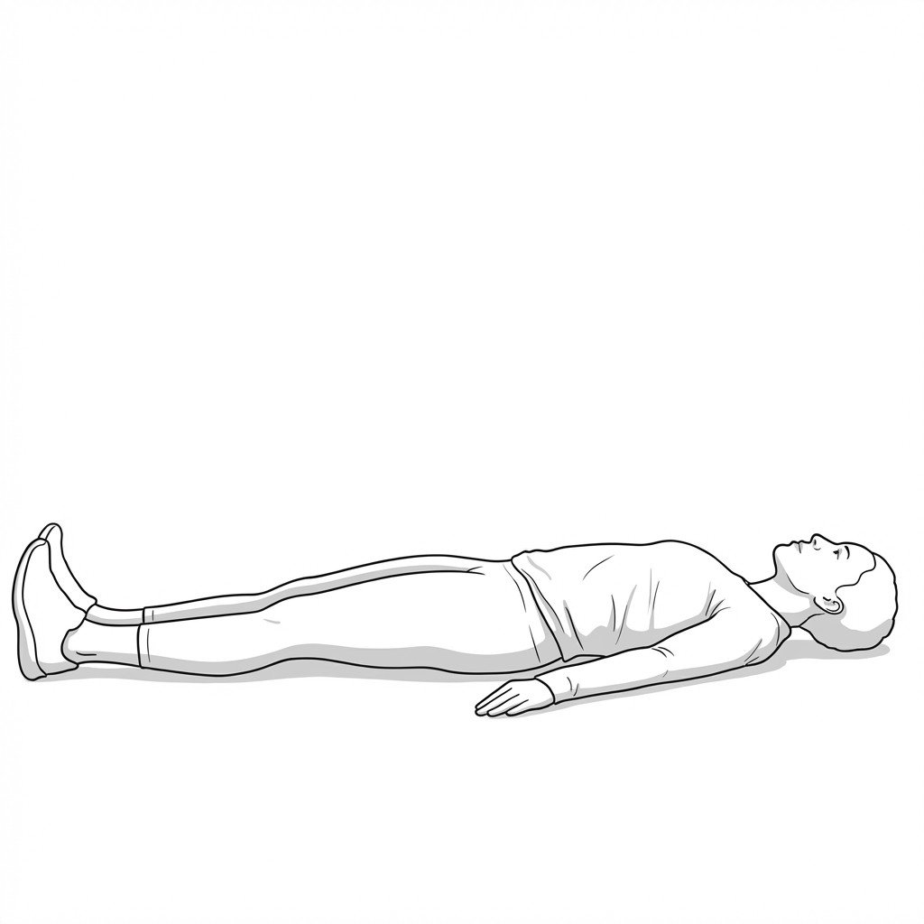
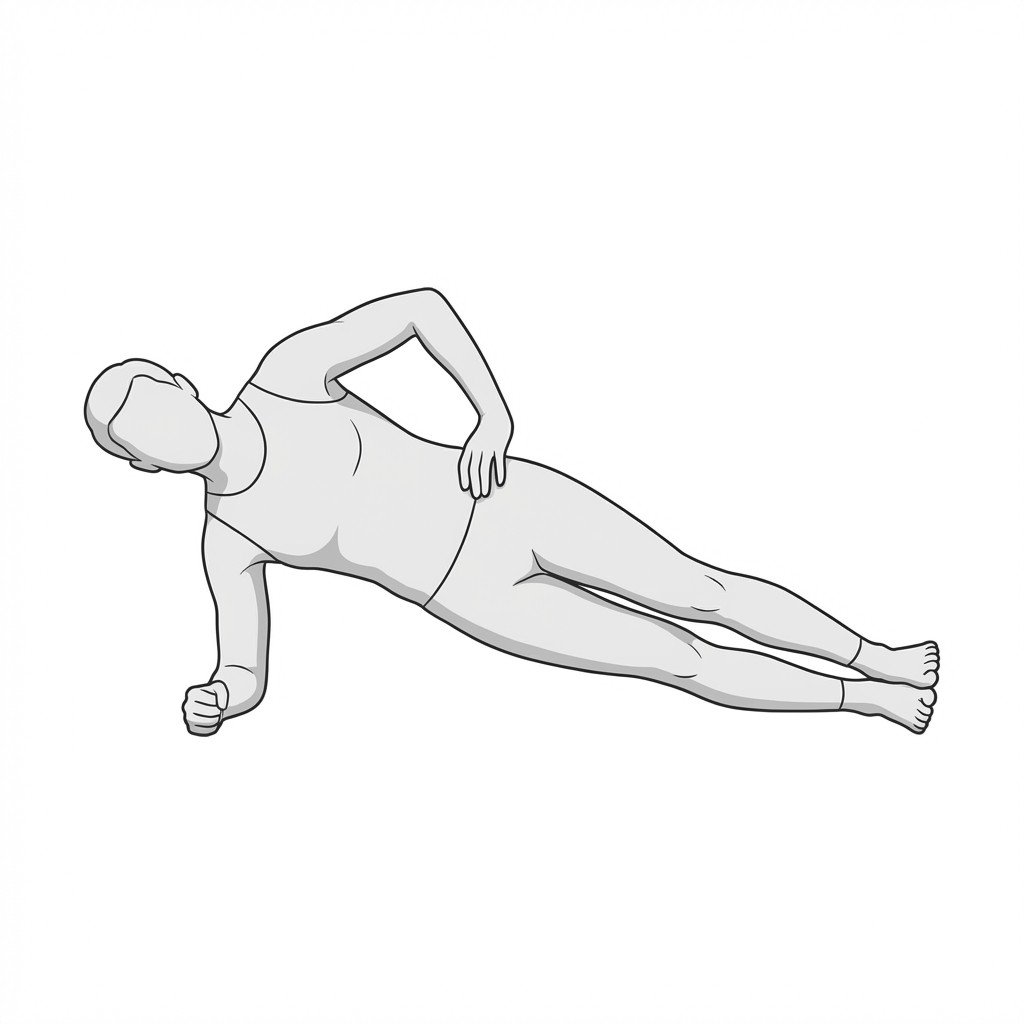
Exercises for Diastasis Recti
Diastasis recti rehabilitation prioritizes transverse abdominis recruitment and pelvic floor coordination before introducing any loading that increases intra-abdominal pressure — these exercises address the inter-recti gap without straining it. Start with 2–3 and progress as tolerated.