Understanding Thoracic Outlet Syndrome & Myofascial Overlap
Thoracic outlet syndrome (TOS) refers to a group of conditions involving compression or irritation of the neurovascular bundle as it passes from the neck into the arm. Neurogenic cases are by far the most common in general practice, while venous and arterial cases are less common but much more urgent.
A major clinical challenge is that many upper-quarter pain presentations look like TOS without being true structural thoracic outlet compression. Scalene, pectoralis minor, and related myofascial dysfunction can reproduce pain, paresthesia, heaviness, and fatigue patterns that overlap strongly with disputed neurogenic TOS.
The most important clinical question is often not “Is this TOS?” but “What combination of neurovascular, myofascial, cervical, and postural factors is creating this arm pattern?”
The practical goal is not to force a yes/no TOS label too early, but to determine whether the presentation is primarily vascular, primarily neurological, primarily myofascial, or mixed. That distinction changes the urgency, workup, and treatment plan significantly.
Anatomical Compression Sites
The thoracic outlet is not a single space but several related anatomical regions where the brachial plexus and subclavian vessels may be stressed. Myofascial dysfunction may contribute to narrowing or traction at any of these sites without being the only explanation for symptoms.
Scalene Group (Anterior, Middle, Posterior)
The anterior and middle scalenes help form the interscalene region through which the brachial plexus and subclavian artery pass. When these muscles are shortened, overactive, or trigger-point-irritable, they may contribute to symptoms that resemble neurogenic thoracic outlet syndrome or cervical radicular pain. Their involvement is common, but they should not be treated as the sole explanation in every TOS-like presentation.
Pain and altered sensation may spread into the neck, shoulder, medial scapular border, arm, radial forearm, or thumb–index region, depending on the pattern and the structures involved.Pectoralis Minor
The pectoralis minor helps position the scapula and may contribute to neurovascular narrowing in the subcoracoid region when adaptively shortened. It is particularly relevant in protracted-shoulder postures and in patients whose symptoms worsen with arm elevation or prolonged forward shoulder posture.
Pain is often felt in the anterior shoulder, upper chest, or along the ulnar side of the arm and forearm, sometimes overlapping with lower-trunk or ulnar-distribution complaints.Subclavius
The subclavius is a small stabilizer beneath the clavicle. Although it is less often the main clinical focus, local tension here may contribute to discomfort in the clavicular / costoclavicular region and can become relevant in patients with anterior shoulder-girdle compression complaints.
Pain is usually felt under the clavicle and into the anterior shoulder, sometimes blending with upper-pectoral discomfort.Upper Trapezius
The upper trapezius does not directly form the thoracic outlet, but it often becomes involved secondarily through altered shoulder-girdle mechanics, protective elevation, or chronic overuse. In some patients it contributes to the overall traction or guarding pattern around the shoulder girdle.
Posterolateral neck pain that may extend to the mastoid, temple, or angle of the jaw.Levator Scapulae
Levator scapulae often becomes hyperactive when the body is trying to protect or reposition the shoulder girdle. It is a common source of neck-angle pain in patients with upper-quarter overload and may coexist with TOS-like symptoms without being the primary compression source itself.
Pain at the superior medial scapular border and angle of the neck, sometimes extending into the posterior shoulder.Coracobrachialis
The coracobrachialis can contribute to anterior shoulder and upper-arm pain and may complicate the regional picture in patients with pectoralis minor dysfunction or coracoid-region overload. It is usually a co-contributor rather than the main thoracic outlet driver.
Anterior shoulder and upper-arm pain, sometimes extending toward the posterior arm or forearm in a less specific pattern.
Interscalene Region
The brachial plexus and subclavian artery pass between the scalenes and first rib; muscular and postural factors may influence this space.
Costoclavicular Space
The neurovascular bundle passes between the clavicle and first rib, where shoulder position and clavicular mechanics may matter.
Subcoracoid / Pectoralis Minor Space
The bundle may be stressed beneath pectoralis minor, especially with prolonged protraction or hyperabduction.
Double-Crush Consideration
Proximal irritation can make distal nerves less tolerant, which is why carpal or cubital tunnel symptoms may coexist with a TOS-like picture.
Myofascial Referral Patterns
Scalene and pectoralis minor trigger points can create referral patterns that look convincingly neurological. This is one reason upper-extremity symptoms are often more complex than a simple imaging label suggests.
- Scalene Group
- Often produces neck, medial-scapular, chest, arm, or radial-forearm pain that may resemble cervical radiculopathy or upper-plexus irritation. The pattern is clinically important but not specific enough to diagnose TOS by itself.
- Pectoralis Minor
- Commonly creates anterior shoulder and chest discomfort with symptoms radiating down the ulnar side of the arm or into digits 4-5, especially when the shoulder remains chronically protracted.
- Subclavius
- Usually causes more focal clavicular or anterior-shoulder discomfort and may be mistaken for local joint or costoclavicular irritation.
Clinical Assessment & Provocation Tests
Provocation tests can be useful, but they have limited specificity. In practice, the most meaningful finding is reproduction of the patient’s familiar symptoms in a pattern that matches the history and differential diagnosis.
Elevated Arm Stress Test (EAST / Roos Test)
Raise the arms with elbows bent and repeatedly open and close the hands. This is a provocative endurance test, not a definitive diagnostic test. It is better used to reproduce the patient’s familiar pattern than to “prove TOS.”
Positive: Reproduction of the patient’s typical pain, heaviness, or paresthesia is more meaningful than fatigue alone. Simple tiredness is a common nonspecific response.
Adson's Test
Neck extension, head turning, and breath hold are used while monitoring the upper limb. Historically this has been used to provoke scalene-related compression, but pulse changes alone are not reliable enough to diagnose TOS.
Positive: The most clinically meaningful finding is reproduction of familiar symptoms, not merely a change in pulse.
Wright's Test (Hyperabduction Test)
The arm is moved into hyperabduction to stress the subcoracoid region. This may be relevant when pectoralis minor compression is suspected.
Positive: Reproduction of familiar symptoms is more important than isolated pulse change, which is not sufficiently specific by itself.
Scalene Cramp / Shortening Test
Active neck positioning is used to shorten the scalenes and determine whether it reproduces the patient’s familiar arm, chest, or scapular pattern.
Positive: Reproduction of the patient’s familiar referred pain pattern suggests a meaningful myofascial contribution, though it still does not distinguish pure myofascial referral from true neurovascular compression on its own.
Evidence-Based Treatment Pathways
For most nonvascular cases, the first-line approach is conservative and focuses on reducing compression load, improving shoulder-girdle mechanics, improving breathing and posture, and treating clearly relevant myofascial contributors.
Self-Care
- Ergonomic changes to reduce chronic shoulder protraction and depressed-girdle load
- Diaphragmatic breathing when accessory-breathing overuse is clearly present
- Gentle symptom-guided scalene and pectoralis minor stretching
- Reducing prolonged overhead positions or carrying loads that clearly reproduce symptoms
- Supportive sleep positioning to reduce sustained traction on the neck and shoulder girdle
Manual Therapy
- Trigger point treatment for scalenes, pectoralis minor, and related upper-quarter muscles when clearly relevant
- First-rib and costoclavicular mobility work when supported by the exam
- Myofascial release of the anterior neck and upper chest when tolerated
- Scapulothoracic and shoulder-girdle mobility where movement restriction is part of the pattern
- Nerve gliding exercises when clinically appropriate and not excessively provocative
Interventional
- Diagnostic or therapeutic muscle injections in selected specialized cases
- Scalene or pectoralis minor procedures only when the diagnosis and indication are clear
- Botulinum toxin only in selected refractory cases, not as a routine first step
- Cervical epidural or spine-directed work only when concurrent cervical pathology is strongly supported
- Surgical decompression reserved for appropriately evaluated refractory or clearly vascular cases
Rehabilitation
- Deep neck flexor training when cervical support is poor
- Lower- and middle-trapezius strengthening for better scapular control
- Serratus anterior activation when scapular positioning is clearly impaired
- Postural re-education focused on reducing sustained shoulder-girdle strain
- Progressive upper-quarter loading only within tolerable symptom ranges
Red Flags & Differential Diagnosis
Vascular or severe neurological presentations require urgent evaluation. The red flags below matter more than any myofascial explanation when they are present.
Unilateral Arm Edema and Cyanosis
Sudden swelling, heaviness, and discoloration of one arm raises concern for venous thoracic outlet syndrome or thrombosis and requires urgent vascular evaluation.
EmergencyAbsent Pulse With Pallor, Coldness, or Ischemic Pain
These features raise concern for arterial compromise and need emergent vascular assessment rather than routine musculoskeletal management.
EmergencyIntrinsic Hand Muscle Atrophy
Visible wasting of the hand muscles suggests a more advanced or alternative neurological process and warrants urgent specialist evaluation.
UrgentHorner's Syndrome or Possible Apical Chest Pathology
Ptosis, miosis, or other signs suggesting sympathetic-chain involvement should broaden the differential immediately toward serious apical or neurological pathology.
UrgentHard Neurological Signs
Profound motor weakness, dense sensory loss, or a strongly dermatomal / myotomal pattern suggests cervical radiculopathy or another neurological lesion rather than simple myofascial referral.
Investigate
Key Clinical Takeaways
Most thoracic outlet presentations are neurogenic rather than vascular, but true vascular cases are the ones that demand the greatest urgency.
Scalene and pectoralis minor dysfunction can strongly mimic or contribute to thoracic outlet symptoms, especially in posture-driven and myofascial presentations.
Provocation tests can support the picture, but no single test is diagnostic by itself.
Electrodiagnostic studies are often more useful for excluding competing diagnoses than for “proving” disputed neurogenic TOS.
A structured trial of conservative care is usually appropriate in nonvascular cases, especially when posture, breathing mechanics, and myofascial pain clearly contribute.
The presence of a cervical rib or anatomic variant does not confirm TOS unless the symptoms and exam fit the finding.
Movement therapy
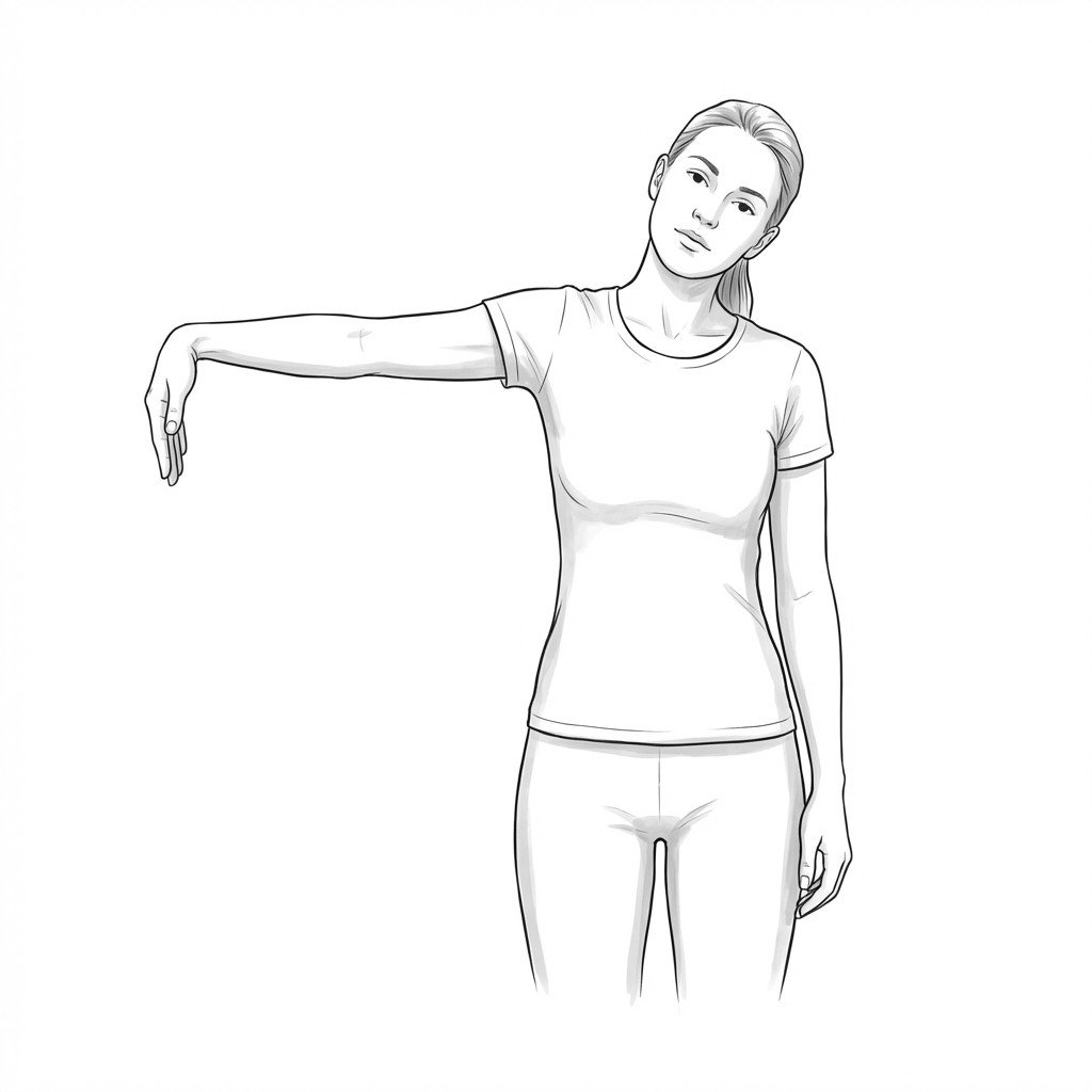
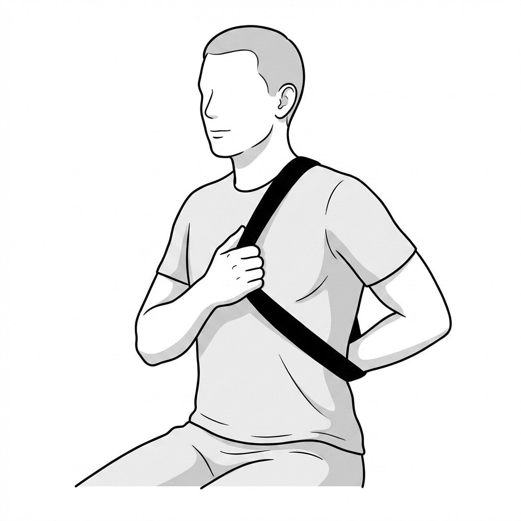
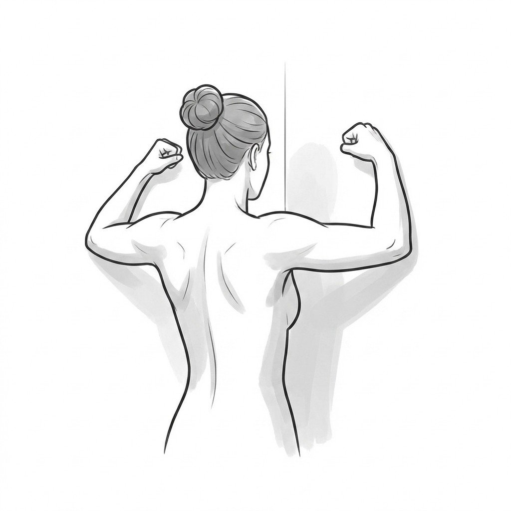
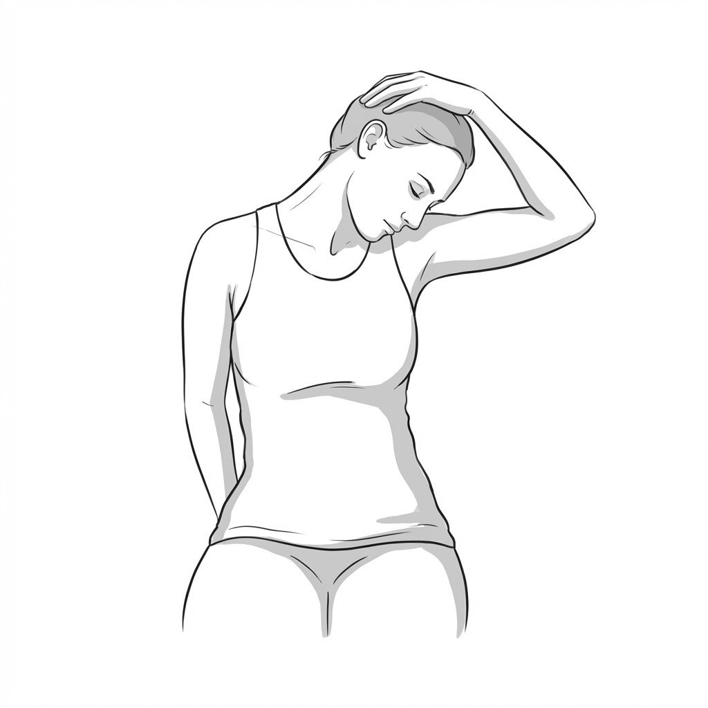
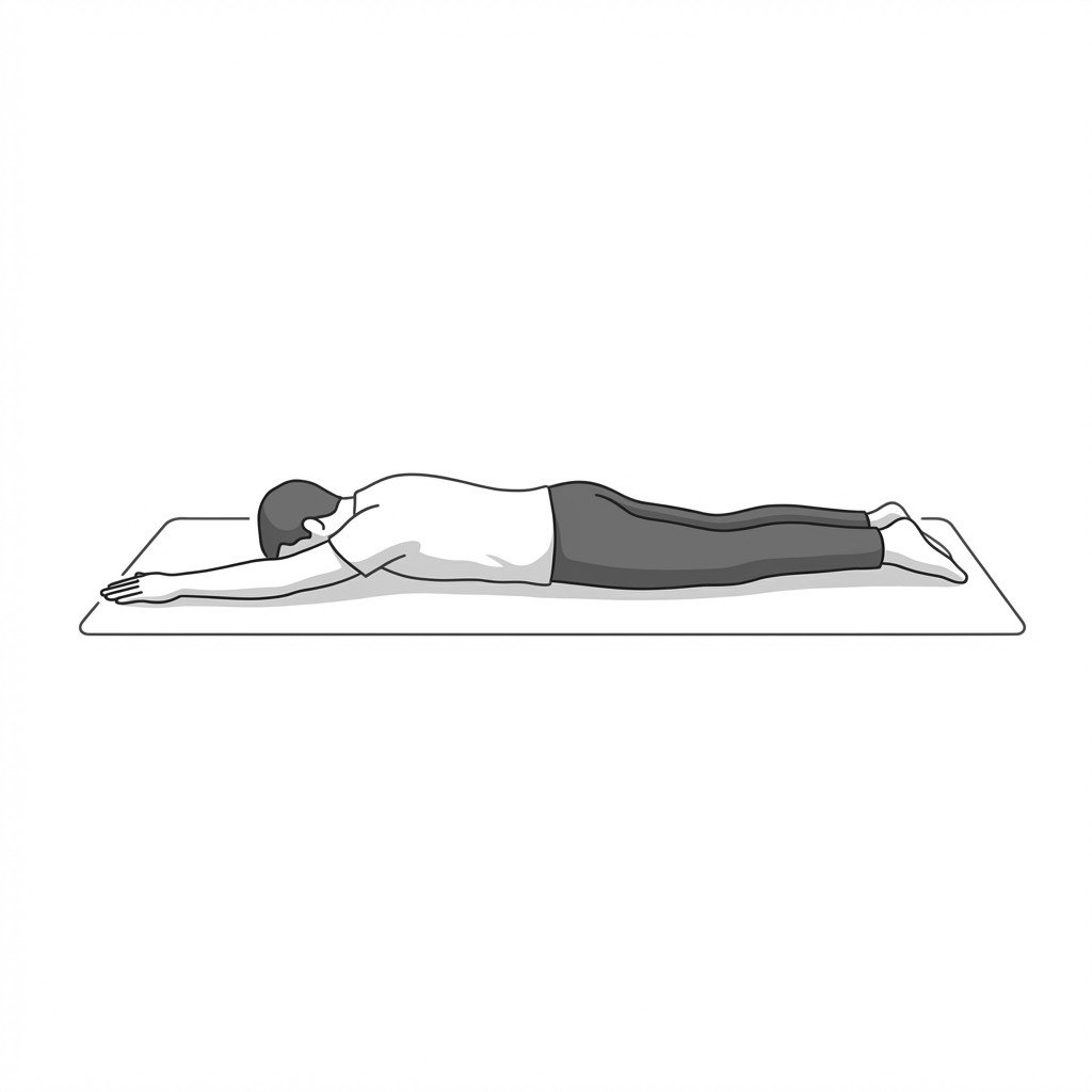
Exercises for Thoracic Outlet Syndrome
TOS rehabilitation targets the structures that compress the neurovascular bundle at the thoracic outlet — these exercises open the costoclavicular space, mobilize the first rib, and build scapular depressor strength to take sustained tension off the scalenes and pectoralis minor. Start with 2–3 and progress as tolerated.