Understanding SIJ & Pelvic Pain
Sacroiliac joint (SIJ) dysfunction is a recognized source of axial low back and posterior pelvic pain, with observational studies suggesting it accounts for 15% to 30% of chronic low back pain cases. The diagnostic challenge lies in distinguishing true articular or ligamentous SIJ pain from adjacent lumbar spine pathology and myofascial pain syndrome (MPS). Presentations frequently emerge postpartum or following pelvic trauma, though insidious onset is also common. Current guidelines generally recommend a comprehensive clinical evaluation, as isolated symptoms rarely provide definitive localization of the pain generator.
The relationship between SIJ dysfunction and pelvic MPS is complex and frequently bidirectional. Muscles such as the quadratus lumborum and piriformis exert direct biomechanical forces on the pelvis; myofascial trigger points within these structures can alter pelvic mechanics, thereby increasing shear forces across the SIJ. Conversely, chronic nociceptive input from an inflamed or unstable SIJ may precipitate secondary, or satellite, trigger points in the gluteal musculature and piriformis. This overlapping presentation often confounds the clinical picture, as both conditions can produce localized posterior pelvic pain with radiation into the buttock and posterior thigh.
Effective management of posterior pelvic pain requires recognizing that sacroiliac joint dysfunction and pelvic myofascial pain are frequently concurrent, mutually perpetuating conditions.
Differentiating primary SIJ pathology from myofascial pain requires a systematic approach. Evidence from systematic reviews indicates that no single orthopedic test is sufficiently reliable; however, a composite of three or more positive SIJ provocation tests (such as the thigh thrust, FABER, and Gaenslen's tests) significantly increases diagnostic specificity. Concurrently, clinicians should palpate for taut bands and reproducible trigger points in the gluteal and piriformis muscles. When conservative measures fail, fluoroscopically guided, dual-diagnostic intra-articular SIJ injections remain the reference standard for confirming the joint as the primary pain generator.
Posterior Pelvic Pain — Three Overlapping Sources
Pain location
Provocation pattern
Active SLR test
Reference standard
First-line treatment
Muscular & Biomechanical Anatomy
The stability of the sacroiliac joint relies on both form closure (bony architecture and ligaments) and force closure (muscular compression). Dysfunction in the surrounding musculature directly impacts this delicate biomechanical balance.
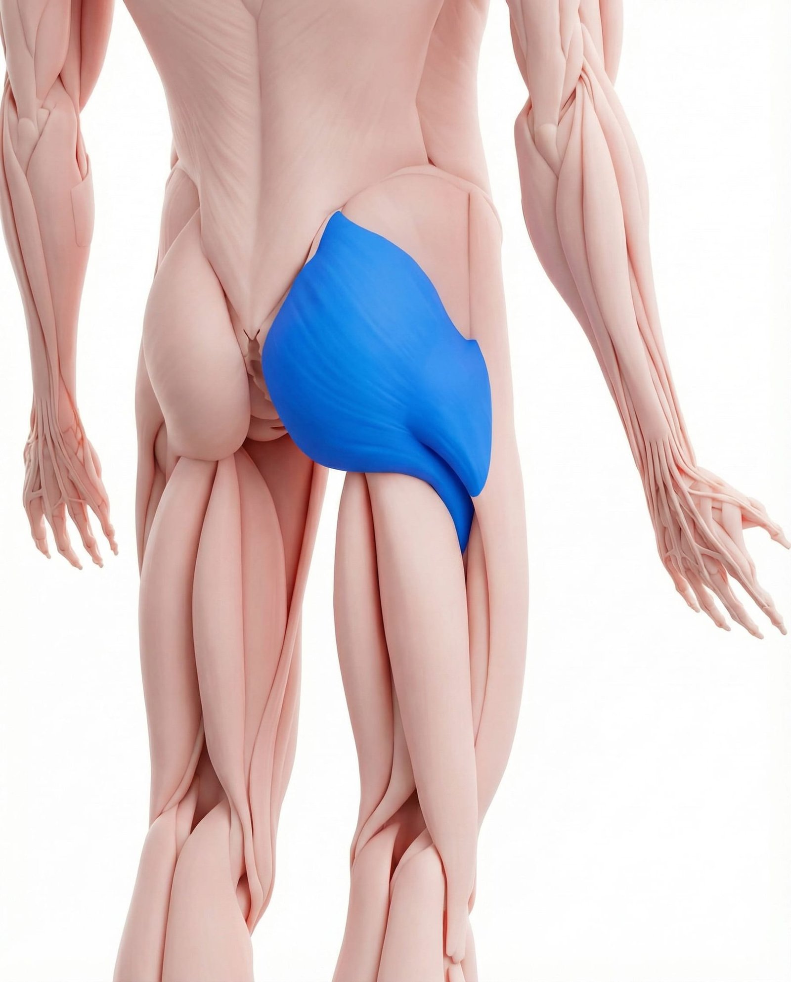
The gluteus maximus provides critical force closure across the sacroiliac joint. Myofascial trigger points in this muscle frequently develop secondary to SIJ instability or inflammation. When active, these trigger points produce a deep, aching pain that closely mimics intrinsic SIJ or sacrotuberous ligament pathology. Clinical evaluation must differentiate between primary gluteal myofascial pain and secondary hypertonicity guarding an unstable joint.
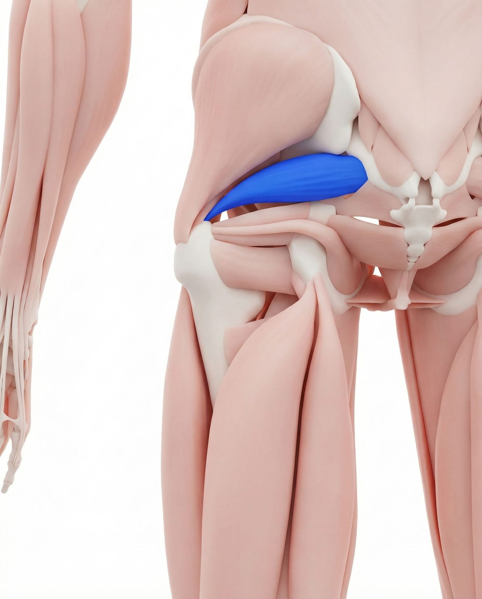
Originating on the anterior sacrum and inserting on the greater trochanter, the piriformis directly influences sacral mechanics. Trigger points here can alter sacral nutation, potentially exacerbating SIJ shear forces. Furthermore, piriformis syndrome and SIJ dysfunction frequently coexist, both presenting with deep buttock pain that may radiate down the posterior thigh. Palpation for taut bands is essential to determine the myofascial contribution to the patient's pain profile.
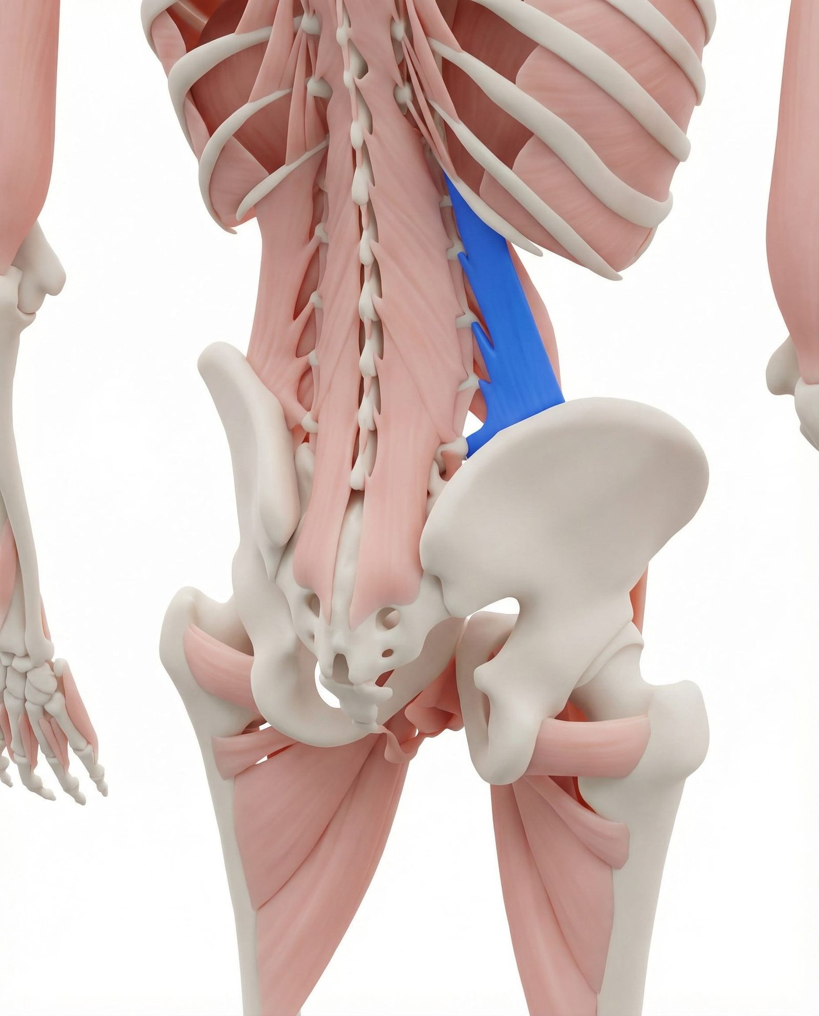
The quadratus lumborum (QL) attaches to the iliac crest and the lower ribs, playing a major role in pelvic positioning. Unilateral QL trigger points or shortening can elevate the hemipelvis, creating a functional leg length discrepancy that abnormally loads the SIJ. Addressing QL myofascial dysfunction is often a prerequisite for achieving lasting relief in mechanical SIJ pain. It is a frequent primary driver of pain that patients misattribute to the SIJ.
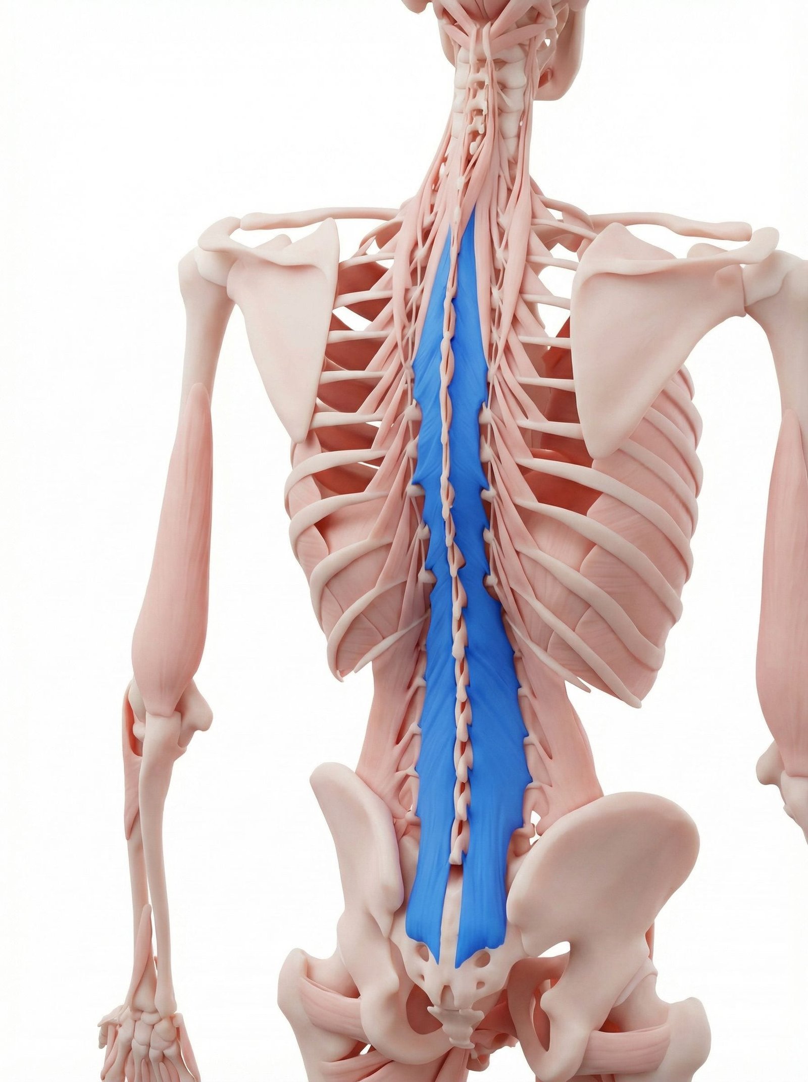
The multifidus muscles are essential for segmental spinal stability and contribute to the stabilization of the sacrum. Chronic SIJ dysfunction is frequently associated with ipsilateral multifidus atrophy and fatty infiltration, observable on MRI. Trigger points in the lower multifidi refer pain directly to the sacral region, complicating the differential diagnosis of posterior pelvic pain.
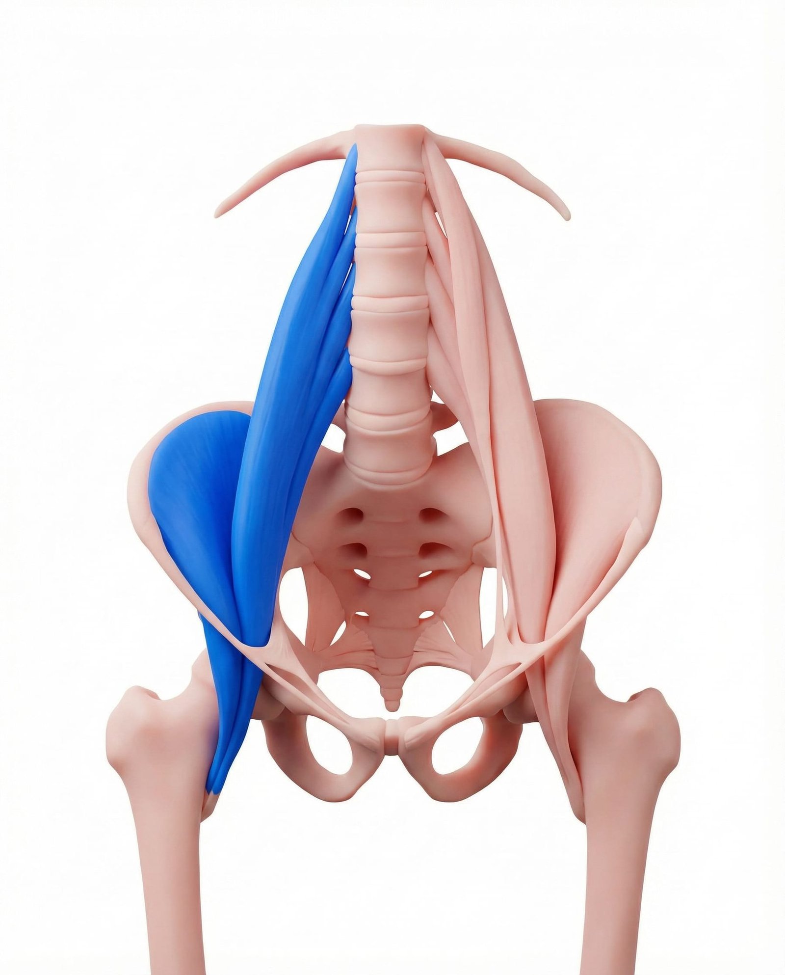
As the primary hip flexor, the iliopsoas significantly influences anterior pelvic tilt. Tightness or trigger points within the iliopsoas can increase lumbar lordosis and alter the mechanical axis of the pelvis, thereby increasing stress on the anterior SIJ ligaments. While it does not typically refer pain to the posterior SIJ, its biomechanical influence makes it a critical consideration in comprehensive pelvic rehabilitation.
Articular Inflammation
True sacroiliitis or intra-articular pathology generating localized nociceptive signals from the joint capsule.
Ligamentous Strain
Microtrauma or laxity in the sacrotuberous or interosseous ligaments, often seen postpartum or post-trauma.
Myofascial Referral
Pain projected to the SIJ region from active trigger points in the gluteals, QL, or piriformis.
Impaired Force Closure
Failure of the stabilizing musculature (multifidus, transversus abdominis) leading to abnormal joint shear.
Myofascial Referral Patterns
Myofascial trigger points in the pelvic and lower back musculature frequently project pain directly into the sacroiliac region. Recognizing these patterns is essential for an accurate differential diagnosis.
Gluteus Maximus
Piriformis
Quadratus Lumborum
Clinical Assessment Tests
Clinical differentiation relies on provocation tests designed to stress the SIJ ligaments and articular surfaces. A cluster of positive tests is required to suggest SIJ involvement over purely myofascial sources.
FABER (Patrick's) Test
Thigh Thrust (Posterior Shear) Test
Active Straight Leg Raise (ASLR)
Piriformis Stretch Test (FAIR Test)
Evidence-Based Treatment Pathways
Current guidelines generally recommend a multimodal approach. Treatment should address both the biomechanical stability of the SIJ and the resolution of associated myofascial trigger points.
Self-Care
- Activity modification to avoid asymmetrical pelvic loading
- Use of a non-elastic sacroiliac belt for temporary stabilization
- Application of heat or ice to the posterior pelvis
- Gentle, pain-free stretching of the gluteals and piriformis
- Ergonomic adjustments to sitting posture and sleep positioning
Manual Therapy
- Ischemic compression of gluteal and piriformis trigger points
- Myofascial release of the quadratus lumborum and thoracolumbar fascia
- SIJ mobilization techniques (if hypomobility is identified)
- Muscle energy techniques (MET) to correct pelvic asymmetries
- Soft tissue mobilization of the sacrotuberous and sacrospinous ligaments
Interventional
- Fluoroscopically guided intra-articular SIJ corticosteroid injections
- Trigger point injections in the piriformis or gluteus maximus
- Radiofrequency ablation (RFA) of the lateral branch nerves
- Prolotherapy or regenerative medicine injections (evidence is emerging)
- Minimally invasive SIJ fusion (for refractory, chronic instability)
Rehabilitation
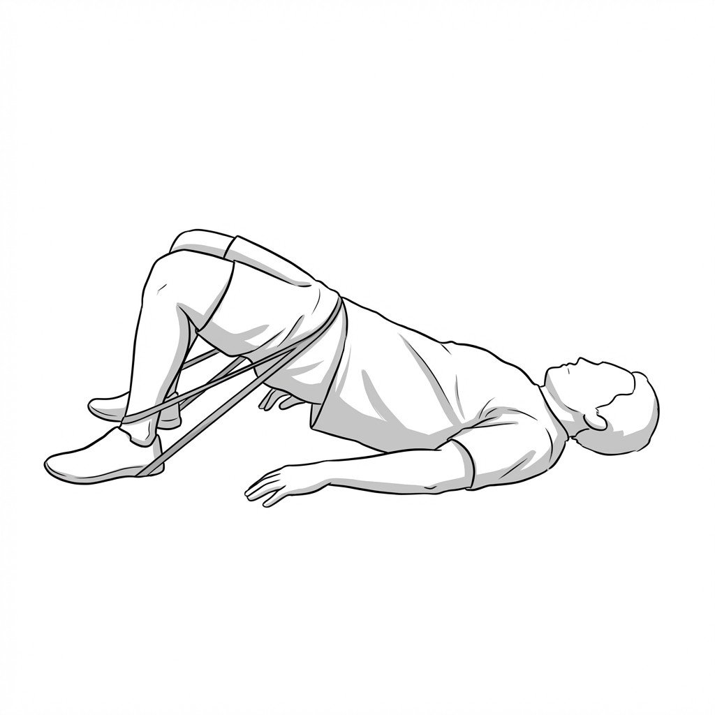
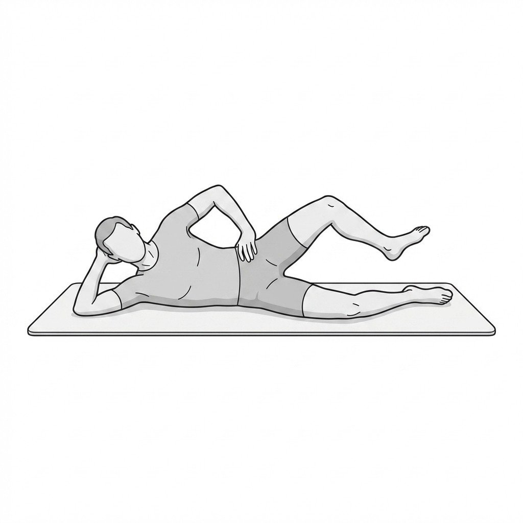
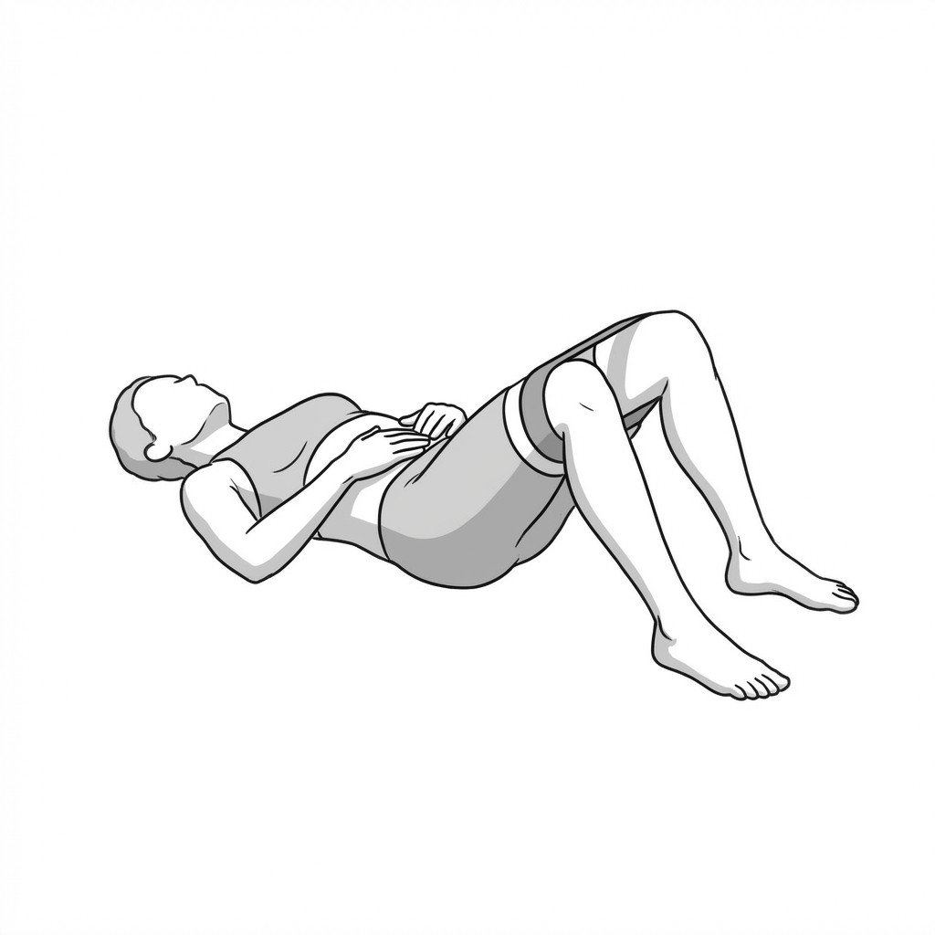
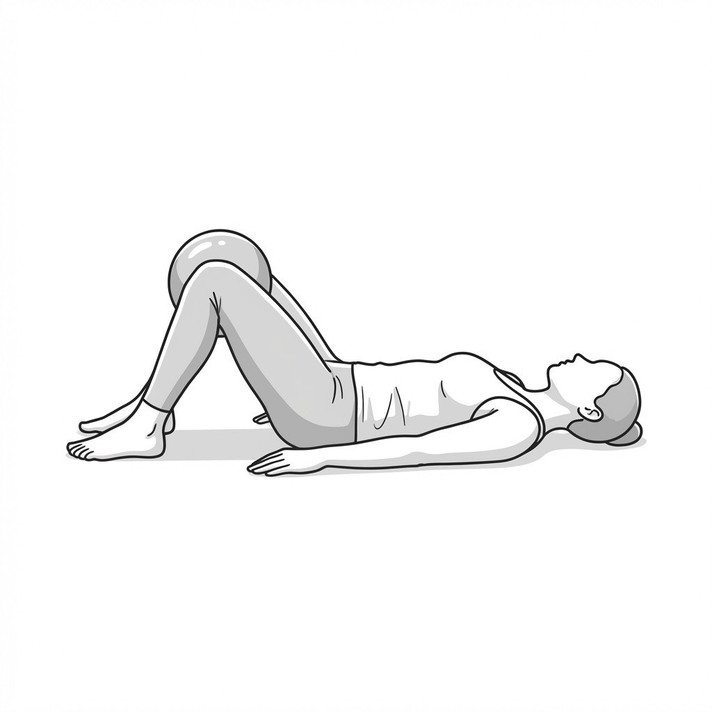
- Motor control training for the transversus abdominis and pelvic floor
- Strengthening of the gluteus maximus and medius for force closure
- Lumbar multifidus activation and endurance exercises
- Correction of functional leg length discrepancies
- Gradual return to symmetrical, load-bearing activities
Red Flags & Differential Diagnosis
While mechanical SIJ dysfunction and myofascial pain are common, posterior pelvic pain can occasionally be the presenting symptom of serious underlying systemic or structural pathology.
Observational studies suggest SIJ dysfunction accounts for 15% to 30% of chronic low back and buttock pain cases.
Myofascial trigger points in the gluteus maximus, piriformis, and quadratus lumborum frequently mimic or coexist with SIJ pathology.
No single physical examination test is diagnostic for SIJ pain; current guidelines recommend a composite of three or more positive provocation tests.
Altered pelvic mechanics from myofascial shortening can increase shear forces across the SIJ, perpetuating joint irritation.
Fluoroscopically guided, dual-diagnostic intra-articular injections remain the reference standard for confirming the SIJ as the primary pain generator.
Conservative management should concurrently address joint mechanics, motor control, and regional myofascial trigger points for optimal outcomes.
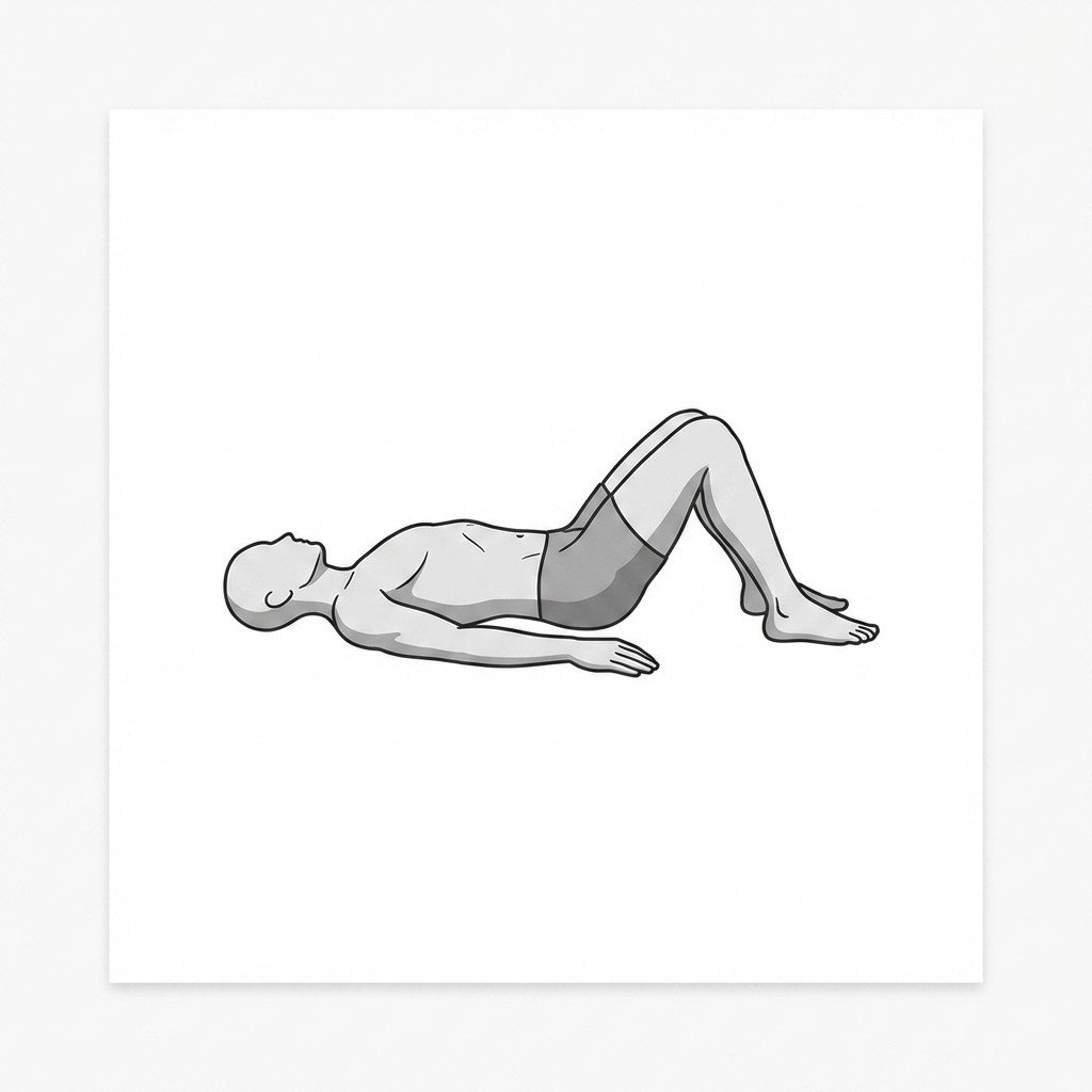
Movement therapy
Exercises for Sij Dysfunction
SIJ dysfunction requires motor control work that closes the joint through coordinated gluteal and deep abdominal activation — these exercises address pelvic load transfer rather than joint mobility. Start with 2–3 and progress as tolerated.