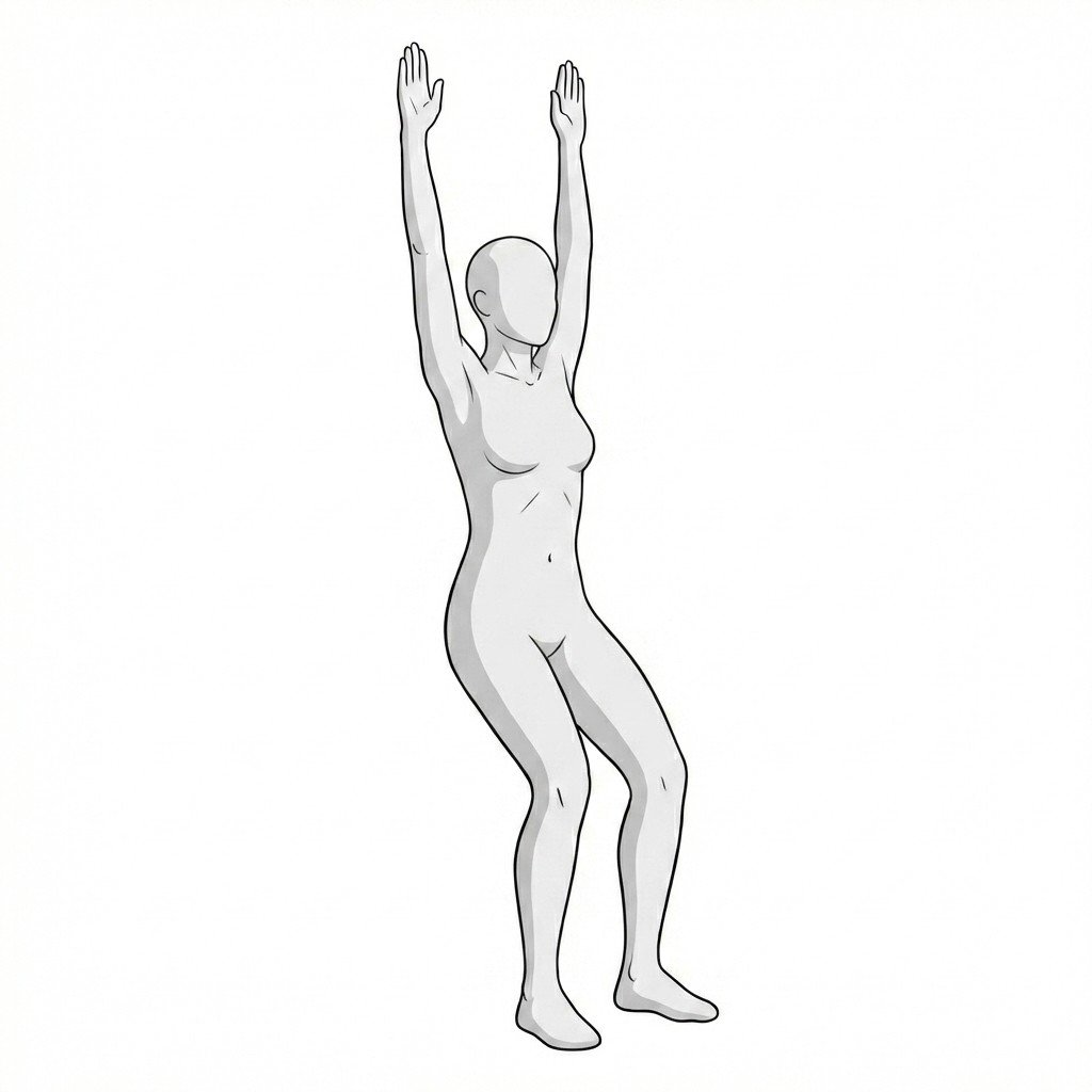
Understanding Thoracic Kyphosis
Thoracic kyphosis refers to the natural posterior convexity of the thoracic spine. Increased kyphosis may reflect a flexible postural pattern, a more rigid developmental pattern, age-related change, vertebral compression, or a mixed presentation.
Myofascial pain often coexists with increased kyphosis because posterior thoracic muscles may work harder to control the body in a flexion-biased posture, while anterior tissues such as pectoralis minor may shorten and reinforce the pattern.
Thoracic myofascial pain can be a consequence of kyphosis, a contributor to worsening posture, or both at the same time.
The key clinical task is not simply to decide whether the curve is “bad,” but to determine how much of the patient’s pain is coming from muscular overload, how much from structural change, and whether any red-flag process is present.
Key Musculature & Biomechanics
The thoracic kyphosis picture is shaped by both passive structures and active musculature. The muscles listed here are clinically useful because they commonly become painful, shortened, lengthened, or overloaded within the broader kyphotic pattern.
Thoracic Erector Spinae
The thoracic erector spinae group helps extend and support the thoracic spine. In people with increased kyphosis, these muscles often work under chronic mechanical disadvantage and may become painful, fatigued, and tender. Trigger points here can contribute to local stiffness and thoracic aching, but they should be understood as one possible pain source within a broader postural or structural pattern.
Pain is usually local or segmental along the thoracic paraspinals, sometimes with some wrapping discomfort around the ribs.Rhomboid Major & Minor
The rhomboids are commonly overloaded in rounded-shoulder, protracted-scapula postures associated with thoracic kyphosis. They often develop superficial interscapular pain and tenderness, especially when scapular control is poor and the thoracic spine remains flexion-biased for long periods.
Pain is usually felt between the medial scapular border and the thoracic spine, often as a dull, nagging interscapular ache.Middle Trapezius
The middle trapezius is a key scapular retractor. In increased kyphosis, it often becomes lengthened and underperforming rather than simply “tight.” Trigger points may develop secondarily when the muscle is repeatedly asked to stabilize a poorly positioned scapula.
Pain is usually local to the interscapular region and may extend somewhat laterally toward the shoulder girdle.Pectoralis Minor
Pectoralis minor often shortens adaptively in kyphotic or rounded-shoulder posture, pulling the scapula into anterior tilt and protraction. It is an important perpetuating factor even when it is not the main painful muscle.
Pain may be felt in the anterior shoulder, upper chest, and sometimes into the arm in a way that can overlap with thoracic outlet or shoulder presentations.Serratus Posterior Superior
This deep upper-thoracic / rib-associated muscle may become symptomatic in patients with restricted rib motion, altered breathing mechanics, or stiff upper-thoracic posture. It is a less common but sometimes relevant contributor in difficult upper-thoracic pain cases.
Pain is often felt as a deeper ache beneath the upper medial scapular region or posterior upper thorax.
Postural Load
Posterior muscles may become chronically overloaded trying to control a flexion-biased thoracic posture.
Structural Deformity
Vertebral shape change or compression may alter alignment and create a different clinical risk picture.
Breathing Restriction
Thoracic stiffness may reduce rib mobility and change breathing mechanics, which can add muscular pain.
Anterior Tightness
Pectoral and anterior fascial shortening may reinforce rounded-shoulder and kyphotic patterns.
Common Pain Referral Patterns
Thoracic muscular pain often stays relatively regional, but overlapping patterns can still mimic rib pain, scapular pain, or even non-spinal complaints if the history is not examined carefully.
- Thoracic Erector Spinae
- Usually produces a vertical or segmental band of paraspinal pain. In some patients the discomfort feels broader and may seem to spread around the rib cage without fitting a clear dermatomal pattern.
- Rhomboid Group
- Typically produces a localized or diffuse interscapular ache between the scapula and the thoracic spine rather than pain far away from that zone.
- Pectoralis Minor
- Can contribute to anterior shoulder or upper-chest discomfort and may complicate scapular mechanics, sometimes making the pain feel more like shoulder dysfunction or thoracic outlet–type strain.
Clinical Screening & Self-Assessment
These screening tests help estimate flexibility, rigidity, breathing restriction, and the possibility of structural involvement. They should be used to guide the next step, not to replace diagnosis.
Occiput-to-Wall Test
Stand with heels, buttocks, and upper back near a wall, then see whether the back of the head can approach the wall without sharply tilting the chin upward. This is a simple screening maneuver, not a diagnosis by itself.
Positive: Difficulty bringing the occiput toward the wall while keeping the gaze level may suggest increased thoracic kyphosis, cervical compensation, or both.
Active Thoracic Extension Test
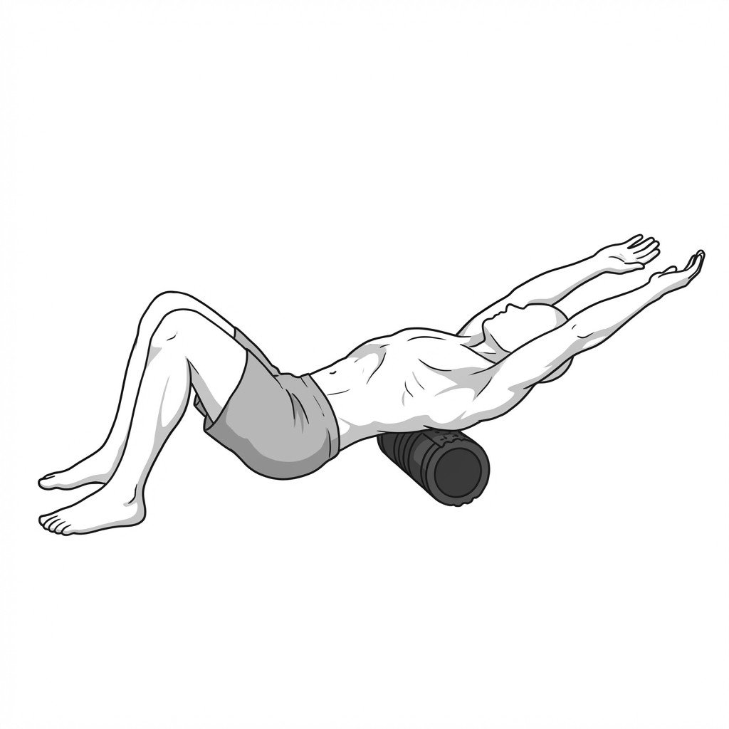
Sit upright and attempt a controlled thoracic extension over the backrest of a chair or in another supported position. Observe whether the thoracic curve can change at all and whether the motion reproduces pain.
Positive: A very rigid curve with minimal change may suggest a more structural pattern, whereas a curve that improves with movement suggests a more flexible or postural component.
Rib Expansion Measurement
Measure chest expansion during full exhalation and inhalation if this is appropriate in the clinical setting. This can help identify whether thoracic stiffness is also affecting breathing mechanics.
Positive: Reduced chest expansion may suggest thoracic wall restriction, breathing dysfunction, or systemic conditions affecting thoracic mobility — it is a clue, not a diagnosis.
Spinal Percussion Screening
A clinician may assess for focal thoracic spinous-process tenderness when fracture or bony pathology is a concern.
Positive: Marked focal tenderness, especially after minor trauma in an at-risk patient, should raise concern for vertebral fracture or other bony pathology and prompt formal evaluation.
Evidence-Informed Treatment Pathways
Treatment is most effective when it matches the dominant problem: flexible postural kyphosis, structural kyphosis, bone fragility, or secondary myofascial overload. Many patients need more than one of these addressed at the same time.
Self-Care
- Postural-awareness changes during sitting and desk work
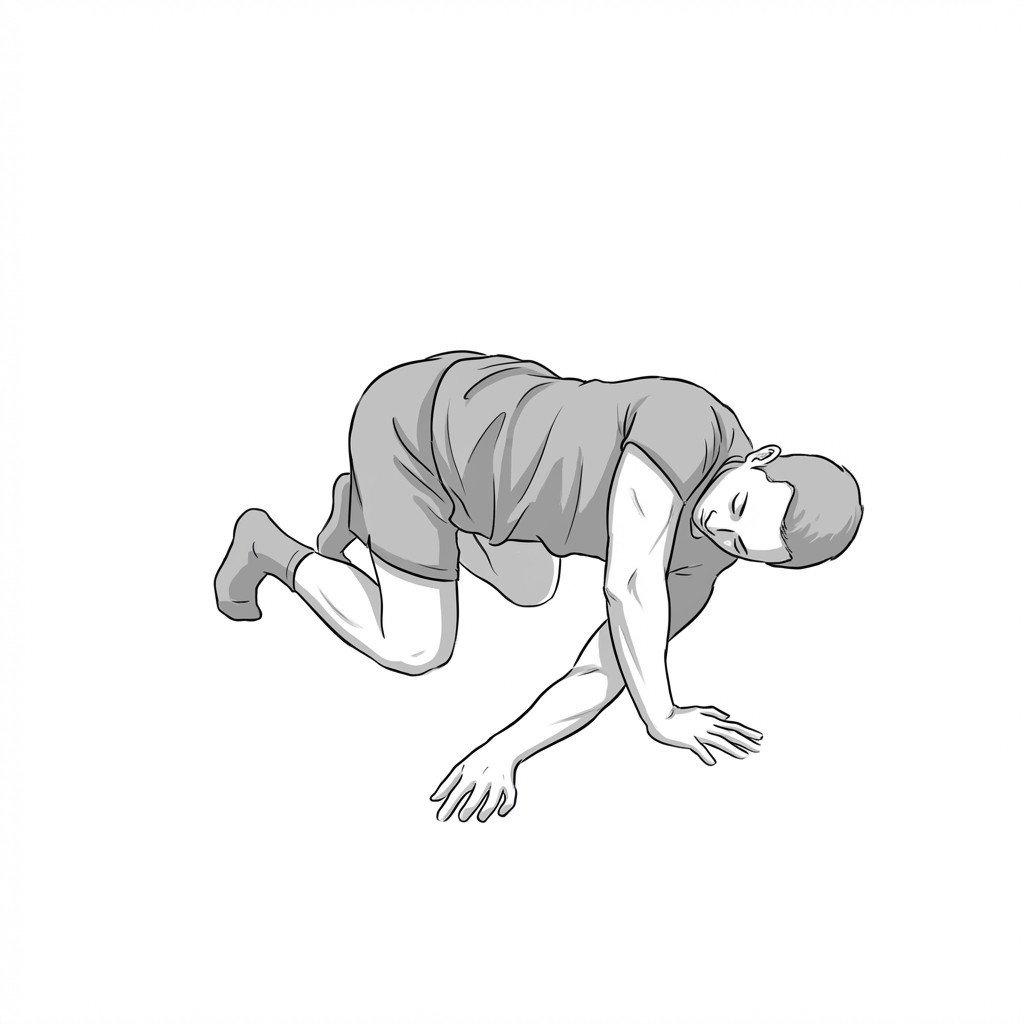
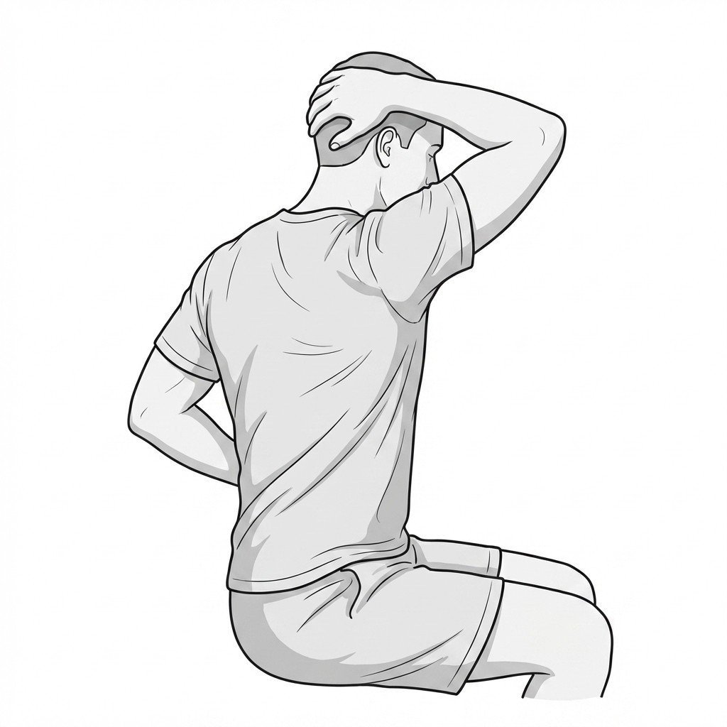
- Gentle active thoracic-extension exercises within a comfortable range
- Heat to posterior thoracic musculature when it improves movement tolerance
- Pectoral stretching or anterior chest opening when clearly indicated
- Diaphragmatic breathing and rib-mobility practice when breathing mechanics are stiff
Manual Therapy
- Myofascial treatment of pectoralis minor, thoracic paraspinals, and interscapular muscles
- Ischemic compression or trigger-point work for rhomboids and erector spinae when clinically relevant
- Thoracic and costovertebral mobilization when movement restriction is a key driver
- Selected soft-tissue techniques for chronically overloaded posterior thoracic tissue
- Reassessment of posture and movement after treatment rather than tenderness alone
Interventional
- Trigger point injections in refractory myofascial cases when clearly indicated
- Bone-density evaluation and bone-health management in at-risk adults
- Fracture-directed procedures only when vertebral compression fracture is actually present and appropriately evaluated
- Targeted spine or facet interventions only when the clinical picture supports them
- Medical management of osteoporosis or osteopenia when part of the underlying problem
Rehabilitation
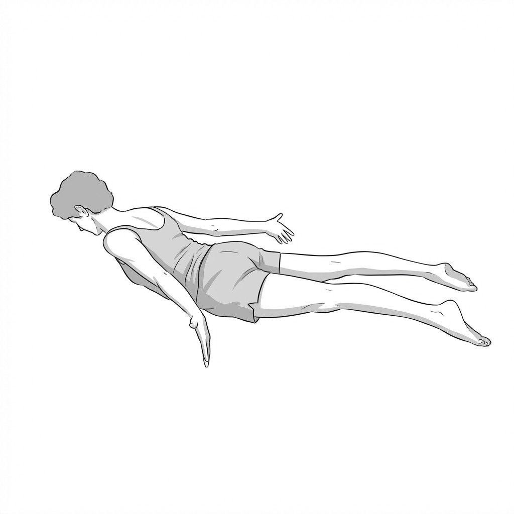
- Endurance work for thoracic extensors and scapular retractors
- Motor-control exercises for trunk alignment and thoracic extension bias
- Breathing work integrated with posture and rib motion
- Postural cueing or external feedback when it genuinely improves carryover
- Bracing only in selected structural cases where it is actually appropriate
Red Flags & Differential Diagnosis
Thoracic pain can signal serious underlying pathology. The red flags below matter more than the myofascial story when they are present.
Sudden Onset Thoracic Pain in Older Adults
Acute thoracic pain after minor strain, lifting, or low-energy trauma in an older or osteoporotic patient should raise concern for vertebral compression fracture until proven otherwise.
UrgentNeurological Deficits
Lower-extremity weakness, sensory change, gait disturbance, hyperreflexia, or bowel / bladder symptoms require urgent evaluation for spinal-cord or major neurological involvement.
EmergencyUnrelenting Night Pain or Systemic Symptoms
Severe night pain, unexplained weight loss, fever, or pain unrelated to movement should broaden the differential toward malignancy, infection, or systemic disease.
InvestigateRigid Deformity in Adolescents
A rigid thoracic deformity in a growing adolescent should prompt formal evaluation for structural causes such as Scheuermann-type deformity rather than being dismissed as “poor posture.”
InvestigateSignificant Morning Stiffness
Marked inflammatory-type morning stiffness, especially in a younger patient with thoracic rigidity, may suggest a systemic inflammatory condition rather than simple myofascial overload.
Investigate
Key Clinical Takeaways
Thoracic kyphosis is common and can be postural, flexible, or structural. Not all increased kyphosis represents the same diagnosis or the same risk level.
Myofascial pain frequently coexists with thoracic kyphosis because posterior muscles are often overloaded and anterior structures may become adaptively shortened.
In older adults, vertebral compression fracture must be considered whenever thoracic pain is sudden, focal, or associated with a clear change in posture.
Trigger point therapy may reduce secondary muscular pain, but it will not correct a fixed structural deformity by itself.
In flexible or posture-dominant presentations, thoracic extensor endurance, breathing work, scapular rehabilitation, and chest opening are often more useful long-term than passive care alone.
Good differentiation requires posture assessment, movement testing, palpation, and — when indicated — imaging or medical evaluation.