Understanding the Differential Diagnosis
Lumbar radiculopathy, commonly referred to as sciatica, is a symptom complex characterized by pain, paresthesia, or weakness radiating down the leg along the distribution of a specific spinal nerve root. It is most frequently caused by disc herniation or foraminal stenosis at the L4-L5 or L5-S1 levels. The natural history of acute lumbar radiculopathy is generally favorable, with observational studies suggesting that a majority of patients experience significant improvement within 6 to 12 weeks of conservative management. However, accurately diagnosing the source of leg pain is critical, as several musculoskeletal conditions can closely mimic nerve root compression.
Myofascial pain syndrome (MPS) is a primary differential diagnosis and a frequent comorbidity in patients presenting with sciatica-like symptoms. The gluteus minimus is particularly notable; trigger points within this muscle can refer pain down the lateral or posterior leg, creating a "pseudo-sciatica" that mimics L5 or S1 radiculopathy. Furthermore, the piriformis muscle can contribute to symptoms either through direct myofascial referral or by mechanically entrapping the sciatic nerve (piriformis syndrome). In many cases, true nerve root irritation at the lumbar spine leads to altered biomechanics and protective muscle guarding, which subsequently fosters the development of satellite trigger points in the gluteals, hamstrings, and calves.
Because asymptomatic disc herniations are highly prevalent in the general population, the presence of a herniation on an MRI does not definitively rule out myofascial pain as the primary source of a patient's leg symptoms.
Differentiating between true lumbar radiculopathy and myofascial pain requires a meticulous clinical examination. True radiculopathy often presents with sharp, shooting pain, accompanied by hard neurological signs such as diminished deep tendon reflexes, specific myotomal weakness, and true dermatomal sensory loss. In contrast, myofascial pain typically presents as a deep, aching sensation; while patients may report vague tingling or numbness, objective neurological deficits are absent. Provocative neural tension tests, such as the Straight Leg Raise (SLR), combined with targeted palpation of the gluteal musculature, help clinicians determine whether the primary pain generator is neural, muscular, or a combination of both.
Distinguishing L4, L5, and S1 nerve roots
Pain distribution
Sensory pattern
Motor weakness
Reflex affected
Common myofascial mimic
Anatomy & Pain Generators
The lower back and pelvic region contain a dense network of nerves and muscles. Understanding the spatial relationship between the lumbar nerve roots, the sciatic nerve, and the deep gluteal muscles is essential for accurate diagnosis.
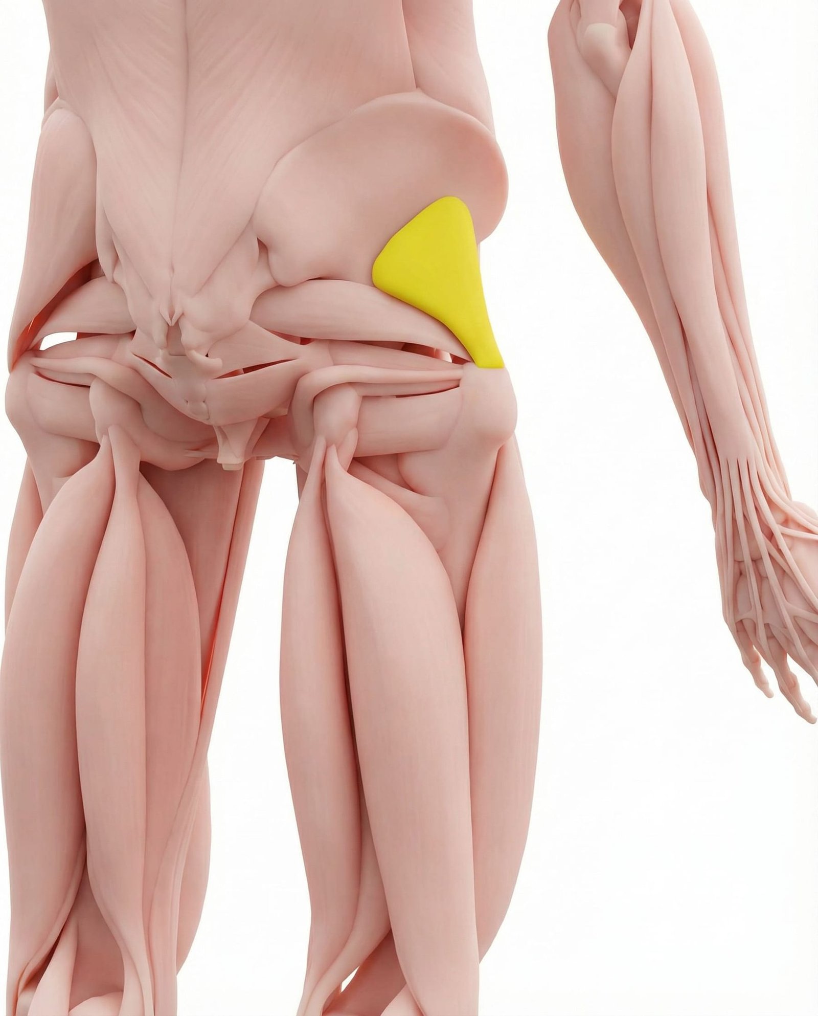
Often referred to in clinical literature as the "pseudo-sciatica" muscle. Trigger points in the posterior aspect of this muscle can project pain down the posterior thigh and calf, closely mimicking an S1 radiculopathy. Anterior trigger points may refer laterally down the leg, mimicking an L5 radiculopathy.
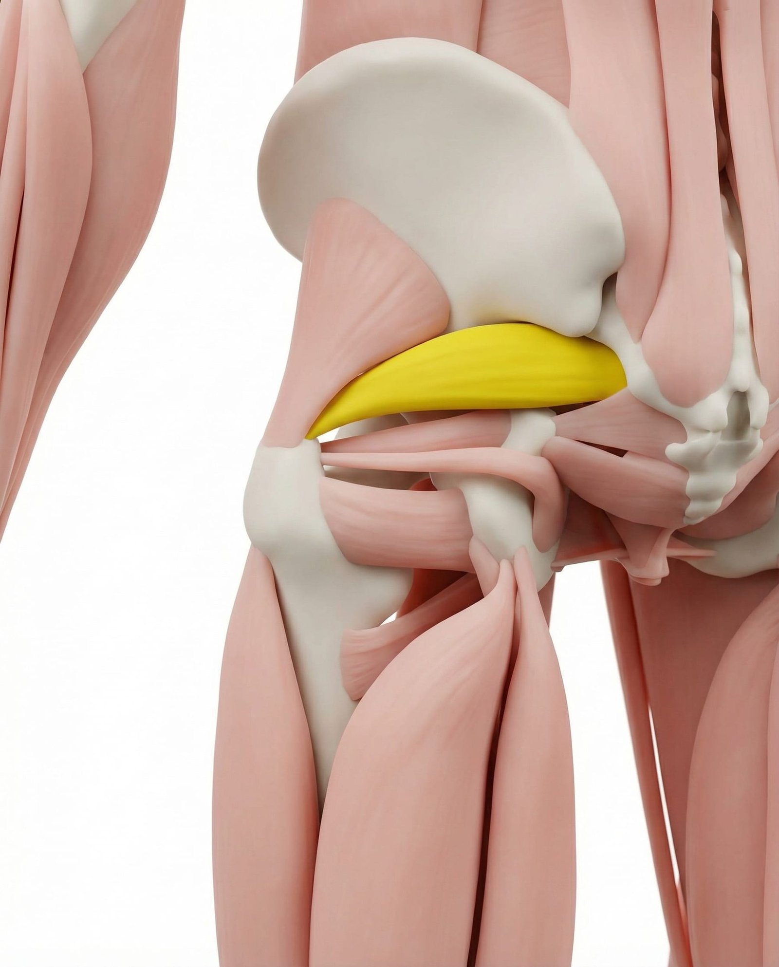
This deep external rotator can contribute to leg pain in two distinct ways. Myofascial trigger points can refer pain to the posterior thigh. Additionally, hypertrophy or spasm of the muscle may mechanically irritate the sciatic nerve (piriformis syndrome), producing true neuropathic pain without lumbar disc pathology.
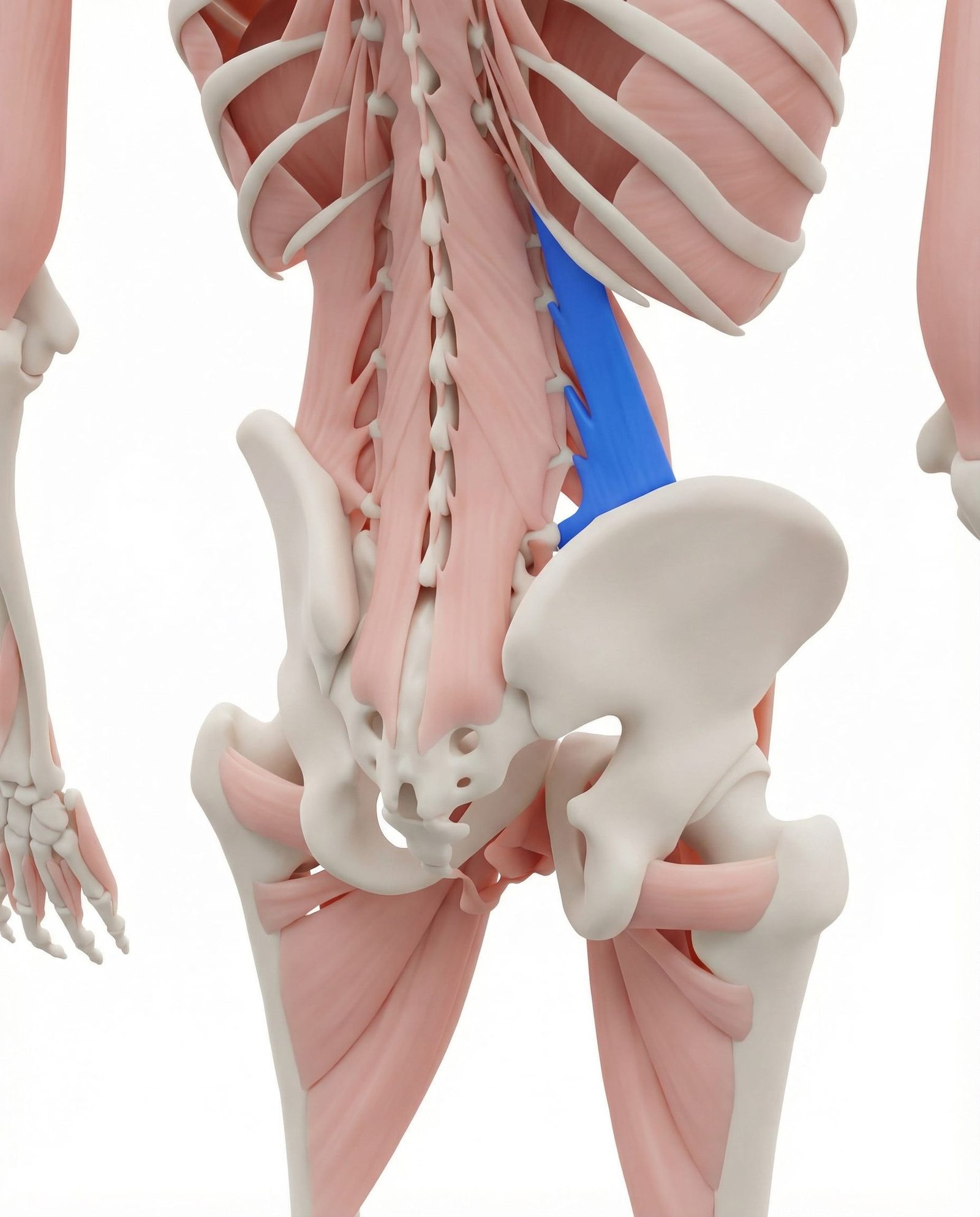
A primary source of severe, acute lower back pain. While it does not typically refer pain below the knee, its referral pattern to the sacroiliac joint and lower buttock can mimic the proximal symptoms of high lumbar (L1-L3) radiculopathy or facet joint pathology.
Often develops satellite trigger points secondary to true L5 or S1 radiculopathy due to altered gait and chronic nerve irritation. When active, these trigger points can perpetuate posterior thigh pain even after the primary lumbar pathology has stabilized.
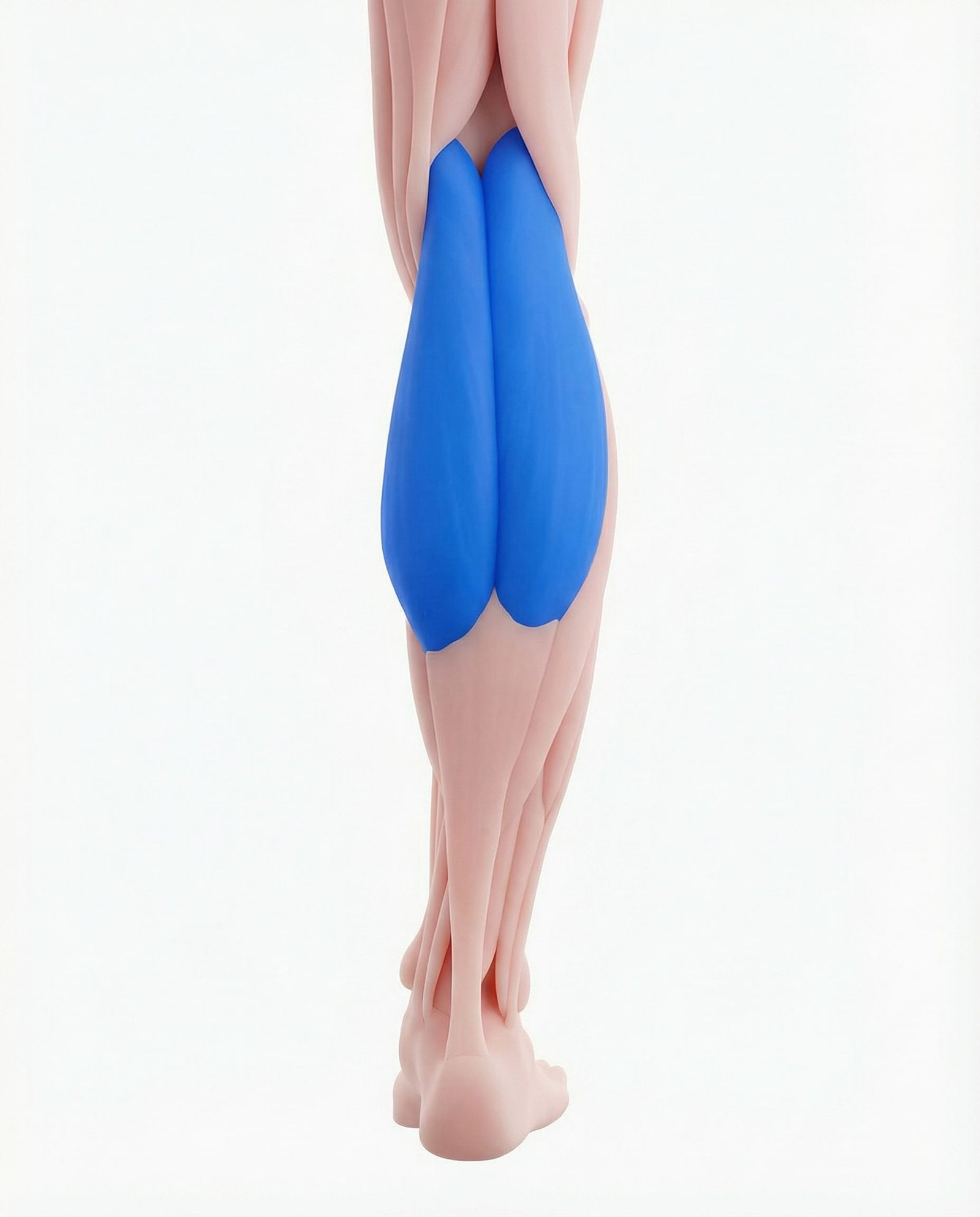
Trigger points in the calf muscles frequently coexist with S1 radiculopathy. They can reproduce deep, aching calf pain and nocturnal cramping that patients often confuse with ongoing nerve root compression.
Paraspinal muscle spasm is a common protective response to lumbar disc herniation. Trigger points here primarily cause localized back pain but can refer to the buttock, complicating the clinical picture when assessing radicular symptoms.
Pain Quality
Radiculopathy often causes sharp, shooting, or electrical pain. Myofascial pain is typically described as a deep, severe ache.
Neurological Signs
True weakness, absent reflexes, and precise dermatomal numbness suggest radiculopathy. Vague tingling without objective sensory loss suggests MPS.
Provocation
Radicular pain is often worsened by spinal flexion or coughing (Valsalva). Myofascial pain is typically reproduced by direct muscle compression.
Imaging Correlation
MRI findings of nerve compression must match the patient's specific symptoms. Incidental disc bulges are common and may not be the pain source.
Referral Patterns vs. Dermatomes
Distinguishing between a nerve root dermatome and a myofascial referral pattern is a cornerstone of physical diagnosis. While they may overlap geographically, their clinical characteristics often differ.
Gluteus Minimus vs. L5/S1 Radiculopathy
Piriformis vs. Sciatic Nerve Entrapment
Quadratus Lumborum vs. High Lumbar Radiculopathy
Clinical Assessment
Clinicians utilize a combination of neural tension tests and muscular palpation to differentiate the source of radiating leg pain. These assessments help determine the mechanosensitivity of the nervous system versus the presence of active trigger points.
Straight Leg Raise (SLR)
Slump Test
FAIR Test (Flexion, Adduction, Internal Rotation)
Gluteal Palpation with Hip Range of Motion
Evidence-Based Management
Management of radiating leg pain often requires a multimodal approach. Current guidelines generally recommend exhausting conservative measures—including physical therapy, manual therapy, and pharmacological support—before considering interventional or surgical options, provided no red flags are present.
Self-Care
- Activity modification to avoid prolonged sitting or heavy lifting
- Short-term use of NSAIDs as directed by a physician
- Application of heat to spastic muscles or ice to acute lumbar pain
- Gentle, pain-free nerve gliding (flossing) exercises
- Use of a supportive lumbar roll when seated
Manual Therapy
- Ischemic compression of gluteus minimus and piriformis trigger points
- Lumbar spine mobilization to improve segmental mechanics
- Soft tissue massage targeting the lumbar erector spinae
- Myofascial release of the hamstrings and calves
- Passive stretching of the deep external hip rotators
Interventional
- Lumbar epidural steroid injections for confirmed radicular inflammation
- Trigger point injections for refractory gluteal myofascial pain
- Dry needling of the piriformis and gluteus minimus
- Ultrasound-guided piriformis block for diagnostic differentiation
- Selective nerve root blocks for diagnostic specificity
Rehabilitation
- Directional preference exercises (e.g., McKenzie extension protocols)
- Core stabilization and motor control training
- Progressive loading of the posterior chain
- Postural re-education for sitting and standing mechanics
- Graded return to functional activities and exercise
Red Flags — When to See a Doctor
While most cases of radiating leg pain are benign and self-limiting, certain signs and symptoms may indicate severe neurological compromise or serious underlying systemic pathology requiring urgent medical intervention.
Gluteus minimus trigger points are the most common myofascial mimics of L5 and S1 radiculopathy, capable of referring pain down to the ankle.
Because asymptomatic lumbar disc herniations are highly prevalent, MRI findings must be carefully correlated with clinical examination to avoid misdiagnosis.
Myofascial pain and true lumbar radiculopathy frequently coexist; nerve root irritation can precipitate the development of satellite trigger points in the leg.
True radiculopathy is typically characterized by dermatomal pain, paresthesia, and potential motor/reflex deficits, whereas myofascial pain lacks hard neurological signs.
Piriformis syndrome represents a peripheral nerve entrapment that can cause true sciatic nerve irritation without lumbar disc pathology.
Current guidelines generally recommend a trial of conservative care (including physical therapy and trigger point management) for 6 to 12 weeks before considering surgical intervention for non-emergent radiculopathy.
Movement therapy
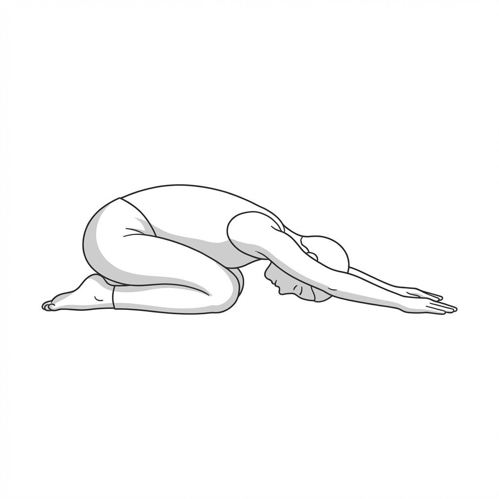
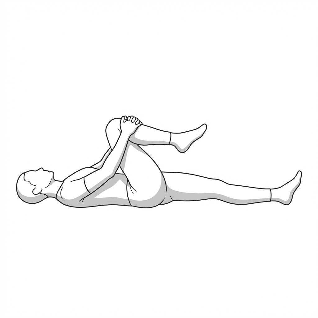
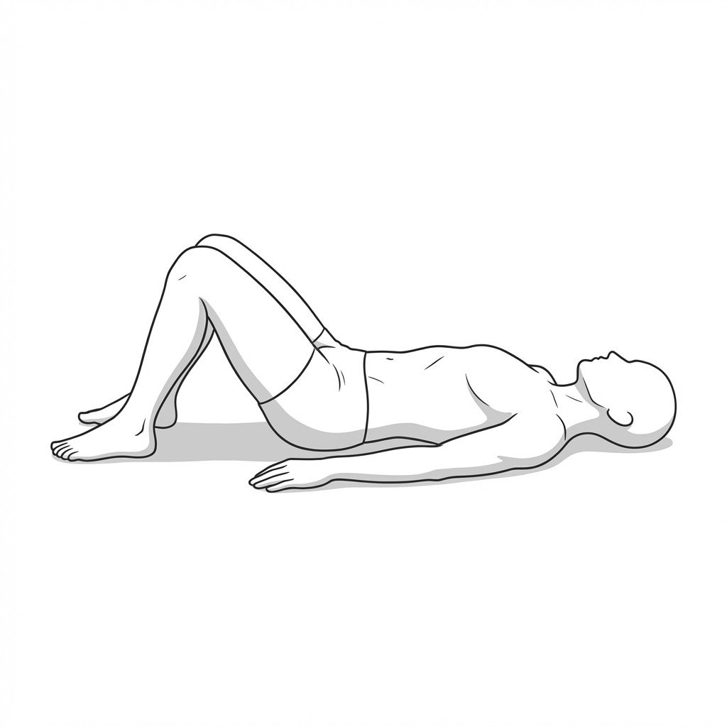
Exercises for Lumbar Radiculopathy
These exercises use lumbar flexion and posterior pelvic movement to create intervertebral space and reduce compressive load on irritated nerve roots. Start with 2–3 and progress as tolerated.