Understanding Posterior Elbow Pain
The distal triceps tendon joins the back of the upper arm to the olecranon, the bony tip of the elbow. Repeated pressing or forceful extension can provoke pain here. A gradual load-related presentation differs from a sudden pop, bruising and loss of extension strength after a fall or heavy effort; suspected rupture needs prompt assessment before rehabilitation.
Posterior elbow pain may also involve the superficial olecranon bursa, the joint, regional muscle pain or neural referral. Swelling directly over the tip, a sharp end-range pinch, locking, or spreading neurological symptoms each changes the examination. Tenderness near the tendon cannot resolve this differential alone.
New weakness after an injury deserves assessment before another loading session.
Record the onset, recent changes in pressing or work volume, swelling and the tasks that have become difficult. Examine active motion and extension strength, then compare tendon, bursal and muscle findings. Add shoulder or cervical assessment when proximal pain, altered sensation or disproportionate weakness supports it. Imaging is useful when the result would clarify a suspected injury or change management.
Muscles and the Posterior Elbow
The triceps supplies elbow extension and the anconeus assists around the posterolateral elbow. The tendon, superficial bursa and posterior joint are close together; examine the symptom behaviour as well as location.
Triceps Brachii
Its three heads converge toward the distal tendon at the olecranon. The long head also crosses the shoulder. Compare painful extension with actual weakness and the onset history; tendon and muscle tenderness can overlap without a separate trigger-point diagnosis.
Posterior arm or elbow pain during loading does not by itself distinguish tendon from muscle.Anconeus
This small posterolateral muscle assists elbow extension and stability. Local tenderness is one examination finding; it cannot rule out joint irritation or identify an isolated source from a referral map.
Posterolateral discomfort should be correlated with motion and joint findings.Infraspinatus
A shoulder external rotator worth examining when shoulder pain or restricted shoulder function accompanies arm pain. It is not a default explanation for pain at the triceps insertion.
A broader shoulder-to-arm complaint may justify proximal assessment.Teres Minor
Another posterior rotator-cuff muscle. Test shoulder movement and strength when the history supports a shoulder contribution; elbow pain alone does not implicate this muscle.
Posterior shoulder symptoms have limited ability to identify one specific muscle.Posterior Deltoid
Helps move the arm backward at the shoulder. A localized upper-arm complaint during shoulder movement differs from focal elbow pain on extension and should be examined in its own context.
Proximal posterior arm discomfort is a regional clue, not a confirmed referral source.Subscapularis
A shoulder internal rotator. Restricted rotation or shoulder pain can justify assessment, but unexplained posterior elbow pain does not require deep subscapularis palpation or release.
Include the shoulder and neck when symptoms extend proximally; do not infer a hidden muscle cause.
Load-related tendon pain
Extension-related pain near the insertion may fit tendinopathy, but pain does not establish the extent of structural change.
Muscle symptoms
Posterior arm tenderness or ache may accompany loading; pressure reproduction alone does not identify a causal trigger point.
Superficial swelling
The olecranon bursa can swell over the elbow tip. Redness, heat or fever changes the urgency of assessment.
Joint or neural signs
Locking, a sharp terminal pinch, altered sensation or disproportionate weakness requires a broader examination.
Interpreting Regional Pain
Regional pain descriptions help decide whether to include the shoulder or neck. They are not diagnostic contours and do not replace testing elbow function.
- Distal triceps region
- Tenderness near the olecranon and pain with extension can fit tendon loading, while bruising or new weakness after trauma raises concern for a tear. Muscle tenderness does not exclude either.
- Long-head triceps region
- Because the long head crosses the shoulder, arm position changes its load. Posterior arm aching during overhead or pressing tasks should be compared with both elbow and shoulder movement.
- Anconeus and posterolateral elbow
- Localized pain may overlap muscle and joint regions. A terminal pinch, catching or loss of motion calls for a joint assessment rather than increasingly forceful massage.
- Shoulder and cervical contributors
- Proximal pain, restricted shoulder motion, neck-related symptoms or altered sensation justifies a broader examination. No posterior-arm contour reliably identifies subscapularis or a cuff muscle.
Clinical Assessment and Differentiation
These checks describe a clinical examination. Following acute trauma, do not test maximum strength or repeatedly provoke the elbow; establish whether a tear or bony injury is possible first.
History and resisted elbow extension
After injury concerns are addressed, support the arm and apply modest resistance to elbow extension in a comfortable range. A clinician compares sides, pain location and strength at more than one elbow position.
Interpretation: Familiar insertion-region pain is compatible with tendon loading but is not tissue-specific. Marked weakness or a sudden change after trauma prompts structural assessment rather than more testing.
Olecranon and swelling examination
Inspect the elbow tip and compare gentle palpation of the tendon insertion, the superficial bursal area and more proximal muscle. Do not press hard into red or hot swelling.
Interpretation: Superficial fluctuant swelling suggests bursal involvement; focal insertion tenderness may accompany tendon pain. Coexistence is possible, and fever or heat changes the urgency.
Muscle and proximal screen
Check gentle posterior-arm palpation against the familiar complaint. Examine shoulder movement and, when indicated, cervical movement, sensation and neurological strength.
Interpretation: Reproduction with pressure is not proof of a causal trigger point. Symptoms linked to shoulder or neck movement, sensory change or unexplained weakness require broader interpretation.
Active extension and mechanical symptoms
Move the elbow comfortably without forcing the final degrees. Tell the examiner about catching, a sharp terminal pinch or an actual block; do not repeatedly push into it.
Interpretation: Locking or blocked motion raises a joint or bony question. This is different from a tolerable ache during resistance exercise and needs assessment before load progression.
Loading, Reassessment and Treatment Limits
For a non-traumatic load-related presentation after assessment, use an individualized progression toward the task that matters. Direct trials do not establish a best chronic distal-triceps exercise dose here. Holds, controlled lifting and lowering, and later heavier tasks are adaptable loading choices; eccentric skull crushers are not an established primary treatment. Rupture rehabilitation follows a separate clinician-directed plan.
Self-care and exposure
- Reduce the pressing weight, repetition count or painful range that reliably provokes symptoms. Keep comfortable daily movement.
- Avoid direct pressure over a swollen elbow tip; padding may help reduce contact during work.
- Do not use aggressive stretching, deep friction or heavy skull crushers to test a newly injured elbow.
- Heat or cold may be a short comfort trial with skin protection; neither establishes the diagnosis or repairs the tendon.
- Record a repeatable light task and its next-morning response rather than judging recovery by tenderness alone.
Adjuncts with reassessment
- Gentle soft-tissue treatment can be an optional comfort measure; do not assign it as the essential treatment for muscle-dominant pain.
- Use a measured change in movement or task tolerance to decide whether an adjunct is worthwhile.
- A joint restriction needs examination before any mobilization; do not force a locking or sharply pinching elbow.
- Treat shoulder findings only when the examination supports a separate relevant impairment.
- Do not add routine ulnar nerve glides to tendon rehabilitation; tingling or hand weakness requires a nerve assessment.
Imaging and specialist decisions
- Promptly assess suspected partial or complete rupture; ultrasound or MRI may define the injury and guide a specialist plan.
- A hot swollen bursa may need investigation for infection. Aspiration is a clinical decision, not home care or a routine tendon treatment.
- Do not extrapolate results from surgically treated professional players to an individual with chronic tendon pain.
- The sources cited here do not establish routine PRP, shockwave, needling or injection benefit for chronic distal-triceps tendinopathy.
- Persistent weakness, progressive swelling or failure to regain function should trigger reassessment before procedural escalation.
Progressive extension capacity
- Begin with low resistance at a comfortable elbow angle; choose a short hold or controlled short-range extension according to tolerance.
- Build controlled lifting and lowering through more range before adding heavier resistance; change one load variable at a time.
- Monitor later-day and next-morning symptoms. Reduce the last increase if pain remains elevated or daily function deteriorates.
- Reintroduce pressing depth, overhead work, speed or repeated sport efforts gradually once ordinary loading is stable.
- Reassess task tolerance and extension strength using the same setup. A plateau prompts review of diagnosis and exposure, not an automatic eccentric-only prescription.
When to Seek Further Assessment
Sudden weakness after trauma and hot swelling with systemic illness need prompt care. Mechanical locking or progressive neurological symptoms also warrant evaluation before continued exercise.
Sudden weakness, bruising or a tendon gap
After a fall, pop or heavy effort, stop resisted exercise and obtain urgent assessment for rupture or bony injury. Ability to extend the elbow does not exclude a partial tear.
UrgentHot swelling, redness or fever
Rapidly increasing swelling over the elbow tip with heat, redness or systemic illness requires urgent assessment for infection or another acute process.
EmergencyProgressive or disproportionate weakness
New loss of extension strength, spreading sensory symptoms or hand weakness needs assessment for structural or neurological causes; do not treat it as routine soreness.
UrgentLocking or blocked extension
A mechanical block, repeated catching or sharp terminal pain warrants examination for joint or bony pathology before forcing range or increasing resistance.
InvestigateProgressive rest or night pain
Pain that becomes unrelenting or unrelated to movement warrants review, especially with systemic illness or unexplained weight loss.
Investigate
Key Clinical Points
Separate gradual loading pain from a traumatic pop, bruising and new extension weakness; the latter needs prompt injury assessment.
Tendon tenderness, muscle tenderness and familiar pain reproduction can overlap without identifying a unique pain generator.
Superficial swelling, locking and neurological symptoms each require a different assessment from straightforward load-related pain.
Shoulder or neck screening follows relevant symptoms and findings; a hidden proximal trigger point should not be presumed.
Use adaptable graded extension loading with repeatable functional reassessment. Eccentric-only superiority and a fixed triceps dose are not established here.
The cited professional-football rupture series informs injury vigilance, not a prognosis or rehabilitation prescription for chronic triceps tendinopathy.
Movement options
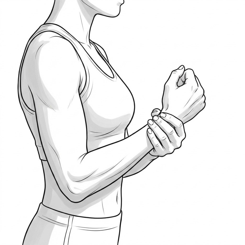
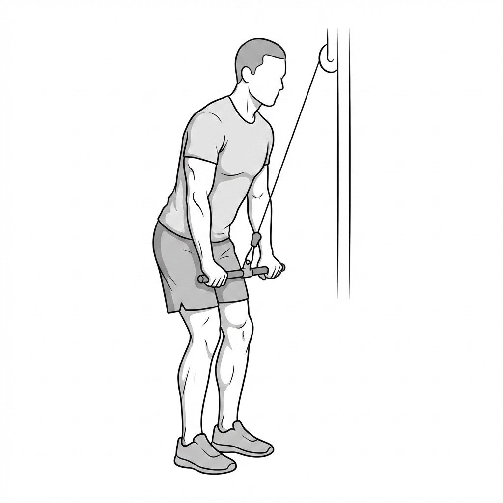
Exercises for Triceps Tendinopathy
These examples belong after suspected rupture and other competing problems have been assessed. A light isometric hold may provide a starting load; controlled push-downs can be adapted as capacity improves. Neither eccentric-only loading nor a particular exercise dose is established as best for distal triceps pain. Track the later-day and next-morning response, and stop for new weakness or sharp pain.