Understanding Lateral Hip Pain
Greater trochanteric pain syndrome (GTPS) encompasses a spectrum of lateral hip pathology, most notably gluteus medius and minimus tendinopathy. Historically termed trochanteric bursitis, current imaging and histological evidence suggests that primary bursal inflammation is rare, with symptoms predominantly arising from tendon degeneration and partial tearing. Epidemiological data indicate this condition disproportionately affects middle-aged and older women, with prevalence estimates in the literature suggesting it affects roughly one in four to one in six women in their fifties and beyond. The primary mechanical driver is currently understood to be excessive compressive loading of the gluteal tendons against the greater trochanter, often exacerbated by specific postures and biomechanical deficits. Accurate differential diagnosis is critical, as lateral hip pain frequently arises from, or is complicated by, overlapping musculoskeletal and neurological conditions.
Myofascial pain syndrome (MPS) frequently coexists with or mimics gluteal tendinopathy, complicating the clinical picture. Trigger points within the gluteus medius and minimus muscle bellies routinely refer pain directly to the lateral hip and greater trochanter, closely simulating tendon-origin pain. Furthermore, myofascial tension in the tensor fasciae latae (TFL) and the superior fibers of the gluteus maximus can significantly increase tension through the iliotibial band. This elevated fascial tension directly amplifies the compressive forces exerted on the underlying gluteal tendons during adduction, potentially accelerating tendon degeneration. Consequently, addressing myofascial trigger points is often a necessary component of managing the compressive load that drives GTPS.
Current clinical guidelines emphasize that lateral hip pain is rarely an isolated bursitis; it is predominantly a compressive tendinopathy frequently complicated by overlapping myofascial pain.
Differentiating primary tendinopathy from isolated myofascial pain requires a systematic clinical approach focused on load provocation and precise palpation. Tendinopathic pain is typically highly localized to the tendon insertions on the greater trochanter and is reliably provoked by sustained compressive and tensile loads, such as prolonged single-leg stance. In contrast, myofascial pain often presents with palpable taut bands within the proximal muscle bellies and reproduces the patient's familiar pain upon sustained ischemic compression of these trigger points. Clinicians must also consider referred pain from the lumbar spine, particularly L4-L5 radiculopathy, which can present as lateral hip pain without local mechanical provocation. Advanced imaging, such as MRI or ultrasound, may assist in identifying structural tendon changes, though findings must always be correlated with clinical presentation due to the high prevalence of asymptomatic tendon pathology.
Anatomical & Biomechanical Context
The lateral hip is a complex intersection of muscular, fascial, and tendinous structures. Understanding the spatial relationship between the gluteal tendons and the overlying iliotibial band is essential for diagnosing compressive tendinopathy.
Gluteus Medius
The gluteus medius is the primary abductor of the hip and a crucial stabilizer of the pelvis during the stance phase of gait. Tendinopathy typically occurs at its insertion on the lateral and superoposterior facets of the greater trochanter. Myofascial trigger points in the muscle belly frequently refer pain to the lumbar region, posterior crest, and directly over the greater trochanter, often mimicking insertional tendinopathy. Dysfunction in this muscle is a hallmark of greater trochanteric pain syndrome (GTPS).
Pain refers primarily to the posterior crest of the ilium, the sacrum, and the posterior and lateral aspects of the buttock and thigh.Gluteus Minimus
Situated deep to the gluteus medius, the gluteus minimus assists in hip abduction and internal rotation. Its tendon inserts on the anterior facet of the greater trochanter, where it is subjected to significant compressive forces. Trigger points within this muscle are notorious for producing severe, radiating pain that mimics L5 or S1 radiculopathy (pseudosciatica). Differentiating its myofascial referral from true tendinopathy or lumbar nerve root compression requires careful neurological and mechanical assessment.
Pain refers intensely down the lateral aspect of the thigh and calf, sometimes reaching the ankle, closely mimicking sciatica.Tensor Fasciae Latae (TFL)
The TFL works synergistically with the gluteal muscles to stabilize the pelvis and tension the iliotibial (IT) band. Increased tone or myofascial trigger points in the TFL significantly increase tension through the IT band. This increased tension directly amplifies the compressive load on the underlying gluteus medius and minimus tendons against the greater trochanter. Managing TFL tension is therefore often a critical component of reducing compressive forces in GTPS.
Pain typically refers to the anterolateral thigh, extending from the greater trochanter down toward the lateral knee.Piriformis
The piriformis is a key external rotator of the hip when extended, and an abductor when the hip is flexed. While not directly involved in lateral trochanteric tendinopathy, piriformis syndrome or myofascial pain can refer laterally, complicating the diagnostic picture. Furthermore, altered biomechanics secondary to piriformis dysfunction can shift load to the gluteal abductors, predisposing them to tendinopathy. Assessment should rule out deep gluteal space pathology when evaluating lateral hip pain.
Pain refers to the sacroiliac region, the buttock, and occasionally down the posterior thigh.Gluteus Maximus
As the largest muscle of the hip, the gluteus maximus is a powerful extensor and external rotator. Its superficial fibers insert into the IT band, meaning increased tension here also contributes to lateral compressive forces over the greater trochanter. Trigger points in the gluteus maximus can refer pain to the gluteal fold and lateral hip. Addressing myofascial restrictions in this muscle is often necessary to fully decompress the lateral hip compartment.
Pain refers locally to the buttock, the gluteal fold, and occasionally to the lateral aspect of the hip.Quadratus Femoris
The quadratus femoris is a short external rotator located deep in the posterior hip. While its primary pathology is ischiofemoral impingement, it is clinically relevant in the differential diagnosis of hip pain. Pain from this region is typically posterior and deep, but patients may struggle to localize it, sometimes describing it as lateral or generalized hip pain. It is generally distinguished from GTPS by provocation during extension and adduction rather than direct lateral compression.
Pain refers deep into the lower buttock and posterior thigh, often worsening with long strides.
Compressive Tendinopathy
Degeneration of the gluteus medius/minimus tendons due to sustained compression against the greater trochanter by the IT band.
Myofascial Referral
Active trigger points in the proximal gluteal muscle bellies referring pain distally to the trochanteric region.
Lumbar Radiculopathy
Irritation of the L4 or L5 nerve roots referring neuropathic pain to the lateral hip and thigh.
Intra-articular Pathology
Hip osteoarthritis or labral tears occasionally presenting with lateral pain, though typically groin pain is predominant.
Myofascial Referral Patterns
Myofascial trigger points in the hip and pelvic musculature frequently refer pain to the lateral hip. Recognizing these patterns is crucial to avoid misdiagnosing myofascial pain as primary tendinopathy or lumbar radiculopathy.
- Gluteus Medius & Minimus
- Trigger points in the gluteus medius refer locally to the lateral hip and buttock, while the gluteus minimus produces a classic "pseudosciatica" pattern radiating down the lateral thigh and calf. Both can perfectly mimic the localized pain of greater trochanteric pain syndrome.
- Tensor Fasciae Latae (TFL)
- Pain concentrates over the greater trochanter and spills down the anterolateral thigh toward the knee. This pattern is frequently mistaken for IT band syndrome or lateral compartment tendinopathy.
- Lumbar Spine (L4-L5 Referral)
- Nerve root irritation at L4 or L5 frequently refers pain to the lateral hip and thigh. Unlike local tendinopathy, this pain is often described as burning or electrical and may be accompanied by neurological deficits.
Clinical Assessment & Differentiation
Clinical differentiation relies on specific provocation tests that selectively load the gluteal tendons, alongside careful palpation to distinguish insertional pain from proximal muscle belly trigger points.
Single Leg Stance Test
The patient stands unassisted on the symptomatic leg for up to 30 seconds. The clinician observes for pelvic drop (Trendelenburg sign) and asks the patient to report any reproduction of lateral hip pain.
Positive: Reproduction of the patient's familiar lateral hip pain within 30 seconds suggests gluteal tendinopathy, as this position places high compressive and tensile loads on the tendons.
Resisted External Derotation Test
The patient lies supine with the hip flexed to 90 degrees and externally rotated. The clinician passively moves the hip into internal rotation while the patient actively resists the movement.
Positive: Pain over the greater trochanter during resisted internal rotation strongly implicates the gluteal tendons; this test has demonstrated good diagnostic accuracy for refractory GTPS in clinical validation studies.
Palpation: Tendon vs. Muscle Belly
The clinician systematically palpates the facets of the greater trochanter (tendon insertions) and then moves proximally into the muscle bellies of the gluteus medius and minimus, feeling for taut bands.
Positive: Exquisite tenderness localized strictly to the trochanter suggests tendinopathy, whereas pain reproduced by compressing taut bands in the proximal muscle belly suggests a primary myofascial component.
FABER (Patrick's) Test
The patient lies supine, and the symptomatic leg is placed in Flexion, ABduction, and External Rotation (ankle resting on the opposite knee). Gentle downward pressure is applied to the flexed knee.
Positive: While traditionally a test for SI joint or intra-articular hip pathology, lateral hip pain during this test can indicate compressive irritation of the gluteal tendons or severe myofascial restriction.
Evidence-Based Treatment Pathways
Management of gluteal tendinopathy has shifted away from passive modalities and corticosteroid injections toward active load management and progressive tendon rehabilitation, supported by trials such as the LEAP study.
Self-Care
- Avoid crossing legs while sitting or standing
- Sleep with a pillow between knees to prevent adduction
- Modify activities to reduce prolonged single-leg loading
- Avoid stretching the IT band, which increases compression
- Apply ice to the lateral hip following aggravating activities
Manual Therapy
- Ischemic compression of gluteus medius/minimus trigger points
- Myofascial release of the TFL and gluteus maximus
- Soft tissue mobilization to reduce IT band tension
- Joint mobilization of the lumbar spine and hip
- Dry needling of proximal gluteal muscle bellies
Interventional
- Corticosteroid injections (short-term relief, potential long-term tendon risk)
- Platelet-rich plasma (PRP) injections (emerging evidence)
- Ultrasound-guided percutaneous needle tenotomy
- Shockwave therapy (ESWT) for recalcitrant tendinopathy
- Diagnostic local anesthetic blocks to confirm pain generator
Rehabilitation
- Progressive tendon loading (e.g., LEAP trial protocol)
- Isometric hip abduction exercises for pain modulation
- Slow, heavy isotonic strengthening of hip abductors
- Core and pelvic stability training
- Gait retraining to reduce excessive pelvic drop
Red Flags & Differential Diagnosis
While lateral hip pain is usually benign and mechanical in nature, clinicians must remain vigilant for signs of severe structural failure, systemic disease, or significant neurological compromise.
Avascular Necrosis (AVN)
Progressive, deep hip or groin pain that worsens with weight-bearing and is present at rest. Risk factors include high-dose corticosteroid use, excessive alcohol intake, or previous hip trauma.
UrgentFemoral Neck Stress Fracture
Deep anterior thigh or groin pain, particularly in endurance athletes or older individuals with osteoporosis. Pain is typically severe with weight-bearing and may present with an inability to hop on the affected leg.
EmergencySeptic Arthritis
Acute onset of severe hip pain, inability to bear weight, accompanied by fever, chills, or systemic malaise. The joint is typically held in slight flexion, abduction, and external rotation.
EmergencyMalignancy
Unremitting pain that is worse at night, history of primary cancer (especially breast, prostate, lung, or kidney), and unexplained weight loss. Bone metastases frequently occur in the proximal femur.
InvestigateAcute Full-Thickness Tendon Tear
Sudden onset of severe lateral hip pain and profound weakness in hip abduction, often following a fall or sudden exertion. Patient may exhibit a pronounced, uncompensated Trendelenburg gait.
Urgent
Clinical Pearls
Greater trochanteric pain syndrome (GTPS) is primarily a tendinopathy of the gluteus medius and minimus, not an isolated bursitis.
Compressive loading—often from adducted postures like crossing the legs—is a primary mechanical driver of gluteal tendon degeneration.
Myofascial trigger points in the gluteus medius and minimus can perfectly mimic the localized lateral hip pain of GTPS.
Increased tension in the TFL and gluteus maximus amplifies compressive forces on the gluteal tendons via the IT band.
Current evidence suggests progressive, heavy-load exercise programs (like the LEAP protocol) are superior to corticosteroid injections for long-term outcomes.
Lumbar radiculopathy (L4-L5) must be ruled out, as it frequently refers pain to the lateral hip and thigh without local mechanical provocation.
Movement therapy
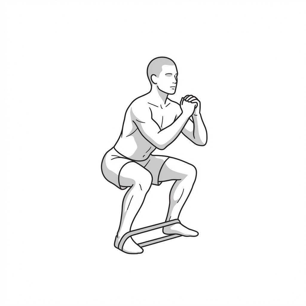
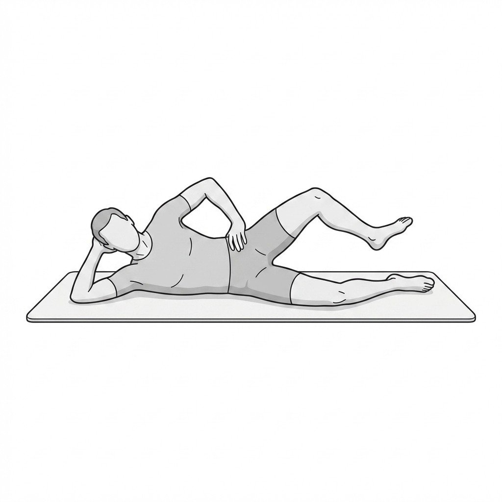
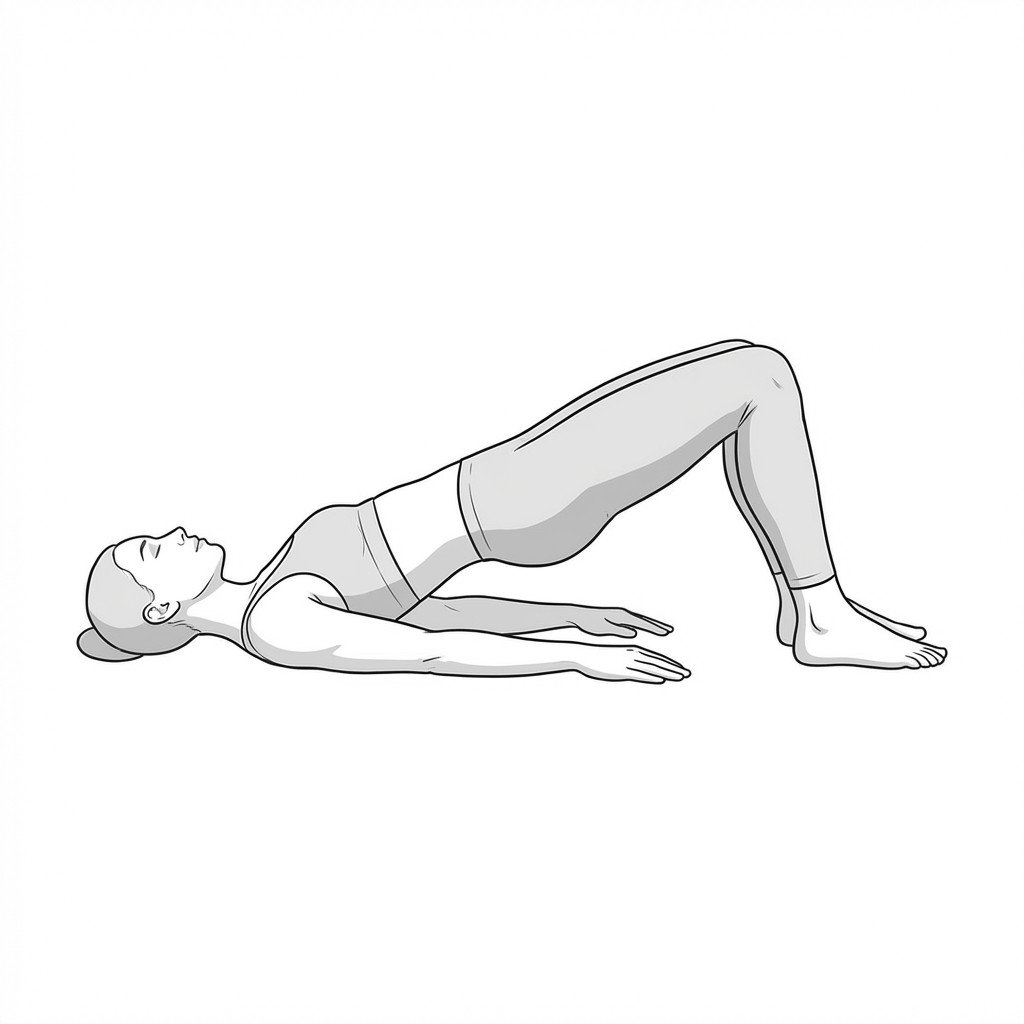
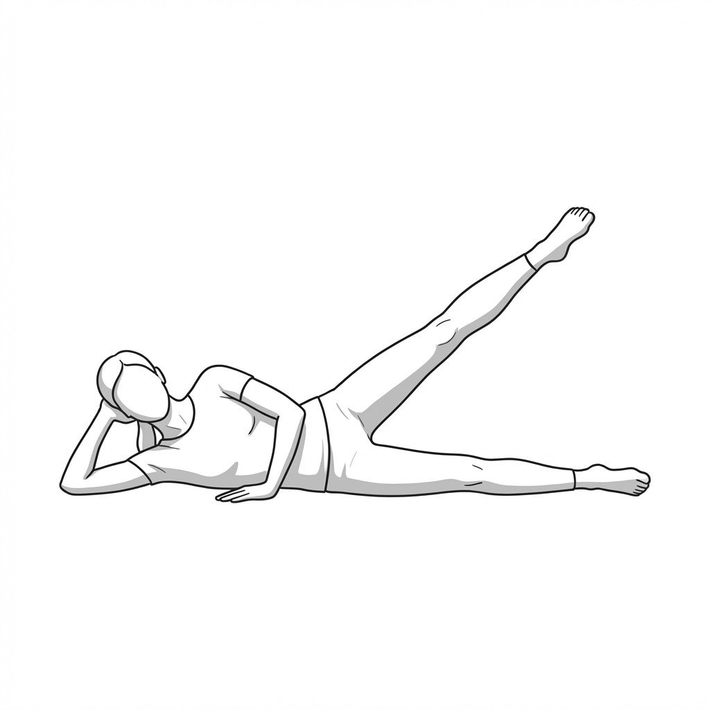
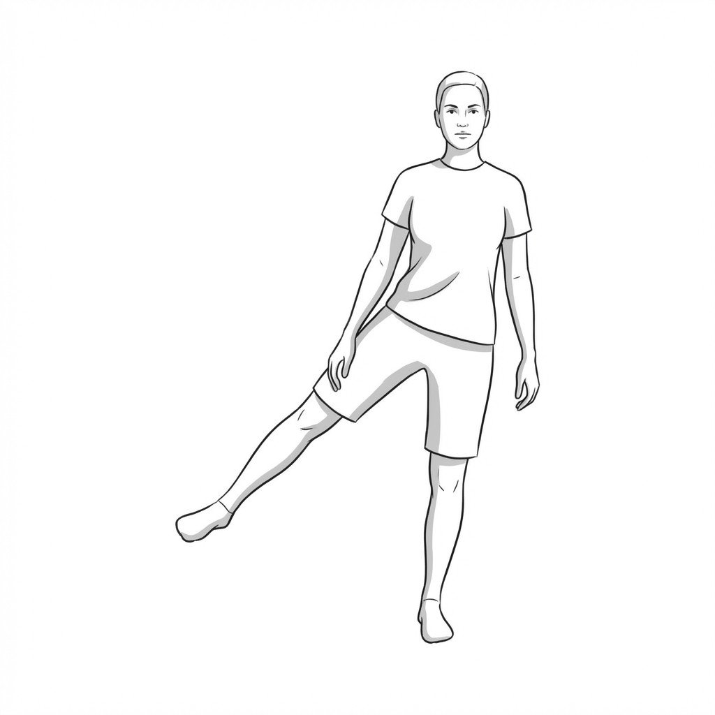
Exercises for Gluteal Tendinopathy
Gluteal tendinopathy responds to progressive hip abductor loading — beginning with pain-free isometrics that build tendon tolerance before advancing to banded dynamic work that challenges hip control in standing. Start with 2–3 and progress as tolerated.