Understanding PTTD & Medial Ankle Pain
Posterior tibial tendon dysfunction (PTTD), often conceptualized within the broader spectrum of adult-acquired flatfoot deformity, represents a progressive failure of the primary dynamic stabilizer of the medial longitudinal arch. Epidemiological data suggest this condition predominantly affects middle-aged to older adults, with a higher reported prevalence in women, though it can occur in any demographic subject to chronic medial column overload. The Johnson and Strom staging system, later extended to include ankle joint involvement, remains a widely used framework — categorizing pathology from early tenosynovitis without deformity (Stage I), to flexible flatfoot (Stage II), rigid deformity (Stage III), and eventual ankle joint involvement (Stage IV). Differentiating early-stage PTTD from other sources of medial ankle pain is critical, as timely intervention may help prevent further structural collapse. A high index of suspicion is reasonable in any patient presenting with insidious onset of medial hindfoot swelling and pain along the tendon's course.
The clinical presentation of early PTTD frequently overlaps with, or is complicated by, myofascial pain syndrome (MPS) affecting the deep posterior compartment of the leg. Trigger points within the tibialis posterior muscle belly can refer pain distally to the medial malleolus, Achilles region, and plantar midfoot, closely mimicking the nociceptive pattern of intrinsic tendinopathy. Furthermore, the flexor hallucis longus (FHL) and flexor digitorum longus (FDL) share the retromalleolar anatomical space; myofascial dysfunction in these synergists often contributes to posteromedial ankle pain and may be misidentified as isolated PTTD. A critical biomechanical link exists between gastrocnemius equinus (calf tightness) and arch collapse, as restricted ankle dorsiflexion forces compensatory midfoot pronation, thereby increasing tensile loads on the tibialis posterior tendon. Consequently, myofascial restrictions in the triceps surae complex are frequently observed as significant contributing factors to the progression of flatfoot deformity.
Accurate diagnosis requires distinguishing the structural failure of the tibialis posterior tendon from the referred pain of deep calf myofascial trigger points, as both present with remarkably similar medial ankle symptoms.
Differentiating myofascial pain from structural tendon failure requires a systematic clinical approach, emphasizing both dynamic functional testing and precise palpation. The single-leg heel rise test is a cornerstone of physical examination; an inability to perform this maneuver strongly suggests Stage II PTTD, whereas patients with isolated myofascial pain typically retain this functional capacity, albeit with potential discomfort. Clinicians should carefully distinguish between focal swelling posterior to the medial malleolus—characteristic of tenosynovitis—and the proximal, deep calf tenderness indicative of tibialis posterior trigger points. Diagnostic ultrasound and magnetic resonance imaging (MRI) are often utilized to assess tendon integrity, fluid accumulation, and spring ligament involvement, providing objective data when clinical findings are equivocal. Recognizing the potential coexistence of tarsal tunnel syndrome, driven by localized edema or structural narrowing, is also essential for comprehensive patient management.
Anatomical & Myofascial Contributors
The medial ankle is a complex anatomical junction where the tendons of the deep posterior compartment navigate behind the medial malleolus. Dysfunction in these structures, whether structural or myofascial, profoundly impacts foot biomechanics.
Tibialis Posterior
The primary dynamic stabilizer of the medial longitudinal arch. In Posterior Tibial Tendon Dysfunction (PTTD), this tendon undergoes progressive degeneration and elongation. Myofascial trigger points within the proximal muscle belly can refer pain distally, closely mimicking the symptoms of early-stage tendinopathy.
Refers pain primarily to the Achilles tendon region, the medial malleolus, and the plantar aspect of the midfoot.Flexor Hallucis Longus (FHL)
A deep posterior compartment muscle whose tendon courses posterior to the medial malleolus alongside the tibialis posterior. FHL tenosynovitis or myofascial dysfunction frequently coexists with PTTD. Trigger points here are a common, often overlooked source of posteromedial ankle pain.
Refers pain to the plantar aspect of the great toe, the first metatarsal head, and the posteromedial ankle.Flexor Digitorum Longus (FDL)
Situated medially in the deep posterior compartment, the FDL assists in plantar flexion and inversion. While less commonly the primary source of pathology than the tibialis posterior, it often develops compensatory myofascial overload as the medial arch begins to fail.
Refers pain to the plantar aspect of the lesser toes and the medial forefoot.Gastrocnemius
The superficial biarticular calf muscle. Gastrocnemius tightness (equinus) restricts ankle dorsiflexion, forcing compensatory pronation through the midfoot during gait. This biomechanical fault is widely considered a primary driver of increased tensile stress on the tibialis posterior tendon.
Refers pain locally to the calf, posterior knee, and occasionally to the instep of the foot.Soleus
The deep, monoarticular calf muscle responsible for postural control and plantar flexion. Soleal trigger points can contribute to overall triceps surae tension, exacerbating the equinus forces that accelerate medial arch collapse in PTTD.
Refers pain to the posterior heel, Achilles region, and the plantar surface of the heel.Peroneus Longus and Brevis
Lateral compartment muscles that act as evertors of the foot. In later stages of PTTD (Stage II and beyond), as the foot falls into a valgus (pronated) position, the peroneals often undergo adaptive shortening and may develop secondary myofascial pain due to chronic contracture.
Refers pain to the lateral malleolus, the lateral aspect of the foot, and the middle third of the lateral leg.
Tendon Degeneration
Chronic tensile overload leads to microtearing, tenosynovitis, and eventual elongation of the tibialis posterior tendon.
Myofascial Overload
Trigger points in the tibialis posterior and FHL refer pain distally, mimicking or exacerbating the symptoms of tendinopathy.
Biomechanical Collapse
Gastrocnemius equinus forces compensatory midfoot pronation, increasing stress on the medial longitudinal arch and spring ligament.
Nerve Entrapment
Swelling from tenosynovitis or structural deformity can compress the tibial nerve in the tarsal tunnel, causing neuropathic pain.
Pain Referral Patterns
Pain perceived at the medial ankle and arch may originate locally from tendon pathology or be referred from proximal myofascial trigger points in the deep calf musculature.
- Tibialis Posterior
- Pain is typically perceived deep within the calf, radiating distally along the tendon's course behind the medial malleolus and inserting into the plantar midfoot. This pattern is frequently indistinguishable from the nociceptive pain of tenosynovitis.
- Flexor Hallucis Longus
- Pain concentrates around the posteromedial ankle and extends along the plantar surface of the foot toward the great toe. It may be exacerbated by resisted great toe plantar flexion.
- Gastrocnemius
- Pain generally localizes to the posterior calf and popliteal space, but distal referral to the instep of the foot can occur, complicating the clinical picture of medial arch pain.
Clinical & Self-Assessment
Clinical evaluation of medial ankle pain involves assessing tendon integrity, dynamic foot posture, and the presence of proximal myofascial restrictions. These tests help differentiate structural failure from functional overload.
Single-Leg Heel Rise Test
While standing unsupported on the affected leg, attempt to rise onto the ball of the foot. Observe the heel from behind; normally, the heel should invert (turn inward) as you rise.
Positive: Inability to perform a single heel rise, or failure of the heel to invert during the rise, strongly suggests Stage II PTTD (tendon incompetence).
Too-Many-Toes Sign
Stand naturally while an observer looks at your feet from directly behind. The observer notes how many toes are visible on the lateral (outside) aspect of the foot.
Positive: Seeing more toes on the affected side compared to the unaffected side indicates forefoot abduction, a hallmark of progressive flatfoot deformity.
Palpation of the Tibialis Posterior Tendon
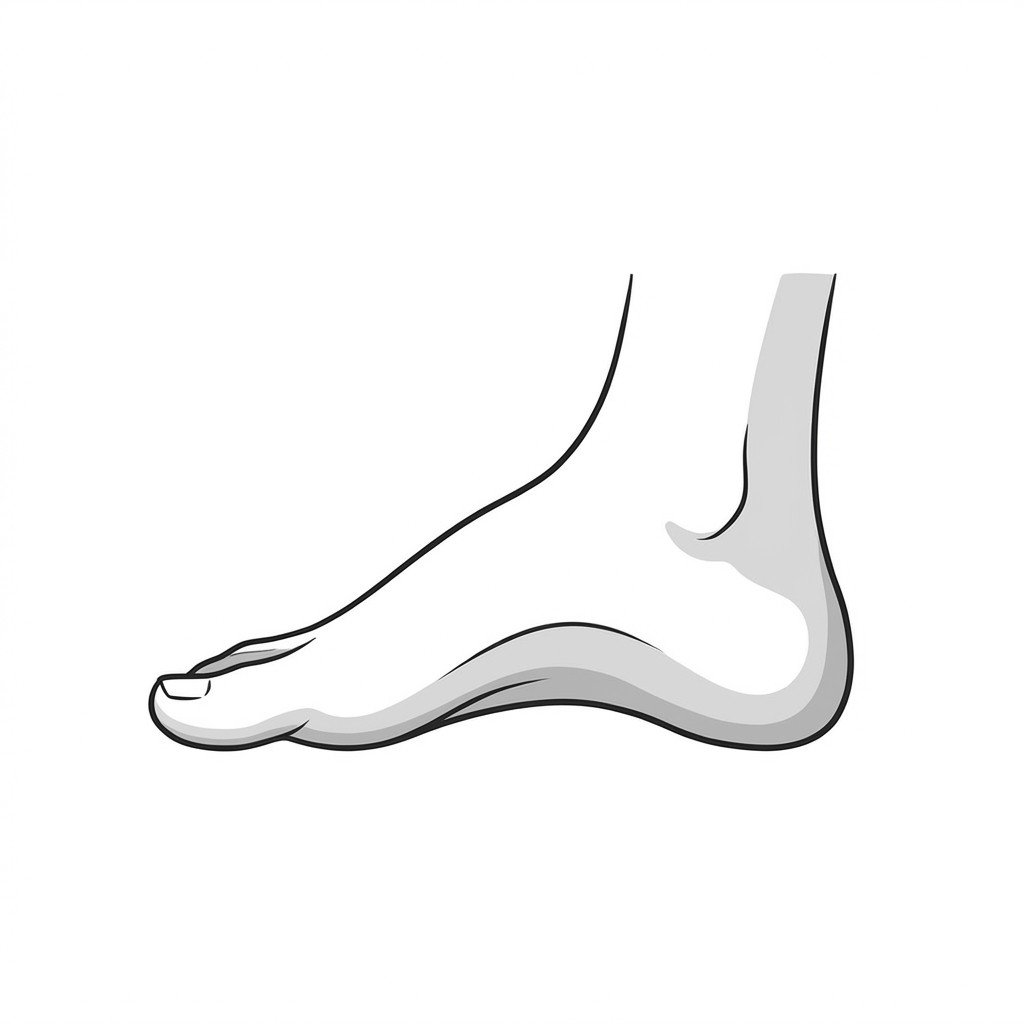
Gently press along the course of the tendon, starting just behind the medial malleolus (inner ankle bone) and following it down to its attachment on the navicular bone in the midfoot.
Positive: Localized tenderness, warmth, or palpable swelling along the tendon sheath suggests tenosynovitis or tendinosis.
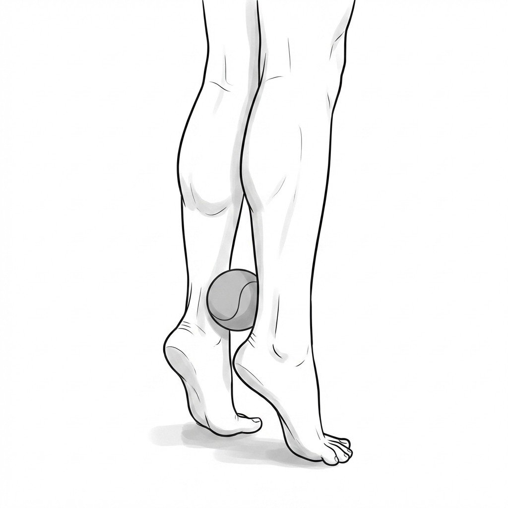
Deep Calf Trigger Point Palpation
Sit with the knee bent. Press deeply into the posterior calf musculature, specifically targeting the medial aspect beneath the gastrocnemius, to locate taut bands in the tibialis posterior muscle belly.
Positive: Reproduction of familiar medial ankle or arch pain upon deep calf compression suggests a significant myofascial component.
Evidence-Based Treatment Pathways
Management of PTTD and associated myofascial pain is stage-dependent. Early intervention focuses on offloading the tendon, addressing biomechanical faults, and resolving myofascial restrictions, while advanced stages may require surgical reconstruction.
Self-Care
- Activity modification to reduce impact loading on the medial column
- Use of supportive footwear with a firm heel counter and medial arch support
- Over-the-counter orthotics to provide temporary arch support
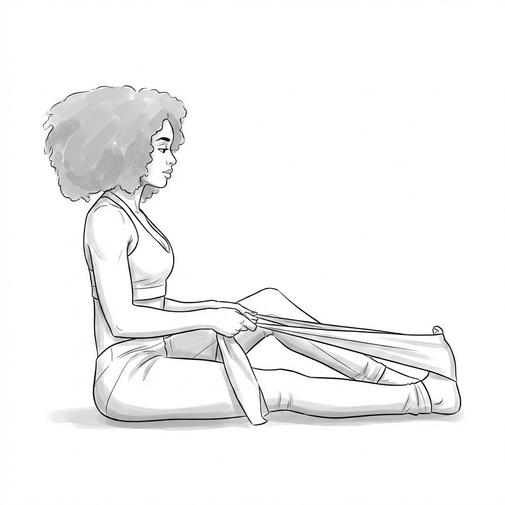
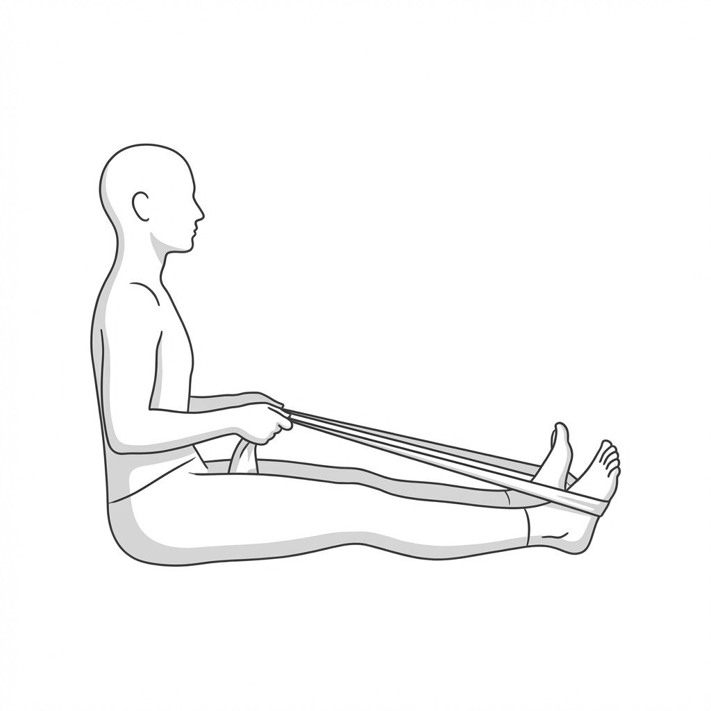
- Gentle, sustained calf stretching (gastrocnemius and soleus) to address equinus
- Ice application to the medial ankle following weight-bearing activities
Manual Therapy
- Ischemic compression of tibialis posterior and FHL trigger points
- Myofascial release targeting the triceps surae to improve ankle dorsiflexion
- Joint mobilization of the talocrural and subtalar joints to restore kinematics
- Cross-friction massage along the tibialis posterior tendon sheath (if tolerated)
- Instrument-assisted soft tissue mobilization (IASTM) for the plantar fascia and medial arch
Interventional
- Custom University of California Biomechanics Laboratory (UCBL) orthoses or articulated ankle-foot orthoses (AFOs)
- Ultrasound-guided corticosteroid injections into the tendon sheath (used cautiously due to rupture risk)
- Trigger point injections into the deep posterior compartment musculature
- Extracorporeal shockwave therapy (ESWT) for chronic tendinopathy
- Surgical consultation for tendon transfer or osteotomy in progressive Stage II-IV disease
Rehabilitation
- Progressive eccentric strengthening of the tibialis posterior and foot invertors
- Intrinsic foot muscle strengthening (e.g., short foot exercises)
- Proprioceptive and balance training on unstable surfaces
- Gradual reloading protocols guided by pain tolerance
- Gait retraining to address compensatory pronation strategies
Red Flags & Differential Diagnosis
Certain signs and symptoms suggest acute structural failure, severe neurological compromise, or systemic conditions that require prompt medical investigation beyond routine conservative care.
Acute Inability to Bear Weight
Sudden, severe medial ankle pain accompanied by an immediate inability to walk or bear weight may indicate an acute rupture of the tibialis posterior tendon.
EmergencyRapidly Progressive Deformity
A noticeable and rapid flattening of the arch over weeks rather than months suggests aggressive tendon failure or potential spring ligament rupture.
UrgentFixed, Rigid Flatfoot
An arch that remains completely flat even when non-weight-bearing, and cannot be manually corrected, indicates Stage III PTTD or arthritic changes requiring specialized orthopedic management.
InvestigateRadiating Numbness or Tingling
Neurological symptoms radiating into the plantar aspect of the foot suggest secondary tibial nerve compression (Tarsal Tunnel Syndrome), which may complicate PTTD.
InvestigateErythema and Systemic Symptoms
Significant redness, warmth, and swelling accompanied by fever or systemic malaise may indicate an infectious process or inflammatory arthropathy rather than mechanical PTTD.
Urgent
Key Clinical Takeaways
Posterior Tibial Tendon Dysfunction (PTTD) is a progressive condition that typically begins with tenosynovitis and can advance to rigid flatfoot deformity.
Myofascial trigger points in the tibialis posterior and flexor hallucis longus can closely mimic the medial ankle pain of early-stage PTTD.
The single-leg heel rise test is a critical clinical tool; inability to perform it suggests structural tendon incompetence rather than isolated myofascial pain.
Gastrocnemius tightness (equinus) is a major biomechanical contributor to PTTD, forcing compensatory midfoot pronation and increasing tendon stress.
Early intervention with orthoses, targeted strengthening, and myofascial release may halt the progression of the deformity.
Surgical intervention is often considered when conservative management fails or when the deformity progresses to a rigid state.