Understanding the Iliopsoas
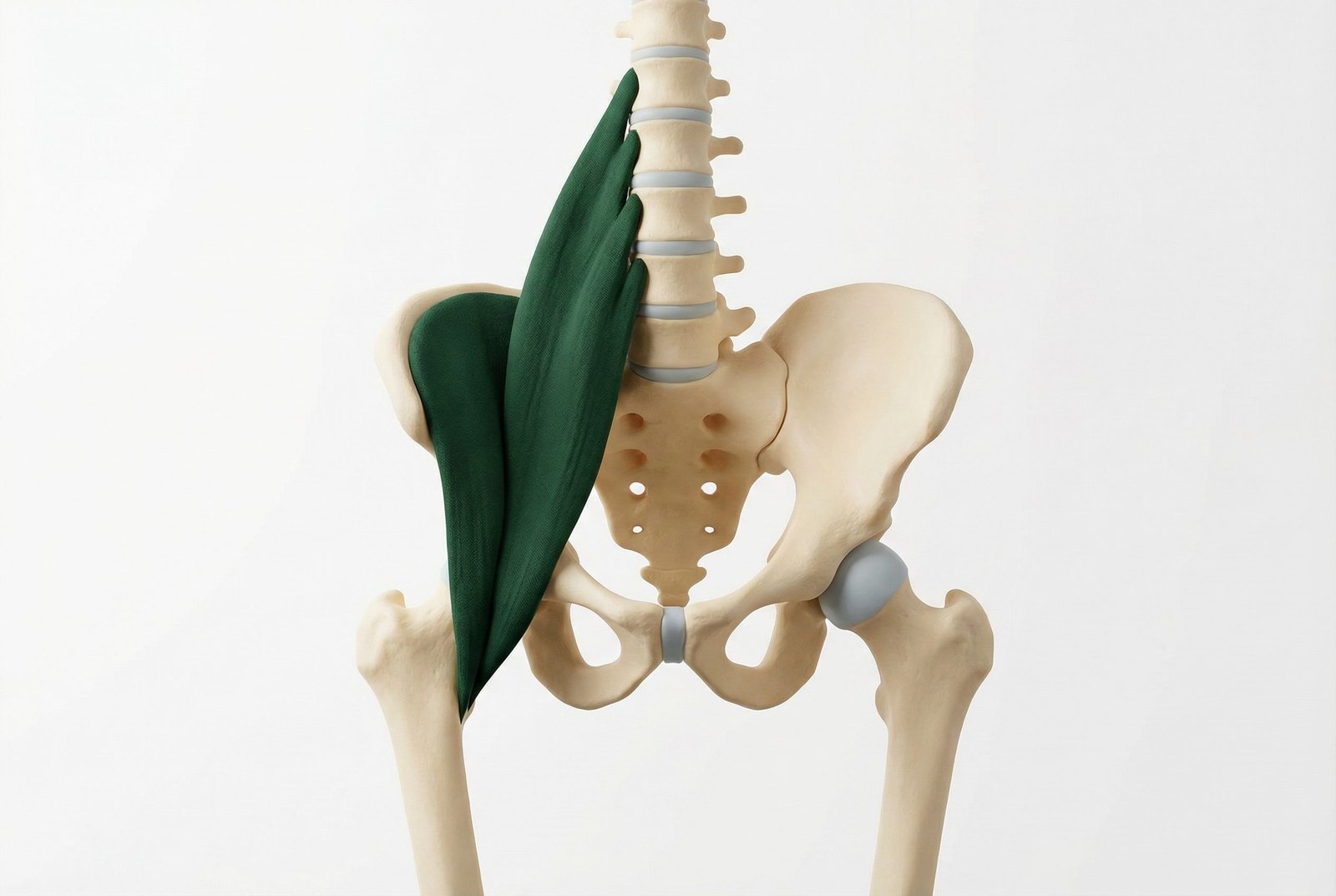
Iliopsoas Anatomy Anterior View Diagram
Two muscles that function as one — the iliacus (arising from the iliac fossa of the pelvis) and the psoas major (arising from the vertebral bodies and transverse processes of T12-L5) join forces at the inguinal ligament and share a common tendon inserting onto the lesser trochanter of the femur. Together they form the iliopsoas, the most powerful hip flexor in the human body.
The primary function is hip flexion, but the psoas plays an equally critical role as a dynamic stabilizer of the lumbar spine. When the iliopsoas is shortened by prolonged sitting, it generates a constant anterior compressive force at L4-L5, pulling the lumbar spine into hyperlordosis and loading the posterior facet joints — a key driver of chronic low back pain that is frequently misdiagnosed as disc disease.
The iliopsoas is a "hidden" muscle — located deep to the abdominal viscera and retroperitoneal fat, it is not palpable from the back and only partially palpable from the anterior abdomen with specific technique. This anatomical inaccessibility creates significant diagnostic confusion, as its referral patterns closely mimic lumbar disc herniation, hip arthritis, and femoral nerve entrapment.
Trigger points in the iliopsoas typically develop from: prolonged sitting (the most common cause), sudden forceful hip flexion under load (sprinting starts, kicking sports), hip or lumbar surgery with prolonged bed rest, psoas abscess and its resolution, and direct trauma to the lower abdomen or anterior hip.
Anatomy & Trigger Points
The psoas major originates from the vertebral bodies and intervertebral discs of T12-L5 and their transverse processes, descending through the pelvis to join the iliacus, inserting on the lesser trochanter of the femur. Its trigger points typically form in the mid-belly region, approximately 2-3cm medial to the anterior superior iliac spine and at the level of L3-L4.
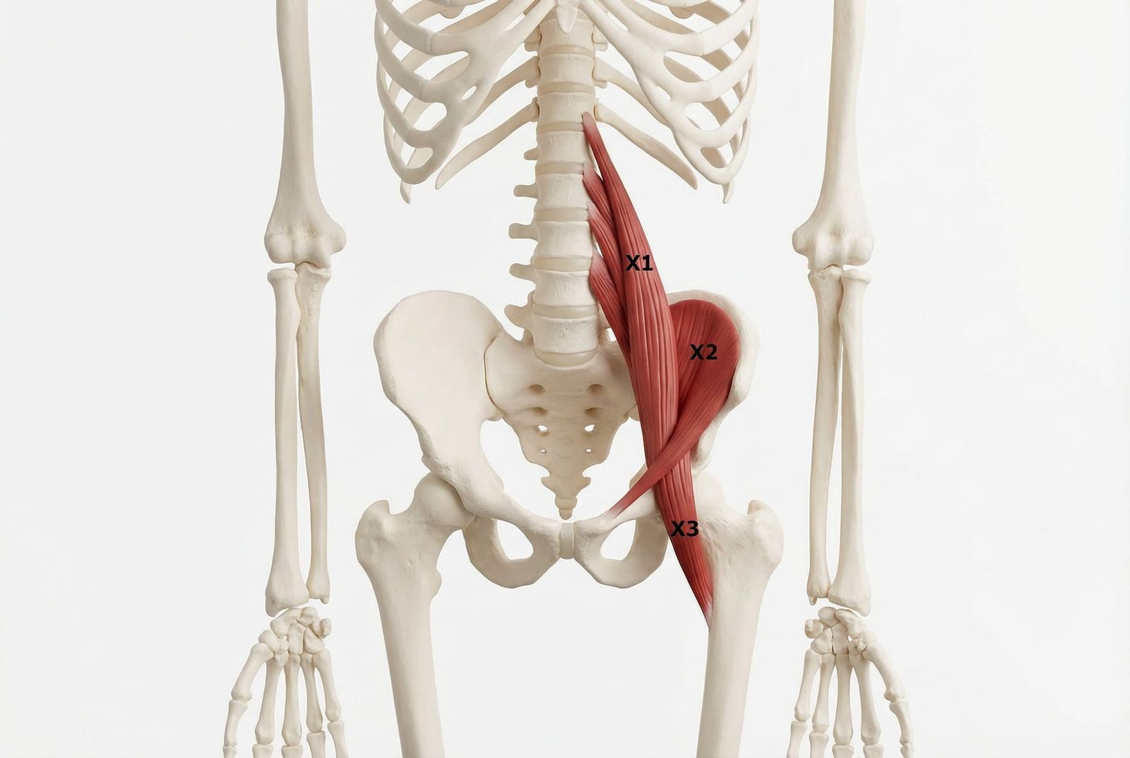
Iliopsoas Trigger Point Locations
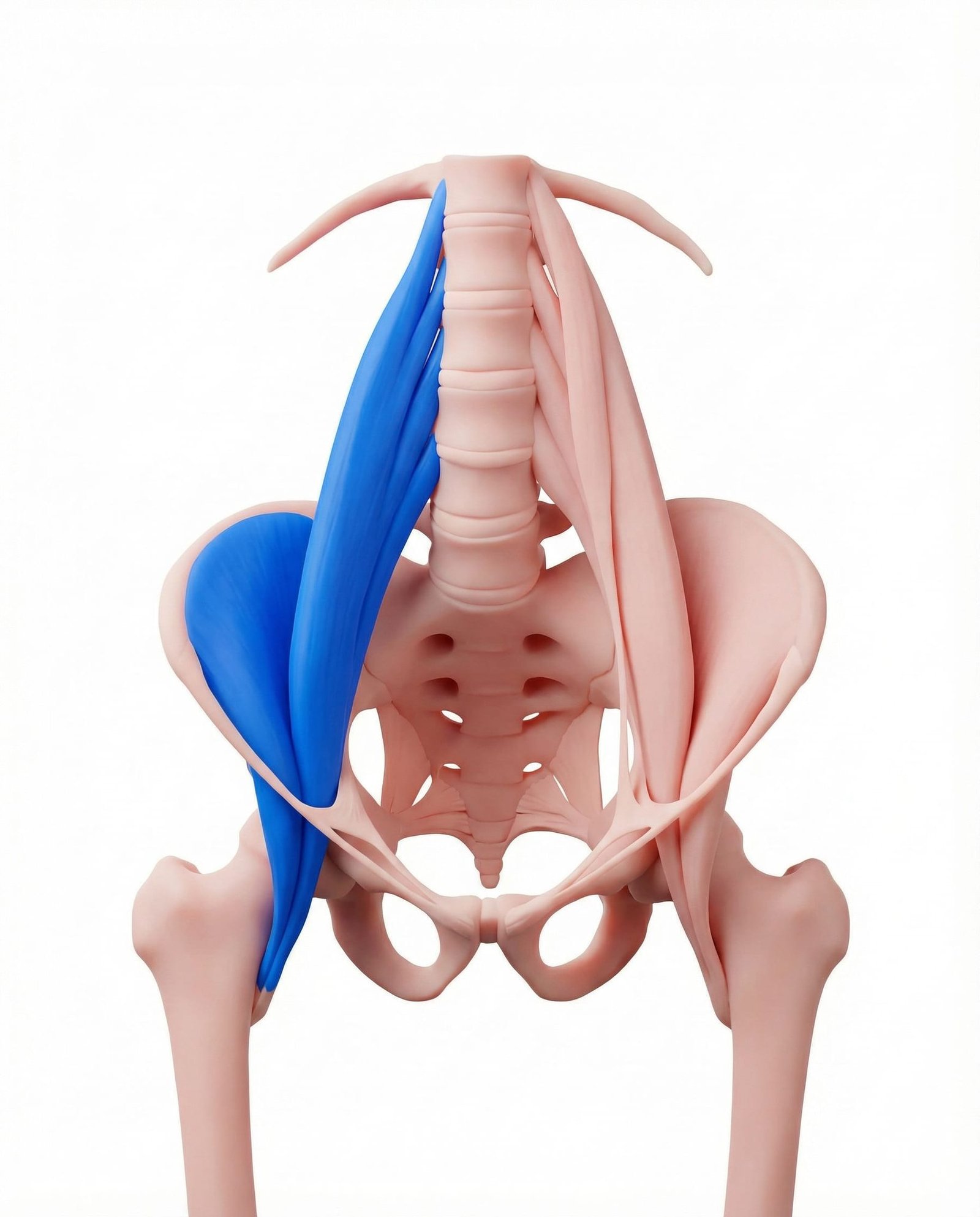
Deep anterior lumbar pain, groin aching, and anterior thigh pain. Keeps the hip in flexion contracture, creating a compensatory lumbar hyperlordosis. A hallmark of prolonged-sitting dysfunction.
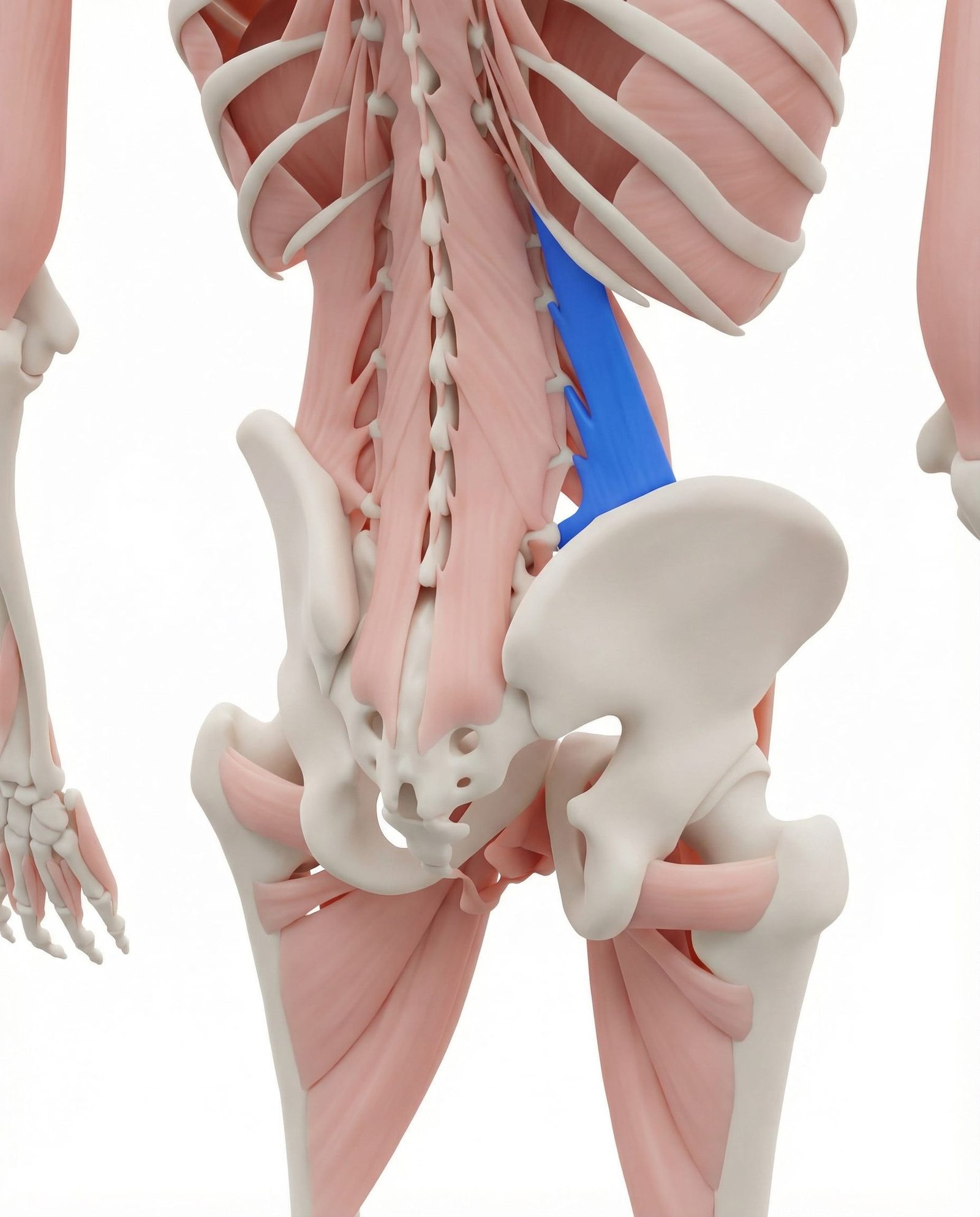
Iliac crest and lateral low back pain, often activated by iliopsoas dysfunction. QL trigger points frequently develop secondary to the anterior pelvic tilt caused by a shortened iliopsoas.
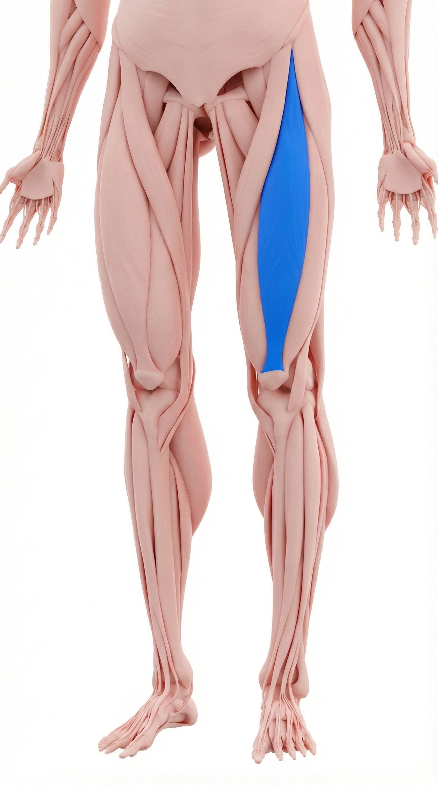
Knee and anterior thigh pain, hip flexion weakness. Often co-shortened with the iliopsoas in patients who sit extensively, contributing to anterior pelvic tilt and hip flexor dominance.
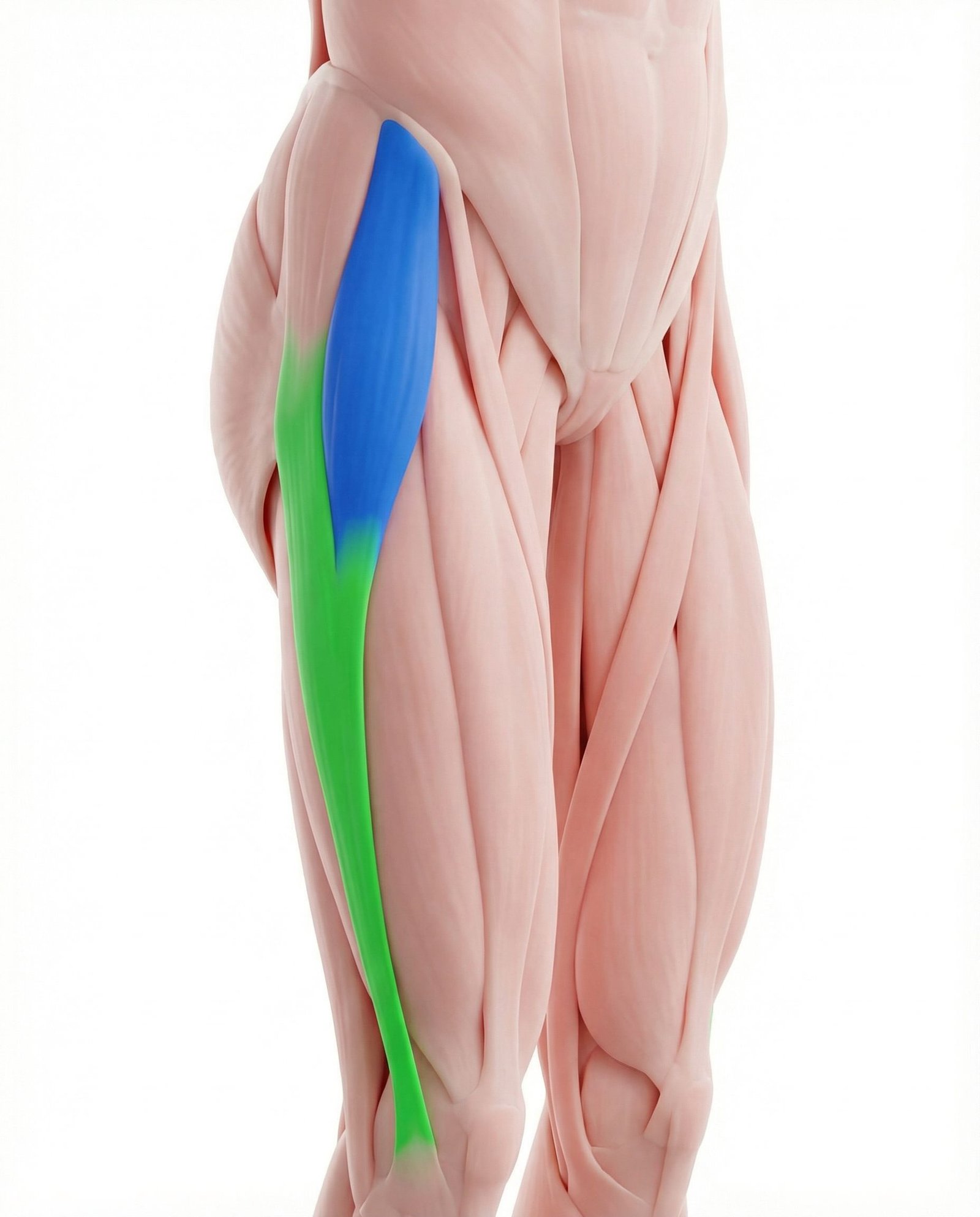
Lateral thigh and hip pain, often co-activated with the iliopsoas in anterior pelvic tilt. TFL overactivity is a compensatory response to weak gluteals and a tight iliopsoas.
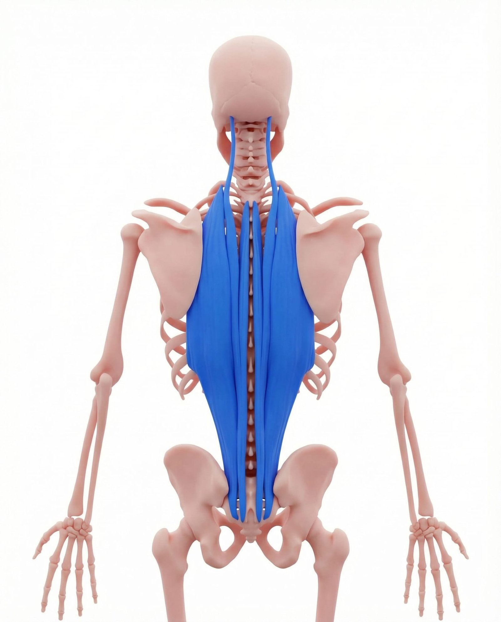
Diffuse lumbar aching, compensating for shortened hip flexors. Lumbar erectors become chronically overloaded when the iliopsoas pulls the pelvis into anterior tilt, forcing the extensors to work constantly against gravity.
Referral Patterns: The Psoas Pain Distribution
Deep anterior lumbar pain that the patient often describes as coming "from inside the spine" — difficult to localize, not clearly on the surface, often worse with standing, walking, or any position that extends the hip. Unlike discogenic pain, it does not clearly worsen with flexion and is not dermatomal in distribution.
Groin and anterior thigh pain that is frequently mistaken for hip arthritis, inguinal hernia, or femoral nerve compression. Patients often report difficulty rising from a low chair, walking up stairs, or bringing the knee toward the chest. Morning stiffness lasting 20-30 minutes after waking is a characteristic feature.
Subtle groin tightness and morning stiffness are common early signs, often preceding the disabling low back pain by weeks to months.
Anterior Lumbar
Groin
Anterior Thigh
Self-Assessment Tests
These tests assess iliopsoas length, strength, and trigger point activity. Note any asymmetry between sides — unilateral tightness is common and causes pelvic obliquity and functional scoliosis patterns.
Thomas Test
Hip Flexor Shortening Assessment
Modified Thomas Test
Edge-of-Table Hip Flexor Length
Hip Flexor Length Standing Test
Lunge Position Psoas Assessment
Active Hip Flexion Compression Test
Seated Resisted Hip Flexion with Deep Palpation
Treatment Pathways
Effective iliopsoas treatment combines direct trigger point release with progressive length restoration and targeted strengthening of the opposing posterior chain. Addressing only one component leads to recurrence.
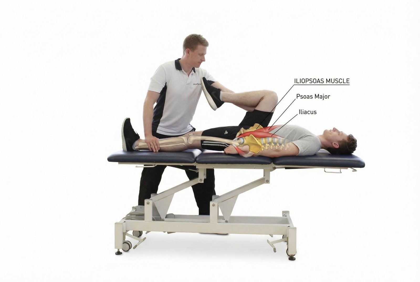
Iliopsoas Kneeling Stretch Treatment
Kneeling Hip Flexor Stretching
First-line approach to restore iliopsoas length. Consistent daily stretching in hip extension positions progressively deactivates trigger points and corrects anterior pelvic tilt over 4-6 weeks.
- Kneeling lunge stretch — back knee down, hips forward, upright torso (60-second holds)
- 90/90 hip stretch — seated on floor, both hips at 90 degrees, lean into the front hip
- Standing psoas stretch — lunge with posterior pelvic tilt, arms overhead to elongate the psoas
- Couch stretch — rear foot elevated on couch, drive hip into extension
- Diaphragmatic breathing during all stretches to neurologically release the psoas
Manual Psoas Release
Professional hands-on techniques targeting the deep iliopsoas. Particularly effective for resolving trigger points that do not respond to stretching alone.
- Direct psoas compression — deep abdominal technique, patient supine with knees bent
- Counterstrain technique — finding and holding the position of ease for 90 seconds
- Iliopsoas fascial release — sustained longitudinal traction through the hip flexor chain
- Soft tissue mobilization of the iliacus along the inner iliac fossa
- Post-isometric relaxation (PIR) for the combined iliopsoas unit
Dry Needling
Targeted interventional technique for deactivating deep iliopsoas trigger points that are inaccessible to manual therapy.
- Direct iliopsoas needling via anterior approach, lateral to femoral vessels
- Ultrasound-guided technique strongly recommended for patient safety
- Iliacus needling from the medial approach through the iliac fossa
- Trigger point injection with local anesthetic for acute spasm or abscess-related contracture
- Series of 3-6 treatments spaced 1 week apart for optimal results
Core & Posterior Chain Strengthening
Long-term reconditioning to balance hip flexor length with hip extensor strength. Essential for preventing recurrence and correcting anterior pelvic tilt.
- Dead bug exercise — alternate arm/leg extension with lumbar spine flat on floor
- Bird-dog — quadruped contralateral arm/leg extension for lumbar stabilization
- Glute bridges and single-leg variations to strengthen hip extensors
- Romanian deadlifts for posterior chain integration
- Lumbar stabilization program — McGill Big 3 (curl-up, side plank, bird-dog)
Red Flags — When to See a Doctor
Most deep hip and groin pain is muscular in origin, but several signs in this region require urgent medical evaluation as they may indicate vascular, infectious, or neurological emergencies.
Key Takeaways
The iliopsoas is the only muscle that directly connects the spine to the lower extremity — dysfunction there affects posture, gait, and pain patterns both above and below.
A tight or shortened psoas creates a constant compressive pull on the lumbar vertebrae (L1-L5), generating a pain pattern frequently misattributed to disc herniation.
Modern sedentary lifestyles involving 8+ hours of daily sitting chronically shorten the iliopsoas, making it one of the most universally problematic muscles in the working population.
The psoas has a direct anatomical relationship with the diaphragm via the medial arcuate ligament — breathing dysfunction and psoas dysfunction frequently coexist and reinforce each other.
True psoas release requires a trained practitioner using the deep abdominal approach — self-release attempts through the abdomen risk injury to the aorta, iliac vessels, and bowel.
Stretching alone is insufficient for lasting correction: the posterior chain (glutes, hamstrings) must be simultaneously strengthened to reverse anterior pelvic tilt and prevent recurrence.