TrP 1
TrP1
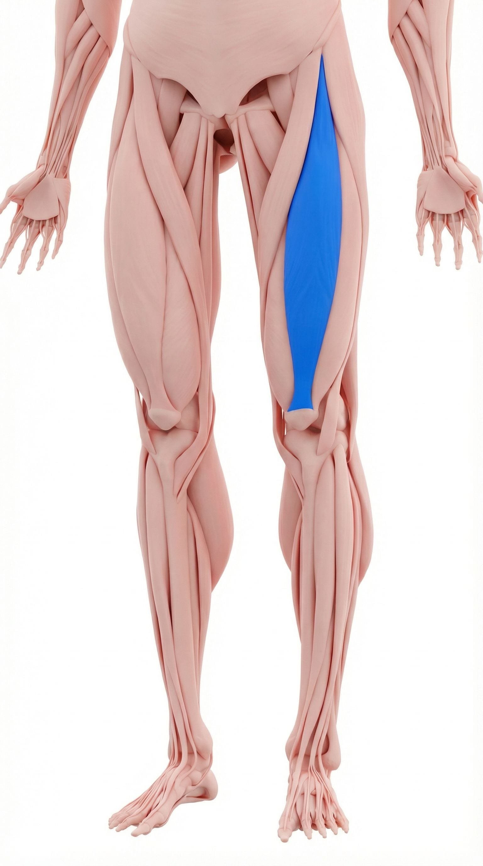
Location. Front of thigh, crosses hip and knee
Pain referral. Front of thigh, knee cap, hip
- Front of thigh
- Knee cap
- Hip
- Lower thigh
Anterior thigh aching from rectus femoris trigger points along the muscle belly
Location. Front of thigh, crosses hip and knee
Pain referral. Front of thigh, knee cap, hip
Location. Upper front thigh, near hip
Pain referral. Front of hip, upper thigh
Location. Lower front thigh, near knee
Pain referral. Knee cap, lower thigh
Location. Mid-belly of rectus femoris
Pain referral. Deep anterior knee pain, especially at night
Location. Distal rectus femoris near patella
Pain referral. Directly over and around patella (kneecap)
Front thigh pain. Anterior thigh aching from rectus femoris trigger points along the muscle belly
Knee pain. Suprapatellar and patellar aching from distal rectus femoris trigger point referral
Hip pain. Anterior hip aching near AIIS from proximal rectus femoris trigger point activation
Difficulty straightening knee. Restricted full knee extension from pain and taut bands in rectus femoris
Knee buckling. Sudden knee giving way from trigger point inhibition of quadriceps extension strength
Upper thigh pain. Referred aching down the anterior thigh from proximal rectus femoris trigger points
Knee discomfort. Distal referred pain at the patella from proximal trigger point in rectus femoris
Knee cap pain. Peripatellar aching from distal rectus femoris trigger points pulling on the patellar mechanism
Lower thigh pain. Anterior distal thigh aching aggravated by active knee extension against resistance
Knee instability. Sensation of knee giving way from quadriceps inhibition caused by active trigger points
Deep knee pain at night. Mid-belly rectus femoris trigger point produces nocturnal deep anterior knee aching disturbing sleep
Anterior knee ache. Referred pain from mid-rectus femoris to anterior knee through quadriceps tendon pathway
Knee stiffness after sitting. Prolonged knee flexion shortens rectus femoris across trigger point creating post-sitting stiffness
Difficulty going downstairs. Eccentric quadriceps loading during stair descent stresses mid-belly rectus femoris trigger point
Nocturnal knee pain disturbing sleep. Trigger point sustained activity during sleep produces deep aching that wakes patient from rest
Kneecap pain. Distal rectus femoris trigger point refers pain directly to patella surface via quadriceps tendon
Pain kneeling. Direct patellar compression during kneeling loads distal rectus femoris trigger point region
Patella tenderness to touch. Referred sensitivity from distal trigger point creates palpable patellar surface allodynia
Crepitus sensation in knee. Altered quadriceps tension from trigger point disrupts patellar tracking creating grinding sensation
Pain with knee extension against resistance. Resisted knee extension directly contracts distal rectus femoris fibers across the trigger point
Running. Repetitive hip flexion and knee extension during running stride overloads rectus femoris
Kicking sports. Forceful hip flexion with knee extension during kicking strains rectus femoris acutely
Squatting. Deep knee flexion under load eccentrically overloads rectus femoris at both joint crossings
Jumping. Explosive knee extension during takeoff and eccentric landing loading fatigues rectus femoris
Prolonged sitting. Sustained hip flexion adaptively shortens rectus femoris creating chronic taut bands
Weak quadriceps. Insufficient quadriceps strength increases rectus femoris compensatory workload during activity
Kicking. Forceful hip flexion with knee extension during kicking strains proximal rectus femoris origin
Kicking sports (soccer, football). Forceful hip flexion with knee extension during kicking maximally loads rectus femoris mid-belly
Cycling. Sustained quadriceps contraction during pedaling fatigues rectus femoris at mid-belly region
Jumping sports. Explosive knee extension for jumping maximally loads rectus femoris mid-belly concentrically
Prolonged sitting with knee bent. Sustained knee flexion maintains rectus femoris in shortened position creating ischemic trigger points
Repetitive kneeling. Direct patellar loading during kneeling transmits compression to distal rectus femoris tendon region
Running downhill. Eccentric quadriceps braking during downhill running maximally stresses distal rectus femoris
Jumping and landing sports. Explosive extension and landing impact overloads distal rectus femoris at patellar junction
Deep squatting. Full depth squat maximally loads distal rectus femoris under stretch creating fiber microtrauma
Cycling with high gear ratio. High resistance pedaling demands excessive distal quadriceps force production per revolution
Direct trauma to knee. Impact to anterior knee damages distal rectus femoris fibers creating post-traumatic trigger points
Lie face down and bend your knee, pulling your ankle toward your buttock with your hand on the same side. You should feel a stretch along the front of your thigh. If you cannot reach your ankle, loop a towel around the foot. Keep your hips pressed against the floor to maximize the stretch.
Lie face down with a foam roller under the front of your thigh. Roll from the hip to just above the knee, pausing on tender spots for 20-30 seconds. Support yourself on your forearms and use the opposite leg to control how much weight you place on the roller.
Kneel on one knee with the other foot flat in front. Shift your weight forward while reaching back to grab the ankle of the kneeling leg. Pull the heel toward your buttock while maintaining forward hip shift. This stretches both the hip flexor and quadriceps components of the rectus femoris simultaneously.
Stand on one leg on a step. Slowly lower yourself over 3-5 seconds by bending the knee, controlling the descent. Use the other leg to push back up. This eccentrically loads the quadriceps, which strengthens them while reducing trigger point sensitivity.
Always warm up with 5-10 minutes of light jogging or cycling before running, kicking, or jumping activities. Stretch the quadriceps thoroughly after exercise when the muscles are warm. Avoid sudden sprinting without preparation.
If anterior thigh or knee pain persists beyond 3-4 weeks, consult a physiatrist or sports medicine physician. They can differentiate rectus femoris trigger points from patellar tendinopathy, patellofemoral syndrome, or hip flexor strain and provide targeted treatment.
Kneel on one knee with the opposite foot flat in front of you in a lunge position. Keep your torso upright and gently shift your hips forward until you feel a stretch at the front of the kneeling hip. Squeeze the glute on the kneeling side to deepen the stretch. Hold without bouncing.
Lie face down with a foam roller under the upper front of the thigh, just below the hip crease. Support yourself on your forearms and opposite leg. Slowly roll from the hip crease down to mid-thigh, pausing on tender spots for 20-30 seconds. Adjust pressure by shifting more or less weight onto the roller.
Stand facing a step or sturdy box (6-8 inches initially). Place one foot fully on the step and press through that leg to stand up, bringing the other foot to the step. Lower back down under control. Focus on driving through the hip and keeping your torso upright. Gradually increase step height as pain allows.
Stand tall holding onto a wall or chair for balance. Slowly lift one knee toward your chest as high as comfortable, hold for 2-3 seconds, then slowly lower. Keep your standing leg slightly bent and your core engaged. Add a light ankle weight as strength improves.
Temporarily decrease the volume and intensity of activities involving forceful hip flexion, such as kicking drills, sprinting, and high-knee exercises. Substitute with lower-impact conditioning like cycling or swimming. Gradually reintroduce kicking and sprinting only after pain-free hip flexion against resistance is achieved.
If anterior hip pain is accompanied by clicking, catching, locking, or significantly limited rotation, consult an orthopedic specialist or sports medicine physician. These symptoms may indicate hip labral pathology, femoroacetabular impingement, or other structural conditions requiring imaging and specialized treatment.
Sit with the affected leg extended. Using your thumbs or a massage tool, apply firm pressure to the quadriceps muscle just above the kneecap. Work in small circular motions, covering the area from 2-4 inches above the patella. When you find a tender nodule, hold sustained pressure for 20-30 seconds until the tenderness decreases.
Stand with your back against a wall and slide down until your knees are bent to approximately 45 degrees — not a full 90-degree squat. Keep your feet shoulder-width apart and press your back flat against the wall. Hold this position, focusing on even quadriceps engagement. Start with shorter holds and progress gradually.
Sit on a chair or table edge with a rolled towel under the knee of the affected leg. Slowly straighten the knee fully by lifting the foot, focusing on tightening the inner quad muscle near the kneecap. Hold the fully straightened position for 3-5 seconds, then slowly lower. Add a light ankle weight as strength improves.
Stand on a low step (4-6 inches) with the affected leg. Slowly lower the opposite foot toward the floor by bending the stance knee, controlling the descent over 3-4 seconds. Lightly touch the heel to the floor, then return to standing. Keep the stance knee aligned over the second toe throughout.
Minimize time spent sitting with knees flexed beyond 90 degrees, such as in low chairs, theater seats, or cars. When seated for extended periods, periodically straighten your legs fully for 30-60 seconds. At work, set a timer to stand and extend your knees every 30-45 minutes.
If anterior knee pain persists beyond 4-6 weeks despite consistent self-care, consult a sports medicine physician or orthopedic specialist. They can differentiate between trigger point pain, true patellar tendinopathy, patellofemoral malalignment, and cartilage issues using clinical examination and imaging.