Understanding Core Instability & MPS
The concept of core stability revolves around the motor control model, which emphasizes the coordinated timing and activation of the deep trunk musculature to manage spinal loads. Clinical evidence suggests that following an initial episode of low back pain, the deep stabilizing muscles—particularly the lumbar multifidus and transversus abdominis—often experience rapid inhibition and subsequent atrophy. This functional instability reduces segmental stiffness, potentially exposing intervertebral discs and facet joints to micro-trauma during routine movements. Differentiating between true structural instability, such as spondylolisthesis, and functional motor control deficits is a critical step in formulating an effective management plan.
There is a complex, bidirectional relationship between functional core instability and myofascial pain syndrome (MPS). When deep stabilizers are inhibited, superficial muscles like the quadratus lumborum, erector spinae, and iliopsoas are forced to overcompensate to maintain upright posture and lumbo-pelvic control. This chronic, low-level overloading creates an ideal environment for the development of active myofascial trigger points. Furthermore, nociceptive input from these active trigger points can reflexively inhibit the deep stabilizers, creating a self-perpetuating cycle of pain, altered motor control, and compensatory muscle spasm.
Treating myofascial pain without addressing underlying motor control deficits often leads to symptom recurrence, while attempting core strengthening in the presence of active trigger points may reinforce dysfunctional movement patterns.
Differentiating primary myofascial pain from secondary pain driven by core instability requires a nuanced clinical approach. If a patient presents with pain that is primarily exacerbated by sustained postures or transitional movements, and exhibits poor performance on load transfer tests, functional instability is highly suspected. Systematic reviews note that imaging studies, such as MRI, may reveal multifidus atrophy, but these findings are often non-specific and must be correlated with clinical examination. A pragmatic diagnostic strategy often involves deactivating key trigger points first; if lumbo-pelvic control remains poor or pain rapidly returns upon functional loading, an underlying motor control deficit is likely the primary driver.
Key Musculature
Understanding the interplay between deep stabilizers and superficial movers is essential. Dysfunction in one layer predictably leads to compensatory pathology in the other.
Lumbar Multifidus
The lumbar multifidus is a deep paraspinal muscle primarily responsible for segmental spinal stabilization rather than gross movement. Clinical evidence suggests that following a first episode of acute low back pain, the multifidus often undergoes rapid atrophy and fatty infiltration. This inhibition reduces segmental stiffness, potentially allowing micro-trauma to facet joints and intervertebral discs. Myofascial trigger points in the multifidus can mimic radicular pain and further disrupt local motor control.
Refers deep, localized aching pain to the adjacent spinous processes, occasionally projecting anteriorly into the abdomen.Transversus Abdominis
As the deepest abdominal layer, the transversus abdominis acts as a corset, increasing intra-abdominal pressure to stabilize the lumbar spine prior to limb movement. Studies utilizing electromyography frequently note delayed firing of this muscle in individuals with chronic low back pain. When inhibited, superficial muscles often overcompensate, leading to myofascial pain syndromes in the abdominal wall and lumbar region. Restoring appropriate timing of this muscle is a common target in motor control rehabilitation.
Trigger points may refer pain broadly across the anterior abdomen, sometimes mimicking visceral pathology or contributing to a sensation of abdominal fullness.Quadratus Lumborum (QL)
The quadratus lumborum connects the pelvis to the spine and lower ribs, functioning as a lateral flexor and a major stabilizer of the lumbar spine. In the presence of deep core inhibition, the QL frequently becomes hyperactive to provide compensatory stability. This chronic overload makes it highly susceptible to developing active myofascial trigger points. QL trigger points are a primary source of severe, aching low back pain that is often misdiagnosed as structural spinal pathology.
Refers a deep, aching pain to the sacroiliac joint, lower buttock, and occasionally the greater trochanter, often exacerbated by coughing or rolling over.Iliopsoas
Comprising the iliacus and psoas major, this powerful hip flexor also exerts significant compressive and shearing forces on the lumbar spine. When the posterior stabilizing musculature is weakened or inhibited, the iliopsoas may adaptively shorten, altering pelvic tilt and lumbar lordosis. Trigger points within the iliopsoas refer pain to the ipsilateral lower back and anterior thigh. Differentiating iliopsoas myofascial pain from true hip joint pathology requires careful clinical examination.
Pain is primarily referred to the ipsilateral lumbar region, running vertically parallel to the spine, and often extends into the anterior thigh and groin.Erector Spinae
The erector spinae group consists of the iliocostalis, longissimus, and spinalis muscles, which manage gross spinal extension and posture. These superficial muscles often exhibit increased resting tone in patients with underlying segmental instability, attempting to splint the spine. This sustained contraction frequently leads to the development of myofascial trigger points. Pain from these trigger points typically presents as a diffuse ache across the lumbar and lower thoracic regions.
Refers pain locally along the paraspinal corridor, sometimes extending upward toward the scapula or downward into the gluteal region depending on the trigger point location.
Multifidus Inhibition
Pain-induced reflex inhibition leads to rapid atrophy of the deep segmental stabilizers, reducing spinal stiffness.
Compensatory Overload
Superficial muscles (QL, erector spinae) become hyperactive to stabilize the spine, leading to myofascial trigger points.
Altered Intra-abdominal Pressure
Delayed firing of the transversus abdominis and diaphragm disrupts the hydraulic cylinder effect needed for core stability.
Central Sensitization
Chronic nociceptive input from overloaded muscles and micro-traumatized joints lowers the threshold for pain perception.
Common Referral Patterns
Myofascial trigger points in compensatory muscles often mimic the mechanical pain associated with lumbar instability. Recognizing these patterns aids in accurate differential diagnosis.
- Quadratus Lumborum
- Active trigger points in the quadratus lumborum typically refer a deep, aching pain to the sacroiliac joint, lower buttock, and occasionally the greater trochanter. This pattern is frequently exacerbated by coughing, sneezing, or rolling over in bed, closely mimicking the mechanical pain of lumbar instability.
- Iliopsoas
- Pain is primarily referred to the ipsilateral lumbar region, running vertically parallel to the spine, and often extends into the anterior thigh and groin. Patients may report difficulty standing fully erect, a symptom that overlaps with both structural lumbar stenosis and functional core weakness.
- Lumbar Multifidus
- Trigger points in the deep lumbar multifidus refer pain locally to the spinous processes and can project anteriorly into the abdomen. This localized, deep ache is often indistinguishable from facet joint arthropathy or discogenic pain without targeted palpation.
Clinical Assessment Tests
Clinical assessment of core instability focuses on load transfer and motor control rather than gross strength. These tests help differentiate functional instability from primary myofascial pain.
Prone Instability Test
The patient lies prone with the torso resting on an examination table and legs resting on the floor. The clinician applies posterior-to-anterior pressure to the lumbar spine to provoke pain, then repeats the pressure while the patient lifts their legs slightly off the floor.
Positive: A reduction in pain when the legs are lifted suggests that active muscle contraction stabilizes the painful segment, indicating potential functional instability.
Active Straight Leg Raise (ASLR)
The patient lies supine and is asked to actively raise one leg about 20 centimeters off the table without bending the knee. The clinician observes for excessive pelvic rotation or compensatory bracing strategies.
Positive: Difficulty lifting the leg, reproduction of lumbar pain, or significant improvement in the effort when the clinician manually compresses the pelvis suggests impaired lumbo-pelvic load transfer.
Single-Leg Stance Test
The patient stands on one leg while the clinician observes the lumbo-pelvic alignment and postural sway. The test assesses the integration of core stabilizers and hip abductors during a dynamic weight shift.
Positive: Excessive pelvic drop (Trendelenburg sign), trunk leaning, or reproduction of low back pain suggests a deficit in lateral pelvic stability and motor control.
Abdominal Drawing-in Maneuver (ADIM)
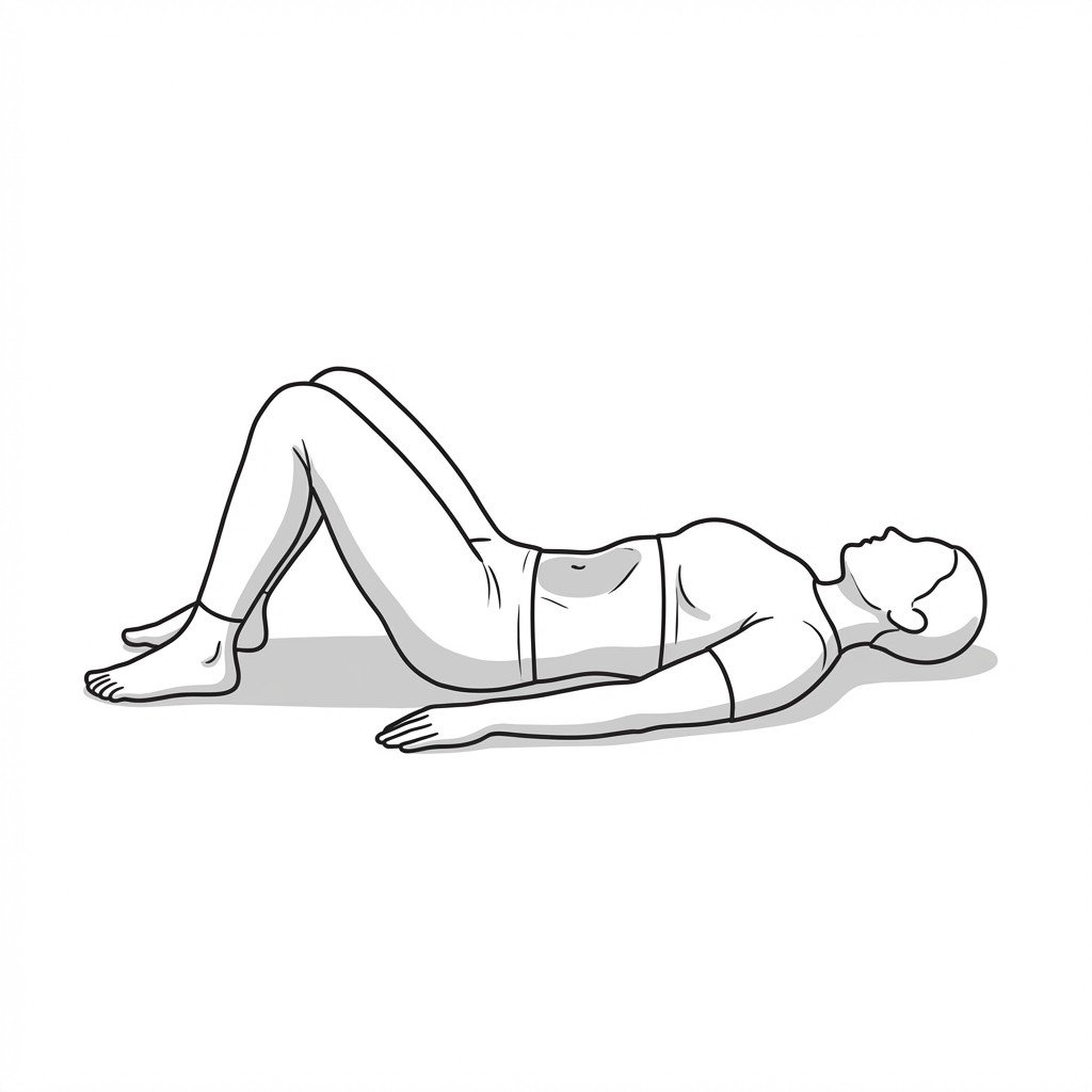
The patient lies supine with knees bent and is instructed to gently draw the lower abdomen inward toward the spine without holding their breath or moving the pelvis. This isolates the transversus abdominis from the superficial rectus abdominis.
Positive: Inability to perform the maneuver without breath-holding, pelvic tilting, or rib flaring indicates poor isolated motor control of the deep abdominal fascia.
Treatment Pathways
Management typically requires a multimodal approach. Evidence from randomized trials suggests that combining manual therapy to address myofascial restrictions with targeted motor control exercises yields optimal long-term outcomes.
Self-Care
- Activity modification to avoid prolonged static postures
- Pacing strategies to prevent fatigue-related compensatory pain
- Gentle abdominal drawing-in maneuvers (ADIM) within pain-free limits
- Application of heat to hyperactive superficial muscles (e.g., erector spinae)
- Ergonomic optimization of seating and workspace
Manual Therapy
- Ischemic compression of quadratus lumborum and iliopsoas trigger points
- Myofascial release targeting the thoracolumbar fascia
- Joint mobilization to address secondary facet joint restrictions
- Soft tissue mobilization of the abdominal wall to reduce guarding
- Proprioceptive neuromuscular facilitation (PNF) for lumbo-pelvic muscles
Interventional
- Trigger point injections for refractory quadratus lumborum or iliopsoas pain
- Diagnostic medial branch blocks to rule out primary facet arthropathy
- Real-time ultrasound imaging for transversus abdominis biofeedback
- Dry needling of the lumbar multifidus to reduce localized spasm
- Epidural steroid injections if concurrent radiculopathy is suspected
Rehabilitation
- Motor control exercises targeting precise timing of deep stabilizers
- Progressive load management focusing on endurance over maximal strength
- Integration of core stability into functional, multi-planar movements
- Diaphragmatic breathing exercises to normalize intra-abdominal pressure
- Lumbo-pelvic proprioception training using unstable surfaces
Red Flags — When to See a Doctor
While functional core instability and myofascial pain are common, clinicians must remain vigilant for signs of serious structural or systemic pathology.
Cauda Equina Syndrome Symptoms
Saddle anesthesia, recent onset of bladder or bowel dysfunction, or severe bilateral neurological deficits in the lower extremities.
EmergencySuspected Spinal Fracture
Severe, localized pain following significant trauma, or minor trauma in individuals with known osteoporosis or osteopenia.
EmergencySpinal Infection
Unrelenting back pain accompanied by fever, chills, unexplained weight loss, or a recent history of bacterial infection or intravenous drug use.
UrgentProgressive Neurological Deficit
Rapidly worsening muscle weakness, foot drop, or progressive sensory loss in the lower extremities.
UrgentSevere Structural Spondylolisthesis
Palpable step-off deformity in the lumbar spine accompanied by severe mechanical pain or neurogenic claudication.
Investigate
Key Clinical Takeaways
Core instability is often a functional deficit in motor control rather than a structural failure of the spine.
Myofascial trigger points in the quadratus lumborum and iliopsoas frequently develop as compensatory mechanisms for deep core inhibition.
Treating myofascial pain without addressing underlying motor control deficits often leads to symptom recurrence.
Conversely, attempting core strengthening while active trigger points are present may exacerbate pain and reinforce dysfunctional movement patterns.
Clinical guidelines generally recommend a multimodal approach combining manual therapy for pain relief with specific motor control exercises for long-term stability.
Real-time ultrasound and specific load-transfer tests are valuable clinical tools for differentiating true motor control deficits from primary myofascial pain.
Movement therapy
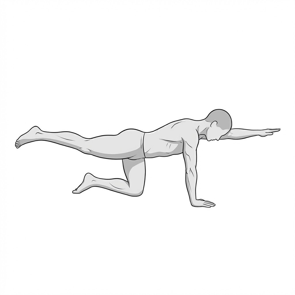
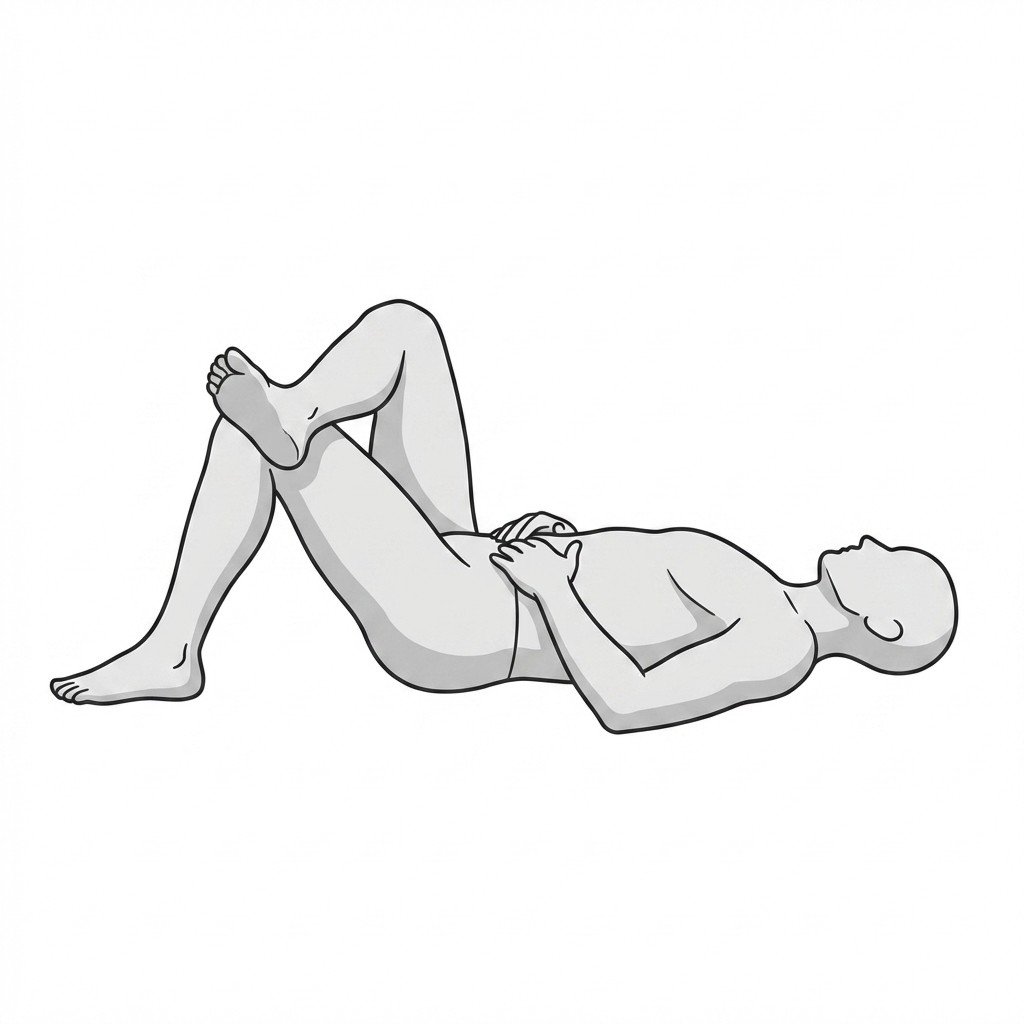
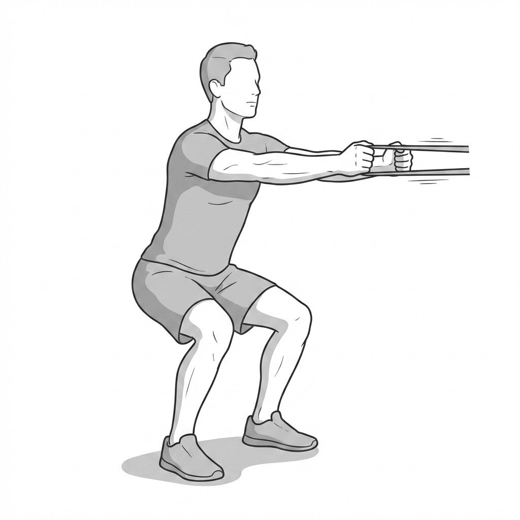
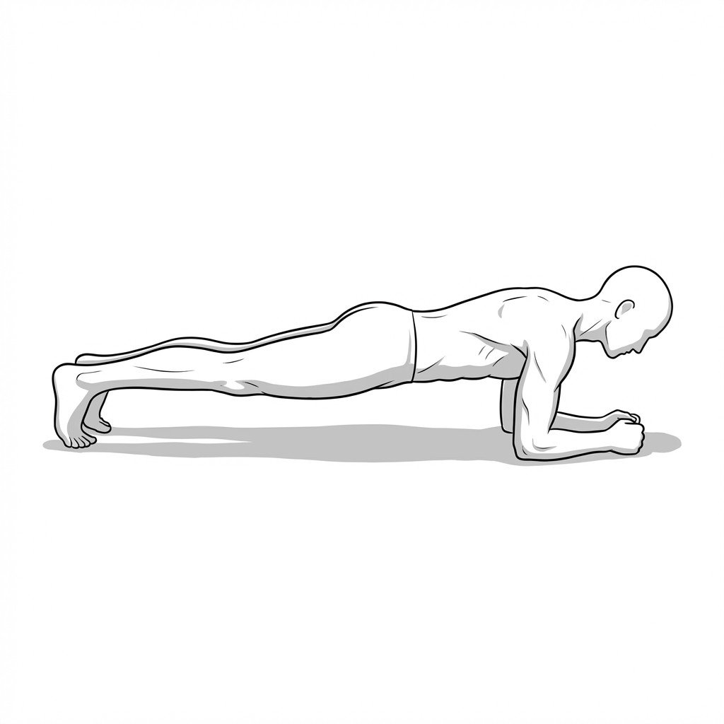
Exercises for Core Instability
These exercises specifically address lumbo-pelvic stability — drawing in the transversus abdominis, reactivating inhibited multifidus, and then building anti-rotation control through bird-dogs, dead-bugs, and the Pallof press. Start with 2–3 and progress as tolerated.