Understanding Adductor Pain
Groin pain presents a complex diagnostic challenge, with adductor muscle strains representing a significant proportion of these injuries in both athletic and general populations. The adductor group, comprising the longus, brevis, magnus, pectineus, and gracilis, functions to stabilize the pelvis and adduct the thigh during dynamic movement. Epidemiological data suggest that the adductor longus is implicated in the majority of acute groin strains, often occurring during rapid changes of direction or forceful kicking. Differentiating between a true musculotendinous tear, chronic tendinopathy, and referred myofascial pain is critical, as the management strategies and prognostic timelines for these conditions diverge significantly.
Myofascial pain syndrome (MPS) frequently overlaps with adductor pathology, either as a primary source of nociception or as a secondary consequence of altered biomechanics following an acute strain. Trigger points within the adductor belly or the adjacent iliopsoas can refer pain directly to the groin crease, closely mimicking the symptoms of an acute strain or tendinopathy. Furthermore, chronic groin pain often involves elements of central sensitization, where persistent nociceptive input from unresolved trigger points or tendinopathic tissue lowers the local pain threshold. Recognizing this overlap is essential, as isolated treatment of the tendon may fail to provide lasting relief if concurrent myofascial pain is not addressed.
Differentiating an acute adductor strain from myofascial referral requires precise palpation; the location of maximal tenderness often dictates the primary pain generator.
A meticulous clinical examination is required to differentiate these overlapping conditions. Palpation plays a central role; tenderness localized precisely at the pubic insertion or musculotendinous junction typically suggests strain or tendinopathy, whereas exquisite tenderness over a taut band in the muscle belly points toward MPS. Clinicians generally utilize provocation tests, such as the adductor squeeze test, alongside hip range of motion assessments to isolate the involved structures. When clinical uncertainty persists, or when conservative management fails, imaging modalities like ultrasound or MRI may be employed to rule out structural tears, osteitis pubis, or intra-articular hip pathology.
Anatomical Considerations
The medial compartment of the thigh contains the adductor muscle group, which originates on the pubis and ischium and inserts along the medial femur. The depth and orientation of these muscles directly affect how reliably each can be reached and distinguished on palpation.
Adductor Longus
The adductor longus is the most frequently injured muscle in the adductor group, particularly during activities requiring rapid changes of direction. In the context of myofascial pain, trigger points within the muscle belly can develop secondary to acute strain or chronic overload. Clinical presentation often involves localized tenderness at the musculotendinous junction during acute injury, whereas myofascial pain typically presents with taut bands in the mid-substance.
Pain typically refers upward deep into the groin and downward along the anteromedial thigh, occasionally extending to the medial knee.Adductor Magnus
As the largest and most powerful of the adductors, the magnus has both an adductor and a hamstring part. Strains here are less common than in the longus but can be more debilitating due to the muscle's role in pelvic stabilization. Trigger points in the adductor magnus are frequently overlooked and can mimic deep pelvic or hip joint pathology.
Refers pain deep into the groin, the anteromedial thigh, and occasionally into the internal pelvic region, sometimes mimicking pelvic floor dysfunction.Adductor Brevis
Situated deep to the adductor longus, the brevis is rarely strained in isolation but is often co-involved in significant adductor injuries. Because of its deep anatomical position, differentiating brevis pathology from longus pathology via palpation can be clinically challenging. Myofascial trigger points here often perpetuate chronic groin aching.
Pain referral is generally localized to the proximal medial thigh and the groin crease.Gracilis
The gracilis is the most superficial medial thigh muscle and the only adductor that crosses both the hip and knee joints. While acute strains are relatively uncommon, it frequently develops satellite trigger points in response to primary adductor longus pathology or medial compartment knee osteoarthritis. Its superficial nature makes it highly accessible for manual assessment.
Pain typically refers along the medial aspect of the thigh, often concentrating as a superficial stinging or aching sensation.Iliopsoas
Though primarily a hip flexor, the iliopsoas is a critical structure in the differential diagnosis of groin pain. Iliopsoas tendinopathy or bursitis can present with symptoms nearly identical to proximal adductor strains. Furthermore, iliopsoas trigger points are a primary source of referred groin pain and are frequently activated by the altered gait mechanics following an adductor injury.
Refers pain strongly to the anterior thigh and groin crease, as well as vertically along the ipsilateral lumbar spine.Pectineus
The pectineus is a short, flat muscle located in the proximal medial thigh. It acts as both a hip flexor and adductor. Injuries to the pectineus are difficult to isolate clinically and are often grouped under the umbrella of "groin strain." Trigger points in this muscle are a potent, yet frequently missed, source of deep groin pain.
Pain is referred deeply into the groin crease, often described by patients as an ache situated directly within the hip joint.
Acute Muscle Strain
Macroscopic or microscopic tearing of muscle fibers, typically at the musculotendinous junction, resulting in sudden onset pain, localized swelling, and weakness.
Myofascial Referral
Referred pain from hyperirritable nodules (trigger points) within taut bands of muscle, often presenting as a deep, poorly localized ache without acute trauma.
Tendinopathy
Degenerative changes within the tendon due to chronic overload, characterized by pain at the bony insertion that warms up with activity but worsens afterward.
Intra-articular Pathology
Issues within the hip joint itself, such as femoroacetabular impingement (FAI) or labral tears, often presenting with mechanical symptoms and restricted internal rotation.
Pain Referral Patterns
Myofascial trigger points in the pelvic and thigh musculature often project pain to the groin. Recognizing these specific referral maps helps differentiate myofascial pain from true joint or tendon pathology.
- Adductor Longus and Brevis
- Trigger points in these muscles frequently refer pain upward into the groin crease and downward along the medial aspect of the thigh. Patients may describe a deep, aching sensation that intensifies during weight-bearing or sudden lateral movements.
- Adductor Magnus
- Referral from the adductor magnus is often experienced as a deep, generalized pelvic or medial thigh ache. Due to its extensive origin along the ischial tuberosity, pain may also radiate toward the gluteal fold, complicating the differential diagnosis with hamstring pathology.
- Iliopsoas and Pectineus
- These muscles typically refer pain directly to the anterior groin and upper thigh. Iliopsoas referral is unique in that it frequently includes a concurrent vertical band of pain in the ipsilateral lumbar spine, which can help differentiate it from primary adductor pathology.
Clinical Assessment
Clinical assessment of groin pain involves a combination of strength testing, flexibility assessment, and precise palpation. These tests help isolate the specific tissues contributing to the patient's symptoms.
Adductor Squeeze Test
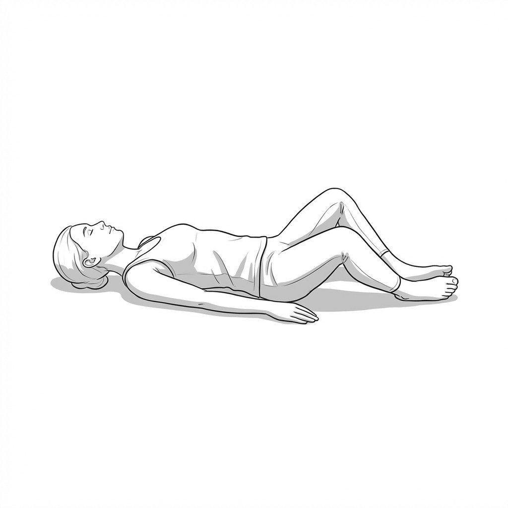
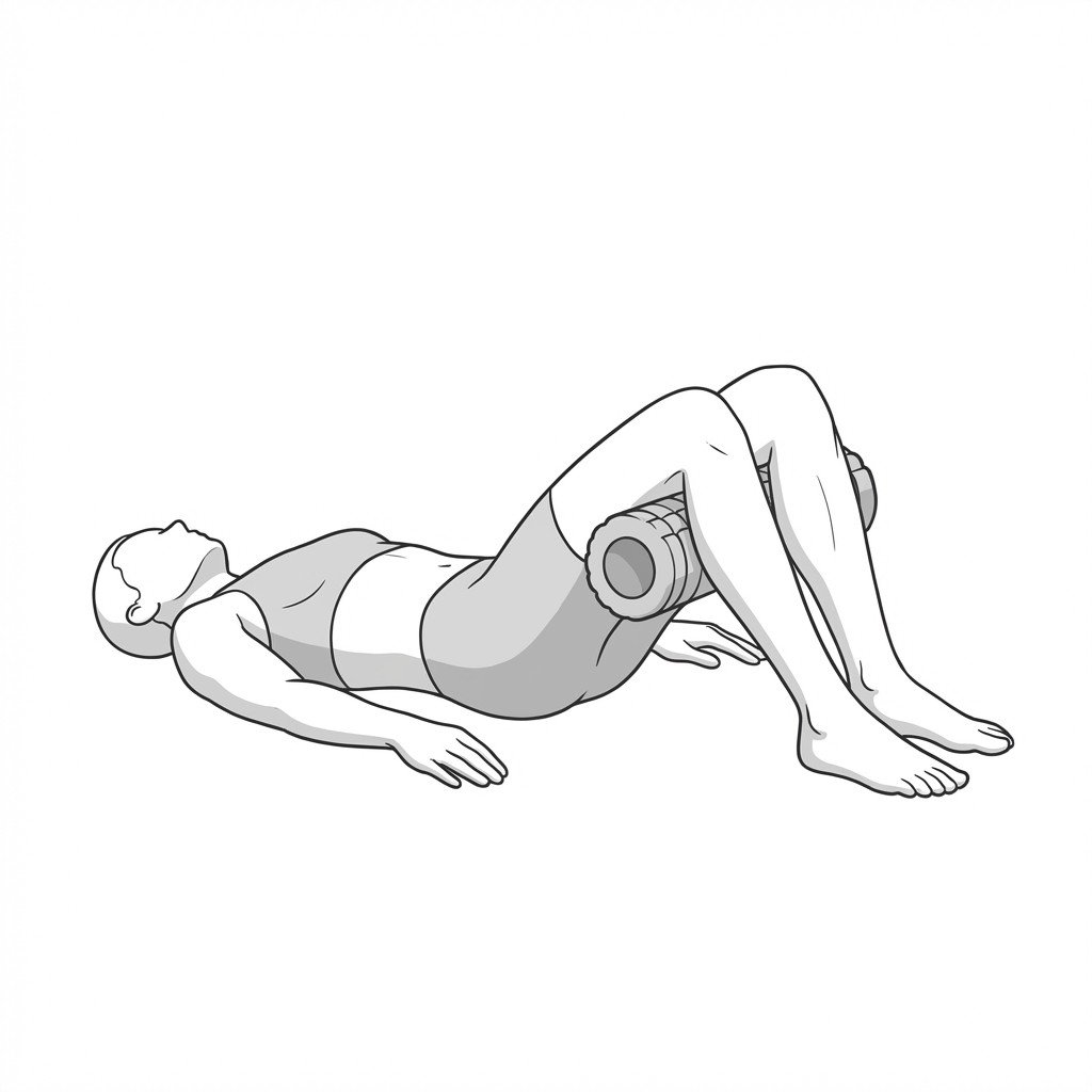
While lying supine with knees bent and feet flat on the floor, place a firm ball or fist between the knees. Gently squeeze inward against the resistance, gradually increasing force. Note the location and character of any provoked pain.
Positive: Reproduction of familiar groin pain, which may indicate adductor tendinopathy, acute strain, or active myofascial trigger points depending on the pain's exact location.
Palpation of the Musculotendinous Junction
In a seated or supine position, gently palpate the inner thigh starting from the pubic bone and moving downward along the prominent tendon of the adductor longus. Compare the sensitivity of the tendon insertion to the softer muscle belly below.
Positive: Sharp, localized pain at the bony insertion suggests tendinopathy or strain, whereas a deep ache provoked by pressing the mid-muscle belly is more characteristic of myofascial trigger points.
FABER (Patrick's) Test
Lie supine and place the ankle of the affected leg just above the knee of the opposite straight leg, forming a "figure-4." Allow the bent knee to gently fall outward toward the table. Do not force the movement.
Positive: Pain in the anterior groin may suggest hip joint pathology (such as osteoarthritis or labral tear) or iliopsoas spasm, while tightness or pain in the medial thigh suggests adductor restriction.
Thomas Test Position
Sit at the very edge of a sturdy bed or table. Pull one knee tightly to your chest and slowly lie back, letting the other leg hang freely off the edge. Observe the resting position of the hanging leg.
Positive: If the hanging thigh remains elevated off the table, it suggests restricted length in the iliopsoas or rectus femoris, which frequently coexists with and complicates adductor pain.
Treatment Pathways
Management of adductor-related groin pain requires a multimodal approach. Current evidence generally supports a progression from acute symptom management to targeted manual therapy and, ultimately, progressive tissue loading.
Self-Care
- Relative rest and modification of pain-provoking activities
- Application of ice during the acute inflammatory phase (first 48-72 hours)
- Gentle, pain-free active range of motion exercises
- Avoidance of aggressive or end-range stretching in the early stages
- Gradual reintroduction of daily activities as tolerated
Manual Therapy
- Ischemic compression of identified trigger points in the adductor belly
- Soft tissue mobilization to address fascial restrictions
- Myofascial release targeting the iliopsoas and pectineus
- Joint mobilization of the hip to ensure adequate capsular mobility
- Friction massage at the musculotendinous junction for chronic tendinopathy
Interventional
- Dry needling of active trigger points in the adductor group
- Ultrasound-guided corticosteroid injections for recalcitrant bursitis or severe tendinopathy (used judiciously)
- Platelet-rich plasma (PRP) injections, which some studies suggest may aid chronic tendinopathy
- Diagnostic local anesthetic blocks to differentiate joint versus extra-articular pain
Rehabilitation
- Progressive isometric loading of the adductor musculature
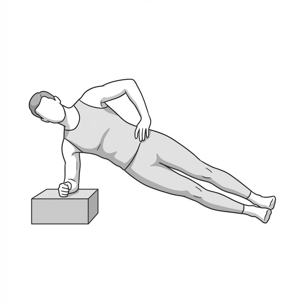
- Implementation of the Copenhagen adduction exercise protocol
- Core and pelvic stabilization exercises
- Eccentric strengthening to improve musculotendinous resilience
- Sport-specific biomechanical retraining and return-to-play progression
Red Flags & Differential Diagnosis
While many cases of groin pain are musculoskeletal in origin, the region shares nerve pathways with the genitourinary and gastrointestinal systems. The following signs warrant prompt medical investigation to rule out severe pathology.
Suspected Avulsion Fracture
In adolescent athletes, sudden, severe groin pain accompanied by a "pop" and inability to bear weight may indicate an avulsion fracture of the ischial tuberosity or pubis. This requires immediate radiographic evaluation.
UrgentComplete Tendon Rupture
A palpable defect or significant bruising (ecchymosis) in the medial thigh following an acute trauma suggests a high-grade tear or complete rupture, necessitating prompt orthopedic assessment.
UrgentIntra-articular Hip Pathology
Mechanical symptoms such as clicking, catching, or locking within the hip joint, especially when accompanied by deep groin pain, may indicate a labral tear or advanced osteoarthritis rather than a simple muscle strain.
InvestigateInguinal Hernia
A visible or palpable bulge in the groin region that increases with coughing or bearing down (Valsalva maneuver) suggests an inguinal hernia, which requires surgical evaluation.
InvestigateGenitourinary Symptoms
Groin pain accompanied by urinary changes, testicular pain, or fever may indicate genitourinary pathology (e.g., epididymitis, kidney stones) and warrants immediate medical attention.
Emergency
Key Clinical Takeaways
Adductor longus strains are among the most common causes of acute groin pain, but myofascial trigger points frequently mimic or complicate this presentation.
Precise palpation is critical; tenderness at the bony insertion suggests tendinopathy or strain, while mid-belly tenderness often indicates myofascial involvement.
The iliopsoas and pectineus muscles are potent sources of referred groin pain and must be evaluated in any patient presenting with adductor symptoms.
Current guidelines generally recommend a graded, progressive loading program (such as the Copenhagen protocol) for the rehabilitation of adductor-related groin pain.
Clinicians must maintain a high index of suspicion for overlapping conditions, including athletic pubalgia, hip joint pathology, and inguinal hernias.
Aggressive stretching during the acute phase of an adductor strain is generally discouraged, as it may exacerbate tissue damage and provoke reactive muscle spasm.
Movement therapy
Exercises for Adductor Strain
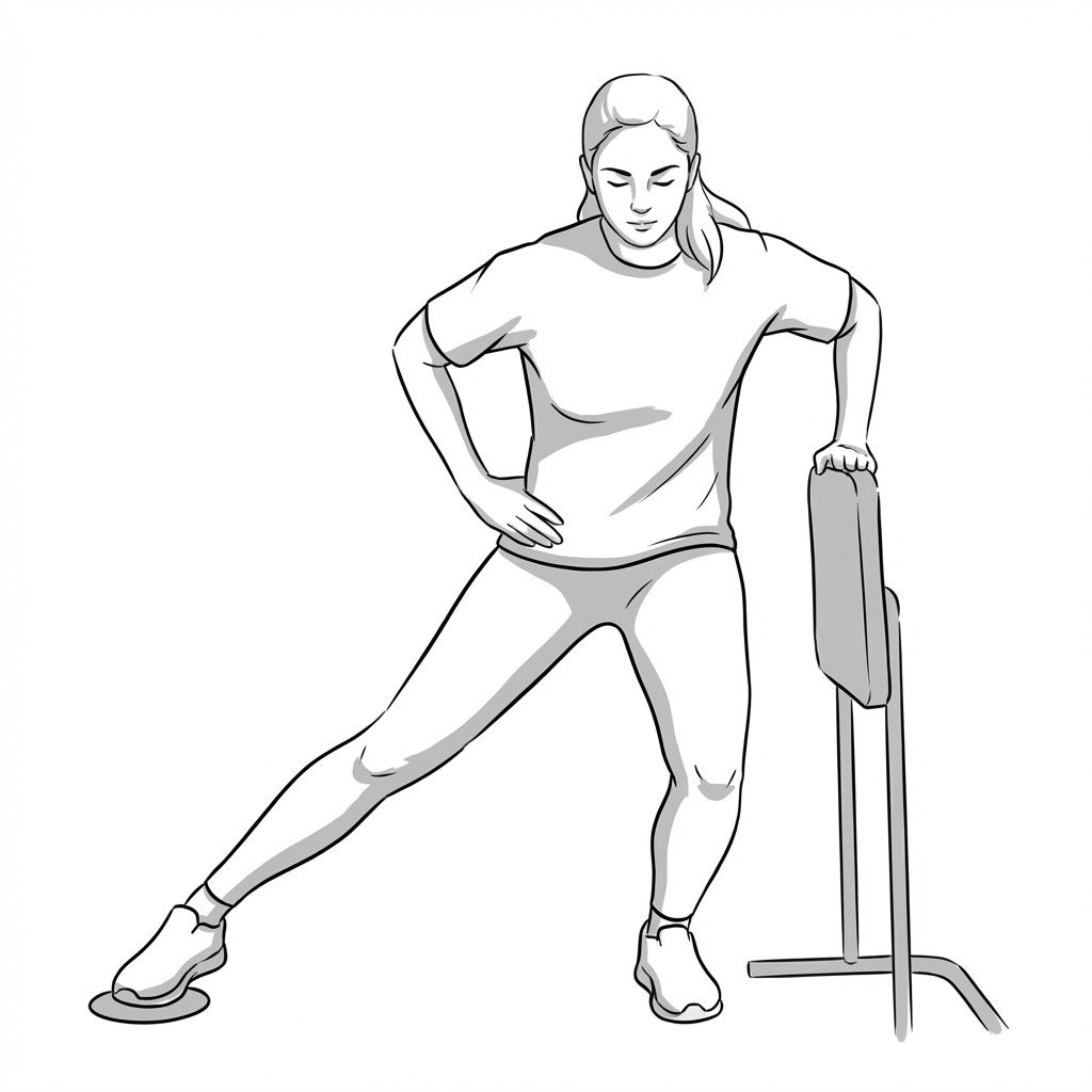
These exercises progress the groin from pain-free isometric holds to the dynamic medial-hip loading needed for return to sport — isometric squeezes reduce pain early, while Copenhagen planks and sliding lunges build the adductor strength that protects against re-strain. Start with 2–3 and progress as tolerated.