Understanding the Suboccipitals
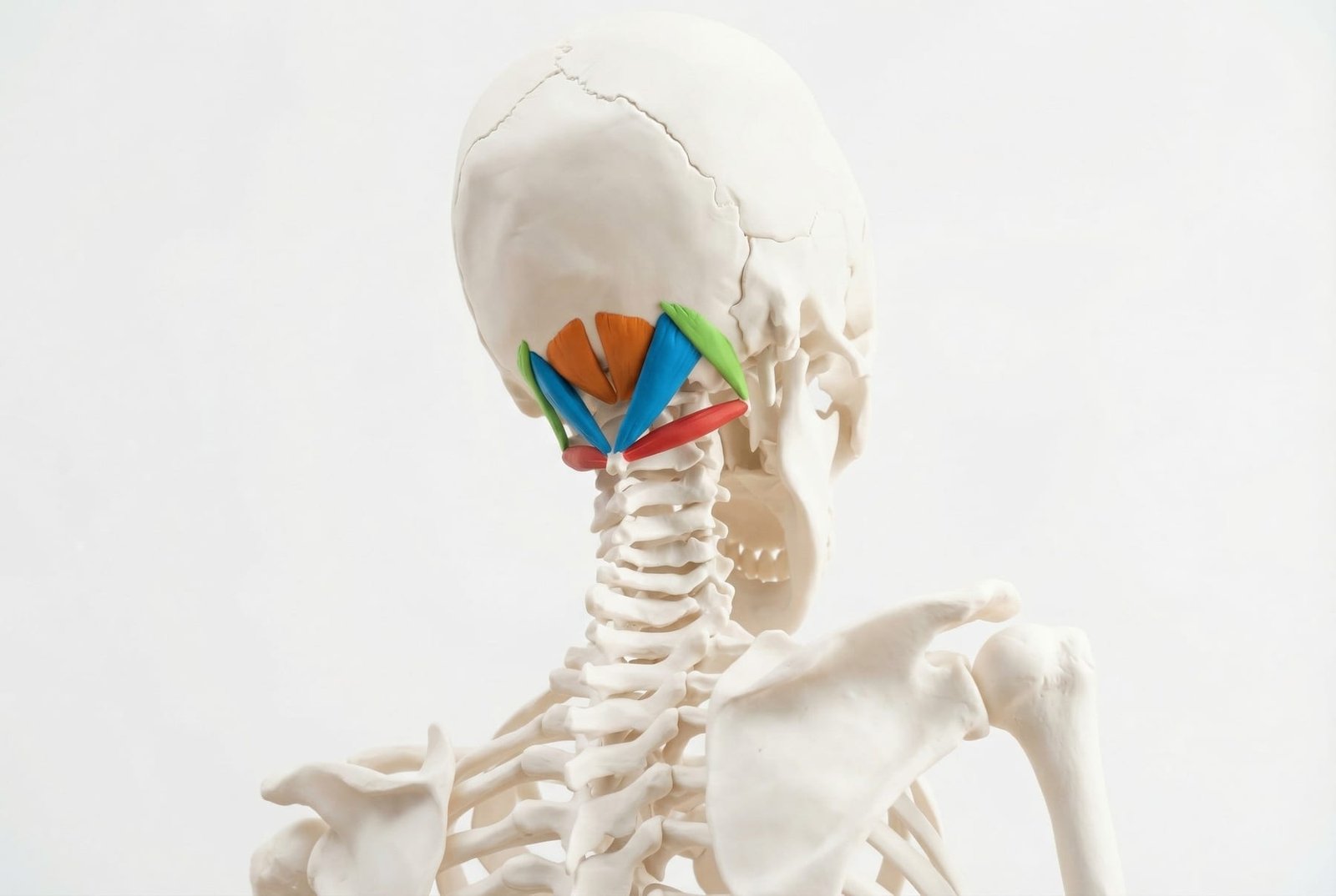
Suboccipital Muscles Anatomy Deep Posterior Cervical View
The suboccipital muscles are a small group of deep muscles at the base of the skull. Their main role is fine control of the upper cervical spine rather than large visible movement.
Because they work constantly to position the head on the neck, they are highly sensitive to postural overload, screen-related forward-head posture, and upper-cervical guarding.
Clinically, these muscles matter because their trigger points can refer pain to the occiput, upper neck, and sometimes deep behind the eye — a pattern that can look surprisingly similar to primary headache disorders.
That does not mean every occipital or peri-orbital headache is suboccipital in origin. It means the suboccipitals should be examined when the symptom pattern, posture, and upper-cervical findings all point in that direction.
Anatomy & Trigger Points
The suboccipital region is small but anatomically dense. This is one reason both palpation and invasive treatment in this area require care and good anatomical judgment.
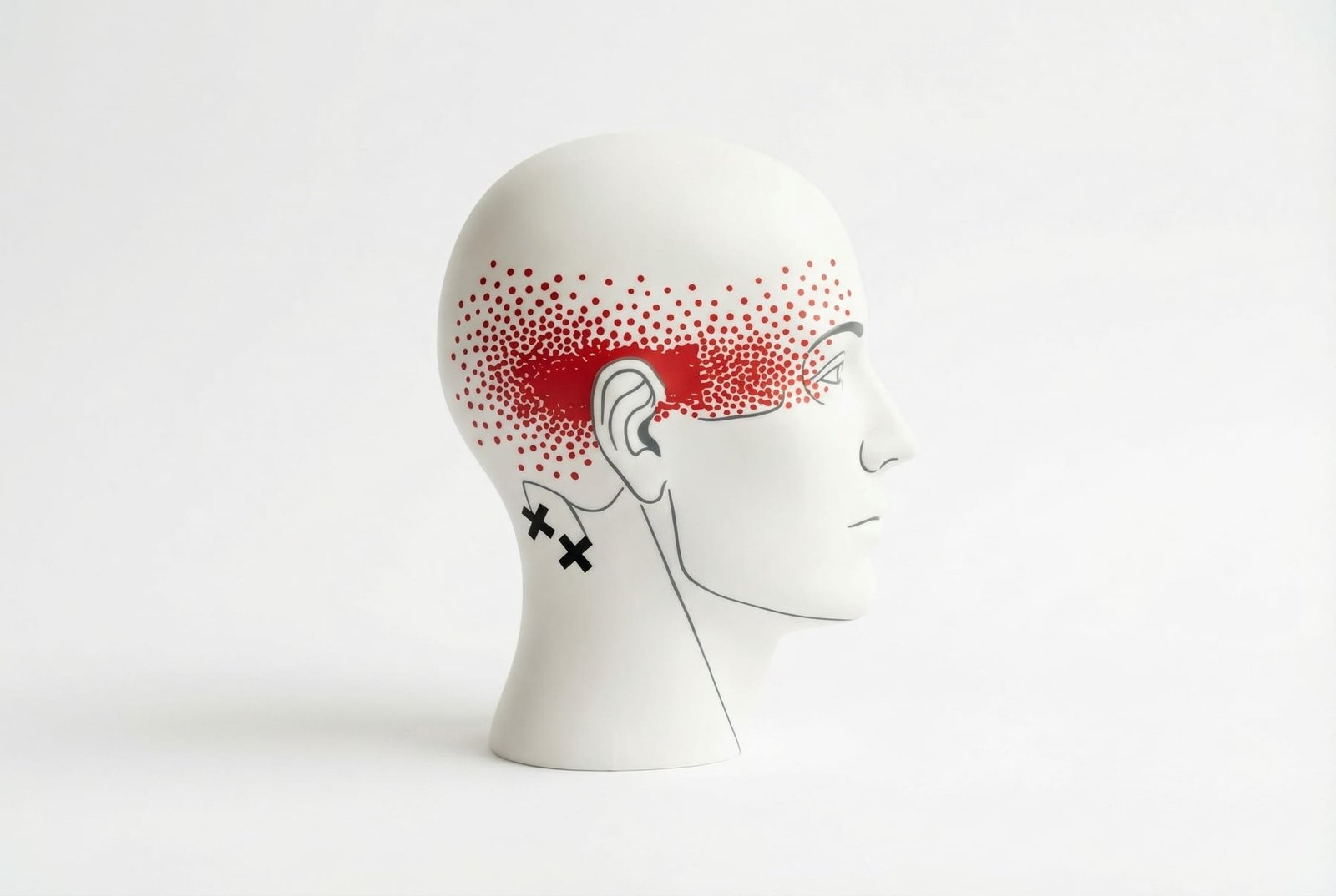
Suboccipital Trigger Point Locations and Referral Zones
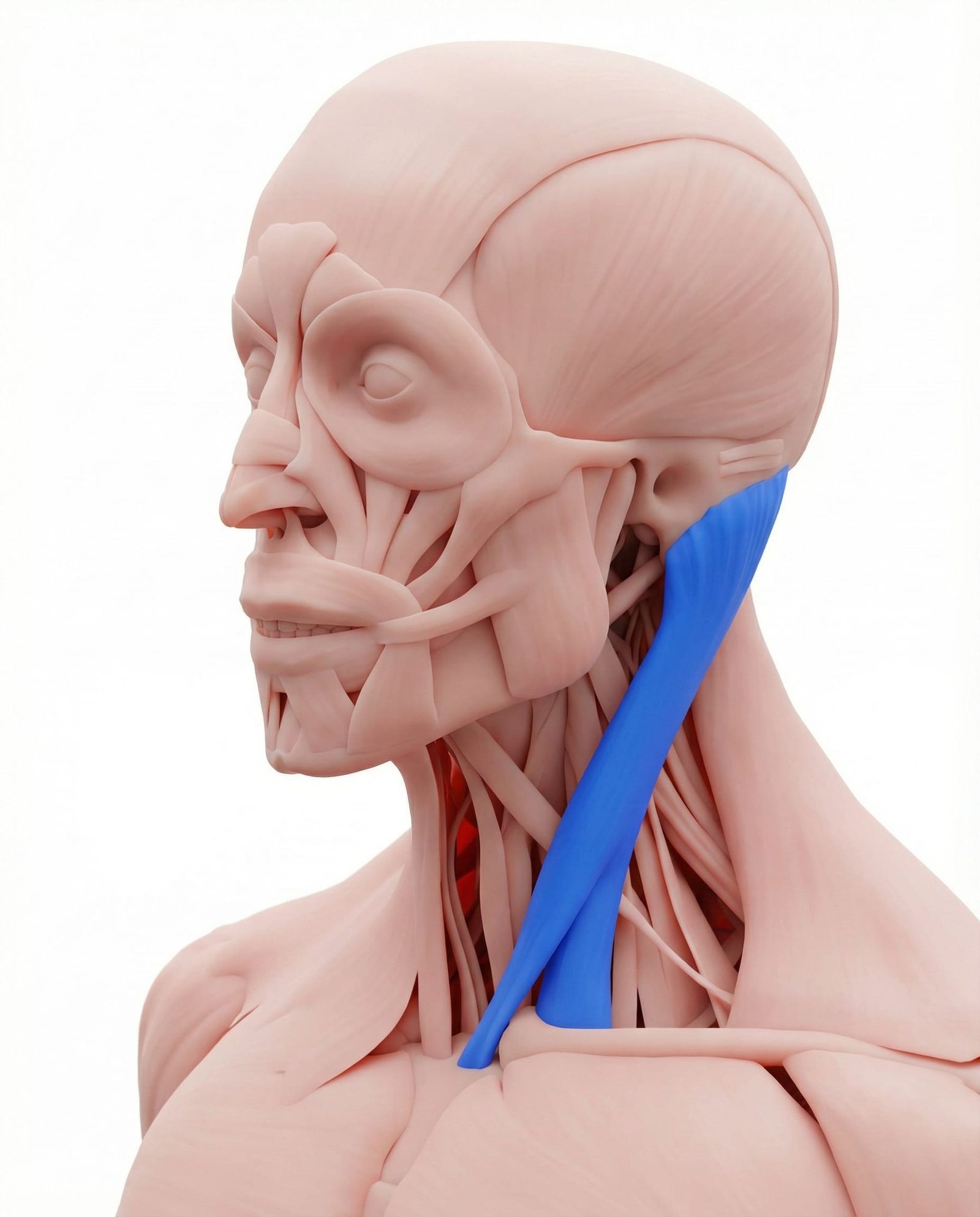
The SCM and suboccipitals commonly coexist in cervicogenic headache patterns. SCM trigger points are more likely to contribute to frontal, facial, ear, or dizziness-related symptoms, while the suboccipitals more often contribute to posterior headache and behind-the-eye referral.
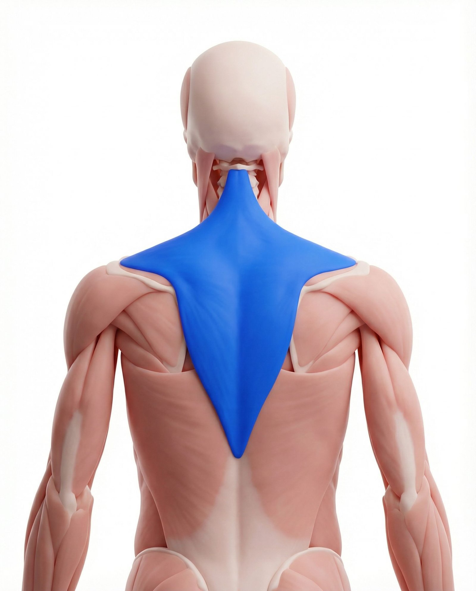
Upper trapezius trigger points often refer toward the neck, mastoid region, and temple. Chronic upper-trapezius overload can also reinforce the postural pattern that keeps the suboccipitals shortened and overworked.
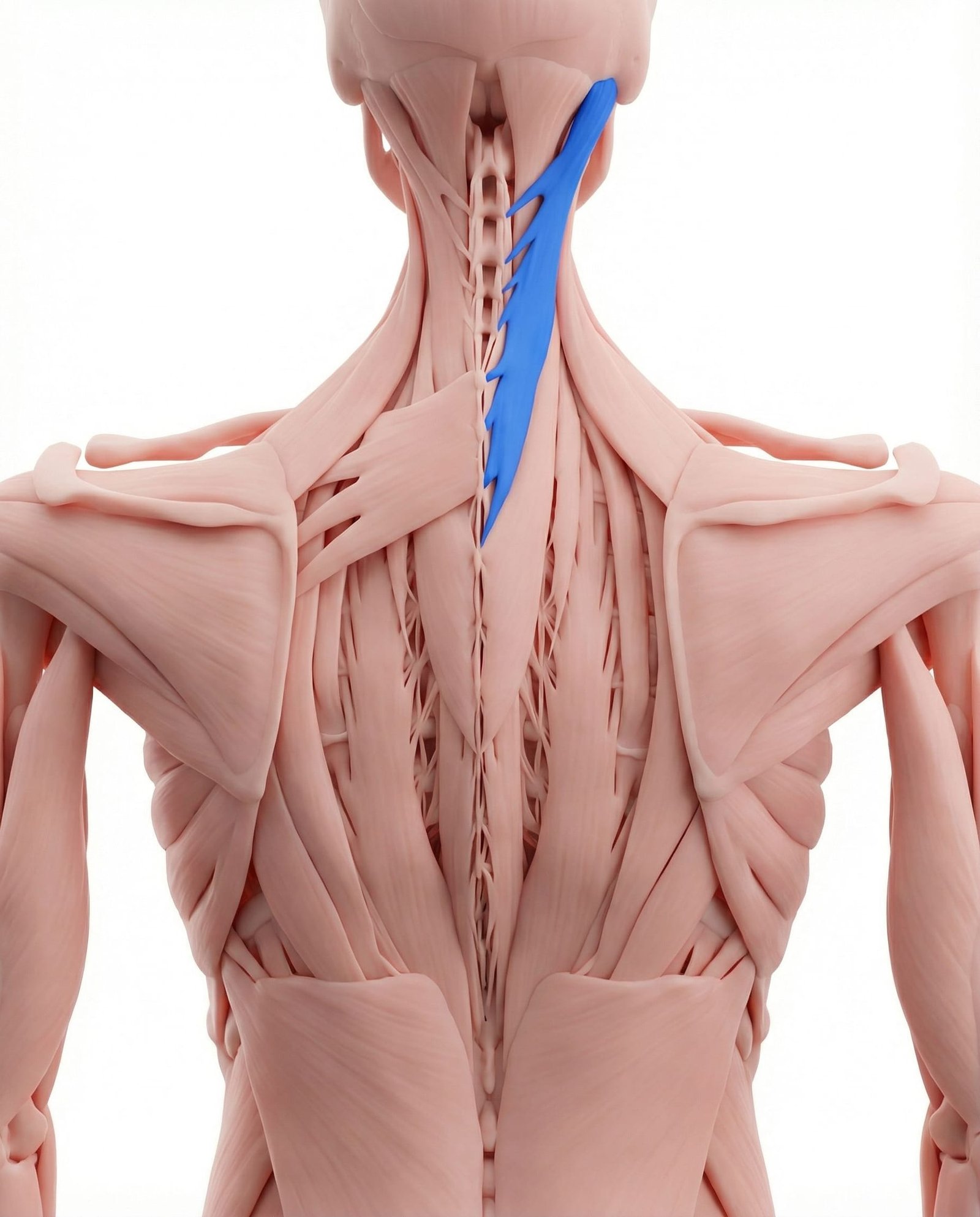
Semispinalis capitis often contributes to broader posterior head and temporal headache patterns. It lies superficial to the suboccipitals, which is one reason the two are often confused on palpation.
Can refer pain toward the top of the head and lateral skull. It commonly coexists with suboccipital dysfunction in forward-head posture and chronic upper-cervical strain.
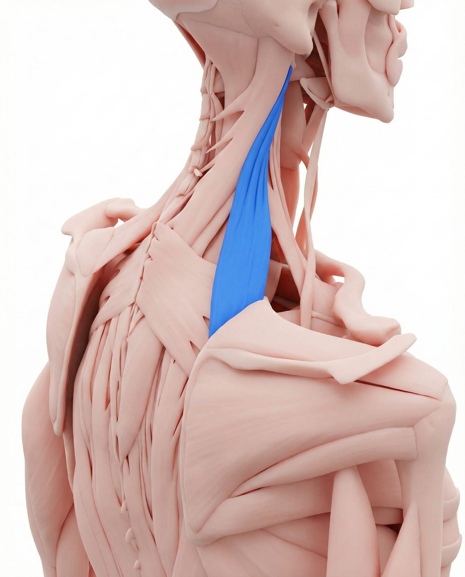
Levator scapulae often contributes to the painful “stiff neck” pattern and commonly overlaps with suboccipital tension because both are overloaded by prolonged forward-head and rounded-shoulder posture.
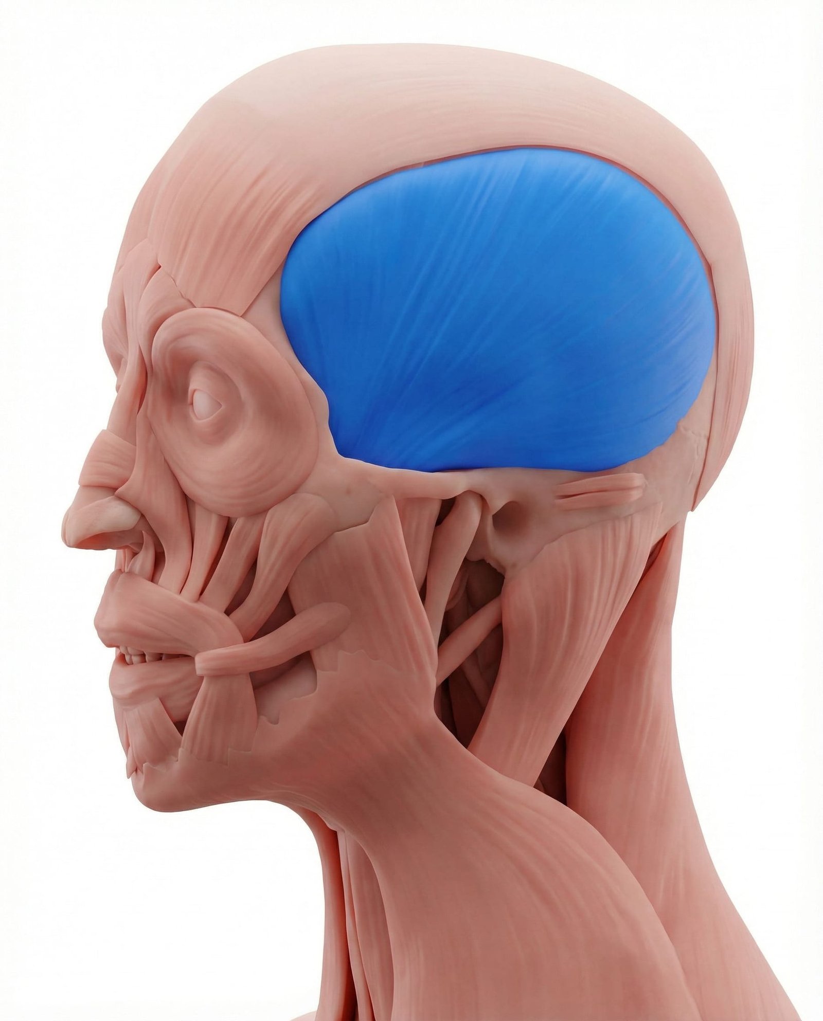
Temporalis trigger points may contribute to temple pain and head pain that overlaps with cervical headache patterns. In some patients, upper-cervical dysfunction and temporalis symptoms reinforce each other clinically.
Referral Patterns: Headache & Eye Pain
A common suboccipital pattern is a posterior headache that wraps forward and concentrates around the eye or upper forehead. In practice, patients often describe it as “coming from the base of the skull” even when the felt pain is more forward.
Because other muscles — especially SCM, semispinalis capitis, and upper trapezius — may create nearby or overlapping patterns, the referral map should always be interpreted together with the physical exam.
The suboccipitals matter because they can create a symptom pattern that feels much larger and more dramatic than the size of the muscles would suggest.
Occiput
Behind the Eye
Neural Overlap
Self-Assessment Tests
These tests help estimate whether the suboccipitals are active contributors. None of them should be used as a stand-alone diagnosis, especially in patients with complex headache patterns.
Suboccipital Palpation
Deep suboccipital trigger point assessment
Flexion-Rotation Test
Upper cervical rotation assessment
Cranio-Cervical Flexion Test
Deep cervical flexor assessment
Greater Occipital Nerve Test
Occipital neuralgia differentiation
Treatment Pathways
The most effective treatment usually combines local muscle downregulation with better upper-cervical mechanics, improved postural control, and reduced reliance on the suboccipitals as constant stabilizers.
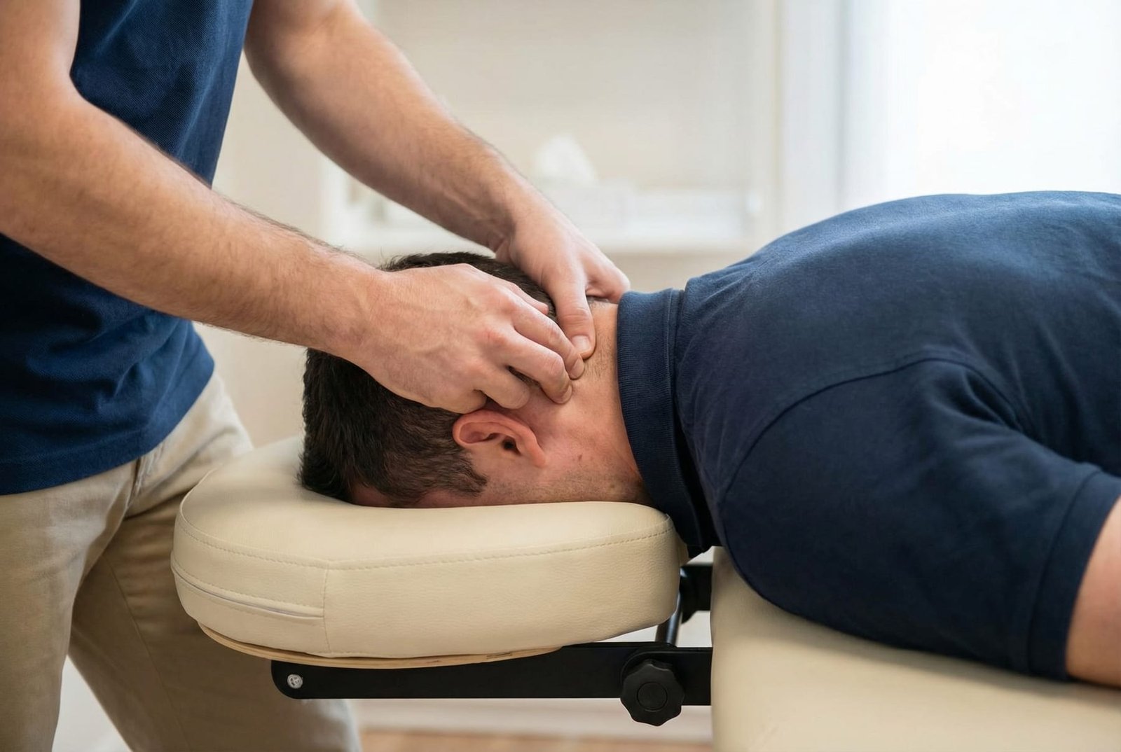
Suboccipital Release Technique and Deep Cervical Flexor Training
Suboccipital Inhibition Technique
Sustained gentle pressure to the suboccipital muscles is one of the most commonly used manual approaches in this region. The effect is usually to reduce guarding and improve tolerance to movement rather than to “fix” the whole problem by itself.
- Supine suboccipital release with gentle sustained contact
- Self-release using supported pressure at the base of the skull
- Low-force, longer-duration holds rather than aggressive pressing
- Slow breathing during the technique to reduce guarding
- Regular repetition in chronic cases when clearly helpful
Upper Cervical Mobility Exercises
Gentle upper-cervical mobility work can reduce mechanical demand on the suboccipitals, especially in patients with prolonged static posture, headaches, and restricted upper-cervical movement.
- Gentle chin nodding and craniocervical flexion work
- Small-range upper-cervical rotation movements
- Eye-head coordination drills where appropriate
- Low-load mobility rather than aggressive end-range stretching
- Frequent short sessions rather than occasional forceful work
Dry Needling: Suboccipital Region
Dry needling in the suboccipital region should be considered an advanced technique because of the density of important anatomy in the area. It should only be performed by appropriately trained clinicians.
- Use only when the anatomy is clear and the indication is strong
- Respect the vascular and neural structures in the upper cervical region
- Use patient feedback and positioning carefully
- Combine with rehabilitation rather than treating needling as a stand-alone cure
- Reassess headache pattern, mobility, and functional change after treatment
Deep Cervical Flexor Rehabilitation
For many patients, long-term improvement depends on reducing overreliance on the suboccipitals by restoring better deep cervical flexor control and better upper-cervical posture.
- Craniocervical flexion / chin-nod training
- Gradual endurance progression of deep cervical flexors
- Simple upright postural drills with minimal effort
- Integration with visual and breathing control when needed
- Consistent practice over weeks rather than expecting rapid change
Rectus Capitis Posterior Major
Often associated with broad upper-cervical and occipital referral that may extend forward.
Obliquus Capitis Inferior
Often clinically important in upper-cervical rotation restriction and posterior headache patterns.
Obliquus Capitis Superior
May contribute to lateral or ear-adjacent symptom overlap in selected cases.
Greater Occipital Nerve Overlap
Neural irritation can coexist with muscular referral, creating a mixed picture rather than a pure trigger point pattern.
Red Flags — When to See a Doctor
Occipital headache is often benign and musculoskeletal, but some headache patterns need urgent medical evaluation before local treatment is considered.
Key Takeaways
The suboccipitals are a small but clinically important group of deep upper-cervical muscles that often contribute to occipital headache, eye-region pain, and upper-cervical dysfunction.
Their trigger points may create headache patterns that strongly resemble migraine, occipital neuralgia, or other headache disorders, which is why careful differential diagnosis matters.
Forward-head posture and chronic upper-cervical overuse are among the most common perpetuating factors.
Suboccipital symptoms often overlap with SCM, upper trapezius, semispinalis capitis, and other cervical pain generators rather than occurring in isolation.
The best long-term management usually combines local release, mobility work, and deep-flexor rehabilitation instead of relying on one treatment method alone.
Because headache is a high-stakes symptom, red-flag screening is essential before assuming a purely muscular explanation.