Understanding the Clinical Overlap
Radial tunnel syndrome (RTS) is a frequently debated clinical entity characterized by deep, aching pain in the lateral forearm. The condition is generally attributed to compression of the posterior interosseous nerve (PIN)—a deep motor branch of the radial nerve—as it traverses the radial tunnel. Current literature suggests that unlike true PIN palsy, RTS typically presents without objective motor weakness or consistent electromyographic (EMG) abnormalities, making the diagnosis primarily clinical. This lack of definitive diagnostic criteria often complicates the differentiation of RTS from other lateral elbow pain syndromes.
The clinical presentation of RTS exhibits significant overlap with myofascial pain syndrome (MPS), particularly involving the supinator and brachioradialis muscles. The arcade of Frohse, a fibrous arch at the proximal edge of the superficial supinator, is considered the most common site of PIN entrapment. Hypertonicity or trigger points within the supinator may not only refer pain to the lateral epicondyle and dorsal web space—mimicking RTS symptoms—but may also mechanically contribute to dynamic nerve compression. Consequently, myofascial dysfunction and nerve entrapment frequently coexist, confounding isolated diagnoses.
The clinical distinction between radial tunnel syndrome and supinator myofascial pain is frequently blurred, as hypertonic muscle fibers may simultaneously refer pain and dynamically compress the posterior interosseous nerve.
Differentiating RTS from supinator MPS and lateral epicondylalgia requires meticulous palpation and provocative testing. Clinical guidelines generally recommend identifying the point of maximal tenderness; in RTS, this is typically located 4 to 5 centimeters distal to the lateral epicondyle, over the mobile wad of Henry, whereas lateral epicondylalgia presents with tenderness directly at or immediately distal to the epicondyle. Diagnostic approaches may include targeted local anesthetic injections or, in refractory cases, advanced imaging to rule out space-occupying lesions, though appropriate clinical uncertainty remains a hallmark of managing this regional pain complex.
Anatomical Context
The radial tunnel is a 5-centimeter anatomical space extending from the radiocapitellar joint to the distal edge of the supinator muscle. Understanding the structures forming this tunnel is essential for identifying potential sites of nerve compression and myofascial overlap.
Supinator
The supinator muscle forms the floor and lateral wall of the radial tunnel. The posterior interosseous nerve (PIN) passes between its superficial and deep heads at the arcade of Frohse. Hypertonicity and trigger points in this muscle are frequently implicated in both direct pain referral and dynamic compression of the PIN.
Primarily refers deep ache to the lateral epicondyle, lateral forearm, and occasionally the dorsal web space of the hand.Brachioradialis
A superficial muscle of the lateral forearm that flexes the elbow, particularly when the forearm is in a neutral position. It forms the lateral boundary of the proximal radial tunnel. Myofascial pain in the brachioradialis often mimics the superficial aching associated with radial nerve irritation.
Refers pain to the lateral epicondyle, radial aspect of the forearm, and the base of the thumb.Extensor Carpi Radialis Brevis (ECRB)
The ECRB is the primary muscle implicated in lateral epicondylalgia (tennis elbow), which frequently coexists with or is misdiagnosed as radial tunnel syndrome. Its medial edge can occasionally contribute to proximal PIN compression. Differentiating ECRB pathology from supinator dysfunction is a critical diagnostic step.
Strongly refers pain to the lateral epicondyle and the dorsal aspect of the forearm and hand.Extensor Digitorum
The primary extensor of the fingers, innervated by the PIN. While trigger points here cause local forearm pain, true weakness in this muscle (finger drop) is a hallmark of PIN palsy rather than radial tunnel syndrome or isolated myofascial pain.
Refers pain distally along the dorsal forearm, often concentrating in the middle and ring fingers.Biceps Brachii
Though primarily an elbow flexor, the biceps is the most powerful supinator of the forearm. Dysfunction in the biceps can alter forearm biomechanics, placing increased secondary demand on the supinator muscle and potentially exacerbating radial tunnel symptoms.
Refers pain to the anterior shoulder, cubital fossa, and occasionally the anterior forearm.
Dynamic Nerve Compression
Intermittent compression of the PIN by the arcade of Frohse or fibrous bands during repetitive pronation and supination.
Myofascial Referral
Active trigger points in the supinator, brachioradialis, or ECRB referring deep, aching pain to the lateral elbow and forearm.
Tendinopathic Overlap
Degenerative changes in the common extensor origin (lateral epicondylalgia) coexisting with or mimicking radial tunnel symptoms.
Cervical Referral
C6 radiculopathy or somatic referral from cervical facet joints presenting as distal arm pain without primary local pathology.
Myofascial Referral Patterns
Myofascial trigger points in the lateral forearm musculature can produce pain patterns that are virtually indistinguishable from radial nerve irritation. Recognizing these patterns is a critical component of the differential diagnosis.
- Supinator
- Pain typically concentrates at the lateral epicondyle and radiates distally along the dorsal-lateral forearm. This pattern is often clinically indistinguishable from the deep ache of radial tunnel syndrome.
- Brachioradialis
- Aching pain is felt along the radial border of the forearm, frequently extending to the base of the thumb and the dorsal web space, mimicking superficial radial nerve sensory distribution.
- Extensor Carpi Radialis Brevis
- Intense, localized pain at the lateral epicondyle that may radiate down the posterior forearm, typically exacerbated by gripping and wrist extension.
Clinical Assessment
Clinical differentiation relies heavily on provocative testing and precise palpation. These assessments aim to stress specific anatomical structures to isolate the primary source of nociception.
Maximal Tenderness Palpation
Carefully palpate the lateral elbow region. Compare the tenderness directly on the lateral epicondyle (bony prominence) with the tenderness in the mobile wad of muscle 4 to 5 centimeters distal to the epicondyle.
Positive: Tenderness maximal 4-5 cm distal to the epicondyle suggests radial tunnel syndrome or supinator myofascial pain, whereas maximal tenderness directly on the bone suggests lateral epicondylalgia.
Resisted Middle Finger Extension (Maudsley's Test)
Extend the elbow fully, pronate the forearm, and extend the wrist. Apply downward resistance specifically to the extended middle finger while the patient attempts to maintain extension.
Positive: Reproduction of pain in the proximal lateral forearm. This tests the extensor digitorum (innervated by the PIN) and the ECRB, often provoking both RTS and lateral epicondylalgia.
Resisted Supination Test
With the elbow flexed to 90 degrees and the forearm fully pronated, the patient attempts to supinate (turn palm up) against the examiner's resistance.
Positive: Pain in the deep lateral forearm. This strongly engages the supinator muscle, potentially compressing the PIN at the arcade of Frohse or provoking supinator trigger points.
Dynamic Compression Test
The patient actively extends the elbow, pronates the forearm, and flexes the wrist simultaneously, holding this position for up to one minute.
Positive: Reproduction of the characteristic deep, aching lateral forearm pain, suggesting dynamic compression of the radial nerve or severe myofascial tension.
Treatment Pathways
Management of lateral forearm pain generally begins with a comprehensive conservative approach. Current evidence suggests that addressing coexisting myofascial dysfunction often yields significant clinical improvement, even when mild nerve compression is suspected.
Self-Care
- Activity modification to reduce repetitive pronation/supination
- Ergonomic assessment of keyboard and mouse positioning
- Avoidance of prolonged elbow extension with wrist flexion
- Gentle, pain-free forearm stretching
- Use of a counterforce brace (applied distal to the radial tunnel)
Manual Therapy
- Ischemic compression of supinator and brachioradialis trigger points
- Myofascial release of the lateral forearm extensor compartment
- Soft tissue mobilization along the radial nerve pathway
- Joint mobilization of the proximal radioulnar joint
- Friction massage over the lateral epicondyle (if co-occurring)
Interventional
- Diagnostic local anesthetic injection into the radial tunnel
- Corticosteroid injection to reduce localized perineural inflammation
- Dry needling of the supinator and brachioradialis muscles
- Ultrasound-guided hydrodissection of the posterior interosseous nerve
- Surgical decompression (reserved for refractory cases or true PIN palsy)
Rehabilitation
- Radial nerve gliding and tensioning exercises
- Eccentric strengthening of the wrist extensors
- Supinator stretching and neuromuscular re-education
- Scapular and proximal shoulder stabilization exercises
- Gradual return-to-load protocols for gripping activities
Red Flags & Differential Diagnosis
While lateral forearm pain is most commonly benign and related to musculoskeletal or mild neuropathic dysfunction, certain signs warrant prompt medical investigation to rule out severe neurological compromise or structural lesions.
True Motor Weakness
Inability to actively extend the fingers or thumb (finger drop/thumb drop) suggests true Posterior Interosseous Nerve (PIN) palsy, a more severe neurological deficit than radial tunnel syndrome.
UrgentPalpable Mass in Proximal Forearm
A distinct, palpable lump in the lateral forearm may indicate a space-occupying lesion, such as a lipoma, ganglion cyst, or tumor compressing the radial nerve.
InvestigateCervical Radiculopathy Signs
Pain radiating from the neck, accompanied by altered reflexes, dermatomal sensory loss, or weakness in the C5-C6 distribution, suggesting a proximal nerve root issue.
InvestigateAcute Trauma or Deformity
Sudden onset of severe pain following a fall or direct blow, potentially indicating a radial head fracture or elbow dislocation.
EmergencyProgressive Atrophy
Visible wasting of the forearm extensor muscles, indicating chronic, severe denervation that requires prompt electrodiagnostic evaluation.
Urgent
Key Clinical Takeaways
Radial tunnel syndrome (RTS) is a clinical diagnosis characterized by lateral forearm pain without the objective motor weakness seen in true PIN palsy.
Myofascial trigger points in the supinator muscle can closely mimic RTS symptoms and are a primary differential diagnosis.
Maximal tenderness in RTS is typically located 4 to 5 centimeters distal to the lateral epicondyle, helping differentiate it from lateral epicondylalgia.
Electromyography (EMG) is frequently normal in RTS, reflecting the condition's nature as a pain syndrome rather than a gross denervation disorder.
Conservative management focusing on myofascial release, nerve gliding, and activity modification is generally recommended as the first-line approach.
True weakness of finger or thumb extension is a red flag for PIN palsy and warrants prompt neurological or surgical evaluation.
Movement therapy
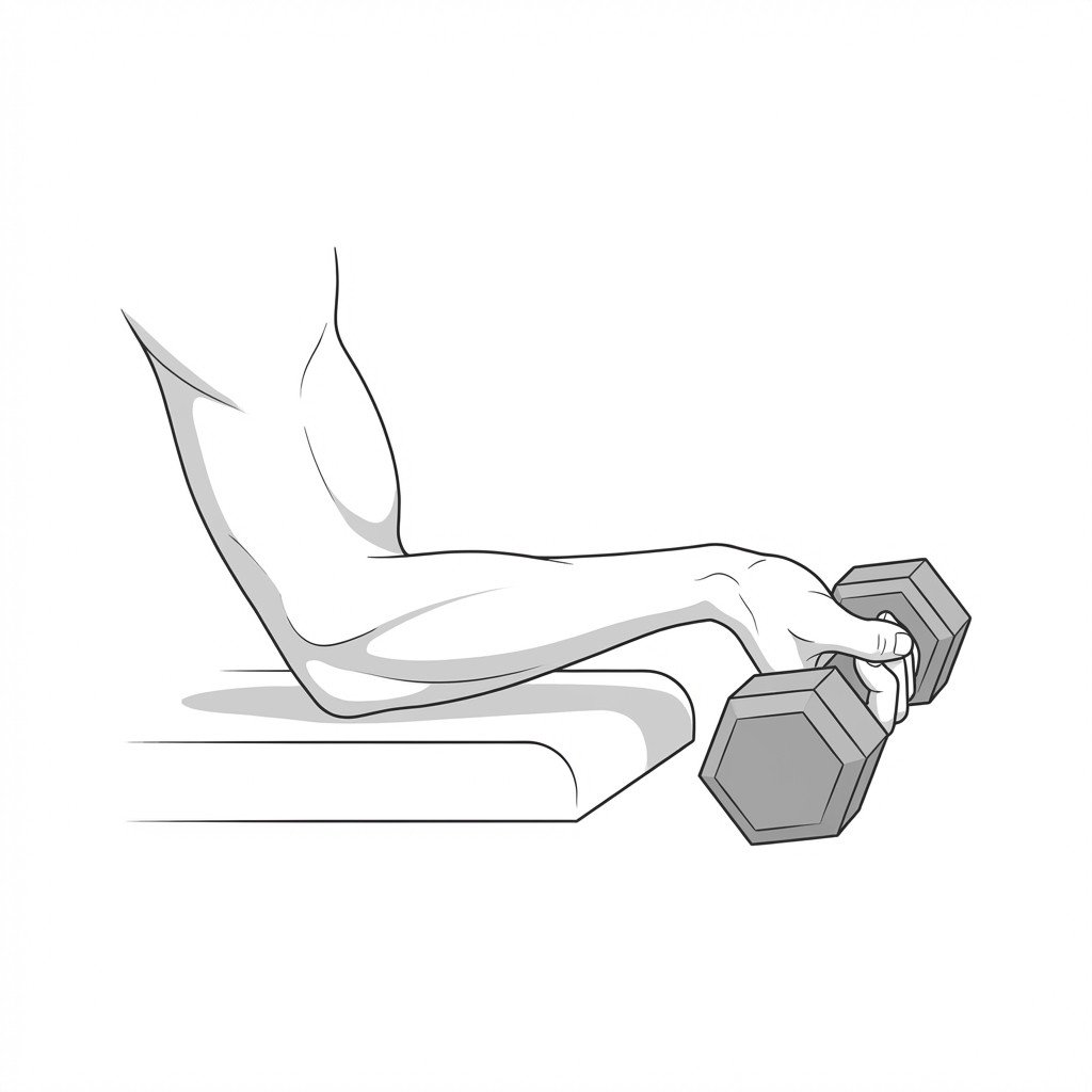
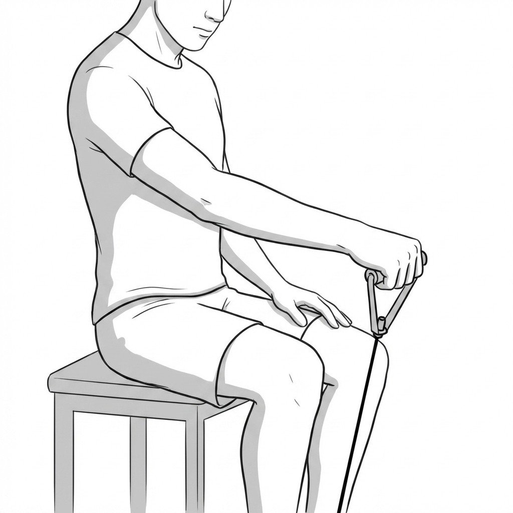
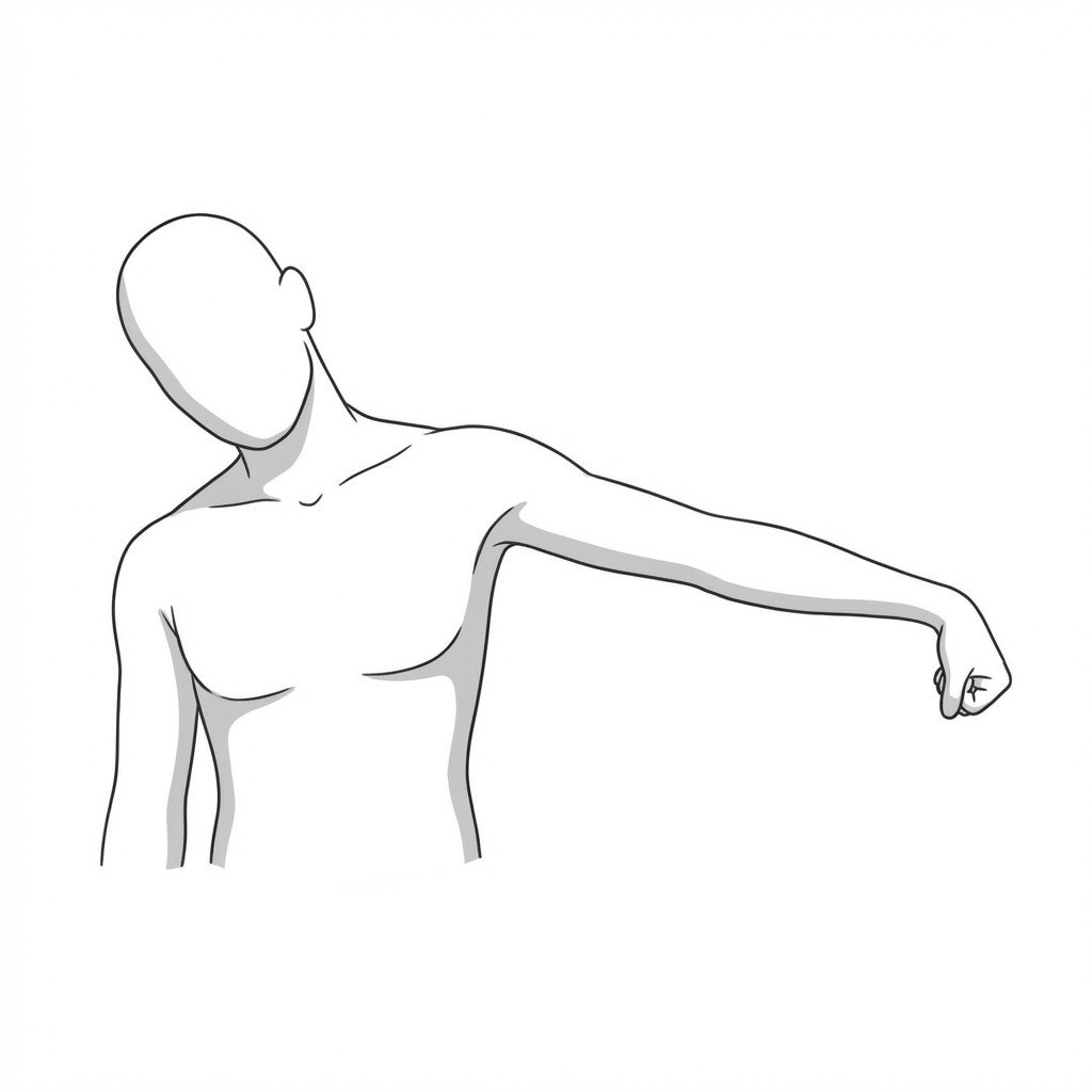
Exercises for Radial Tunnel Syndrome
Radial tunnel pain responds to graded supinator loading and pronator lengthening that reduce compressive force on the posterior interosseous nerve, paired with nerve glides to restore mobility within the tunnel. Start with 2–3 and progress as tolerated.