Understanding the Pectoralis Muscles
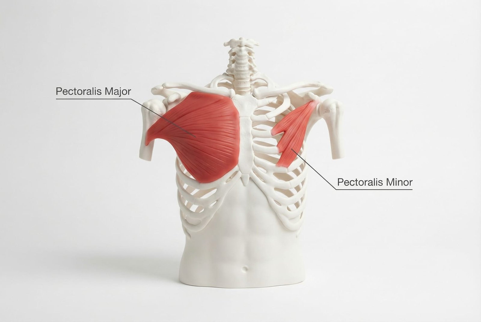
Pectoralis Major and Minor Anatomy Anterior Chest View
The pectoralis major is the large chest-wall muscle that helps with shoulder adduction, internal rotation, and pressing movements. The pectoralis minor lies deeper and attaches from the ribs to the coracoid process, influencing scapular position and upper-quarter mechanics.
Pectoral trigger points can develop with prolonged forward-shoulder posture, repetitive pressing movements, stress-related chest guarding, or sudden overload. In some patients, they contribute to pain that feels alarming because of its chest location.
The clinical importance of the pectoral muscles is not only local pain. They can also contribute to altered scapular mechanics, thoracic outlet-type symptoms, and persistent upper-quarter overload when they remain shortened or irritable.
Because chest pain always carries important medical differentials, pectoral pain should be diagnosed thoughtfully. The clinician’s job is not to assume it is muscular — it is to rule out more urgent causes first and then examine the chest wall appropriately.
Anatomy & Trigger Points
Pectoralis major, pectoralis minor, subclavius, serratus anterior, and the scalenes can all contribute to overlapping chest, shoulder, and arm symptom patterns. In practice, these structures often need to be assessed together.
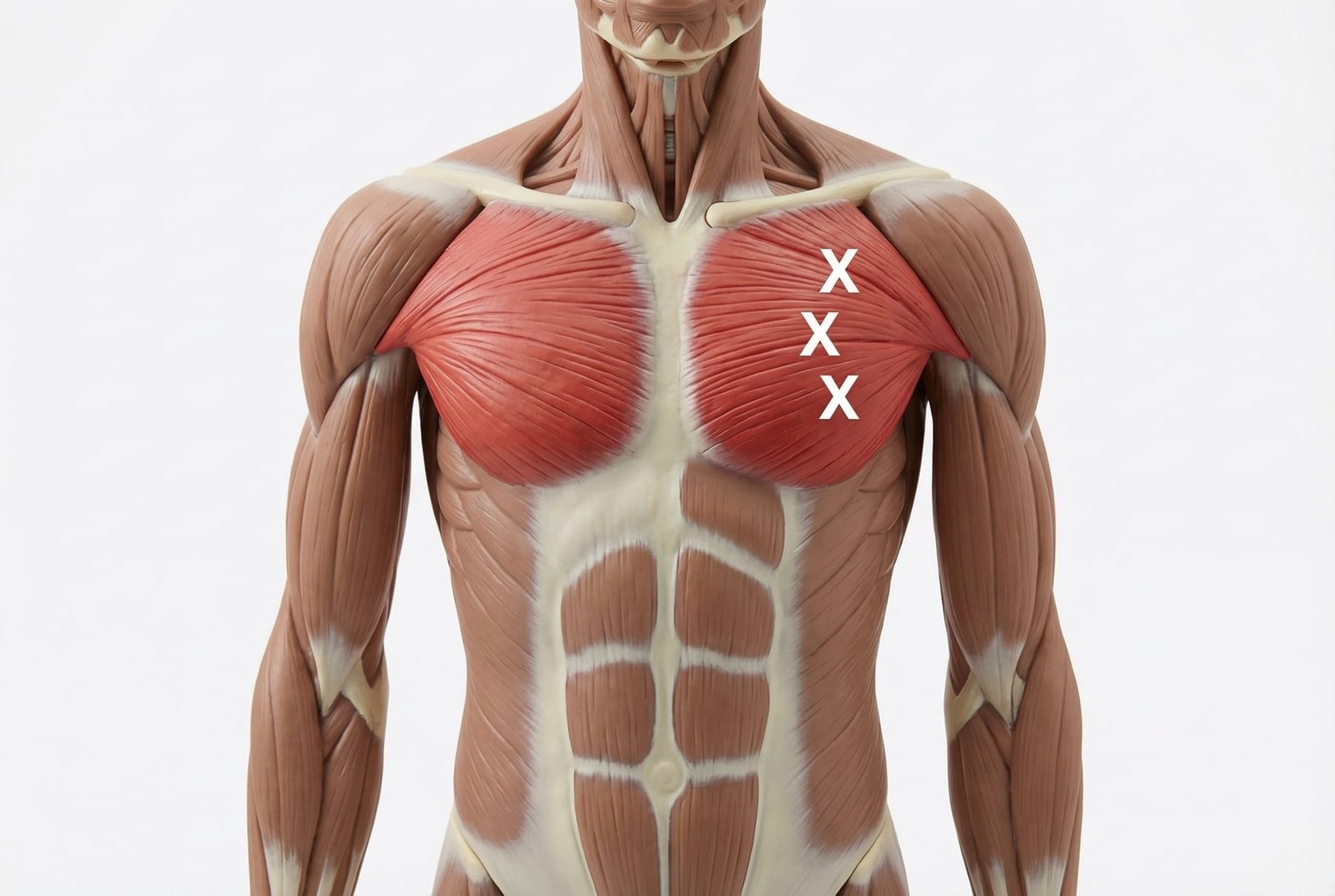
Pectoralis Trigger Point Locations and Referral Zones
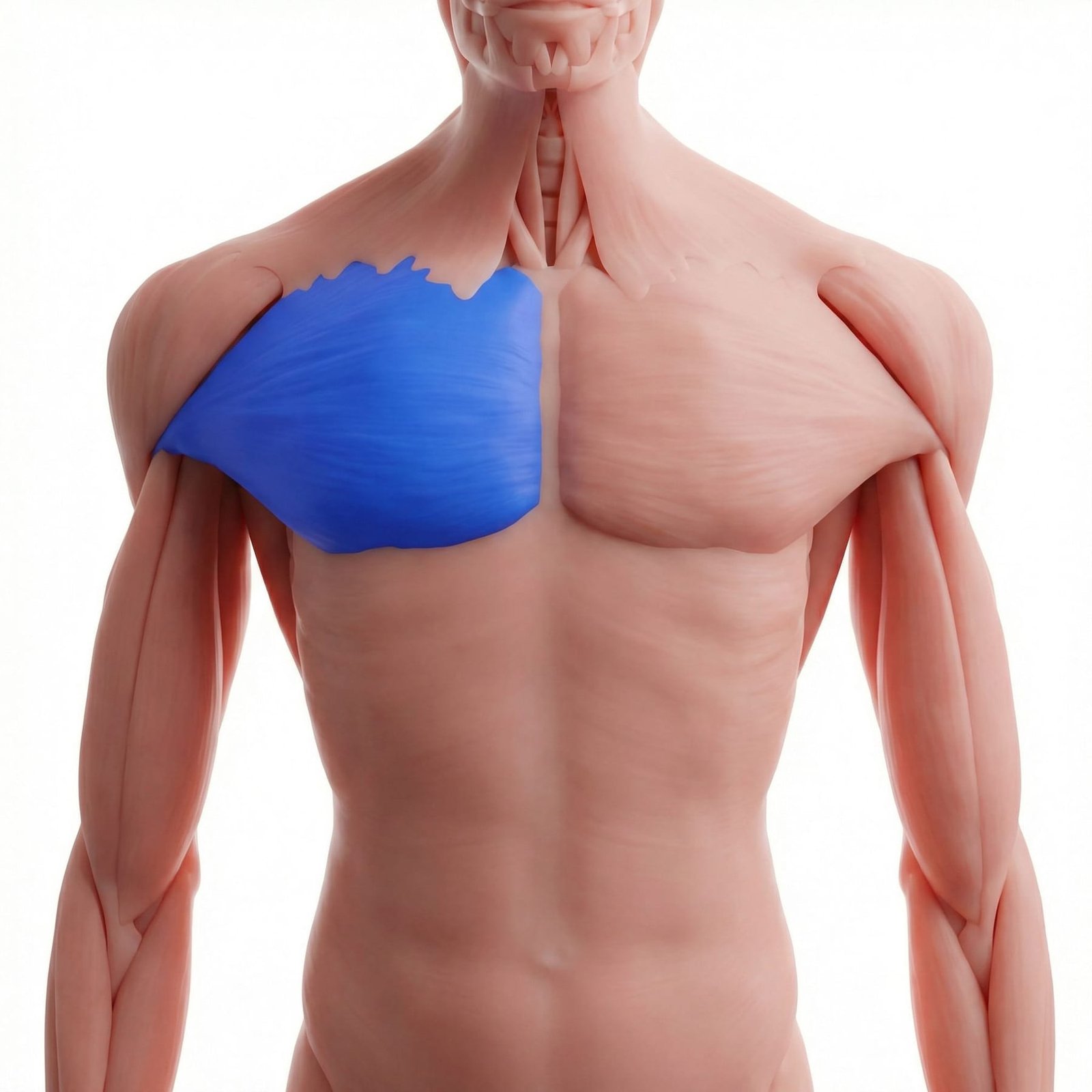
Pectoralis major trigger points can contribute to anterior chest pain, chest wall tightness, and shoulder-region referral. In some patients, this pain can feel alarming and may be mistaken for cardiac pain, which is why acute chest pain always requires appropriate medical assessment first.
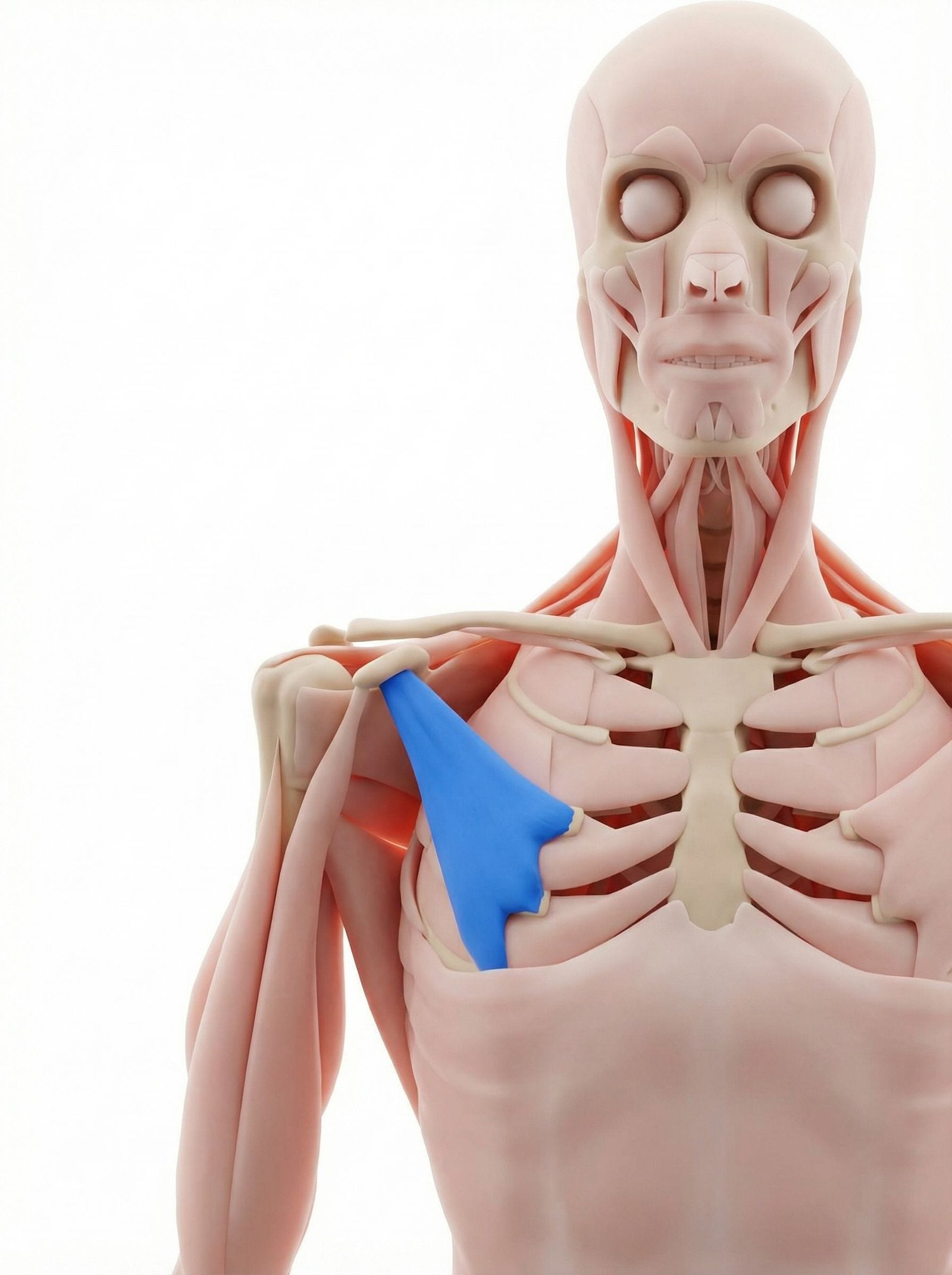
Pectoralis minor dysfunction may contribute to anterior shoulder pain, chest tightness, and symptoms that overlap with thoracic outlet-type presentations, including numbness, tingling, heaviness, or arm fatigue in certain positions. It should be assessed as one possible contributor, not assumed to be the only cause.
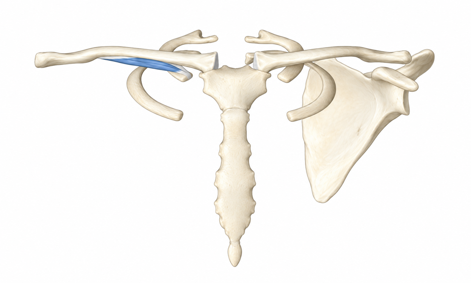
Subclavius-related pain may radiate into the anterior shoulder, upper chest, or arm. Because of its depth and location beneath the clavicle, it is easy to overlook in upper-quarter pain and thoracic outlet-type symptom patterns.
The sternalis is an anatomic variant muscle present in some people. When present and symptomatic, it may contribute to focal sternal discomfort or chest wall pain, though it should never be used to dismiss a necessary cardiac or medical workup.
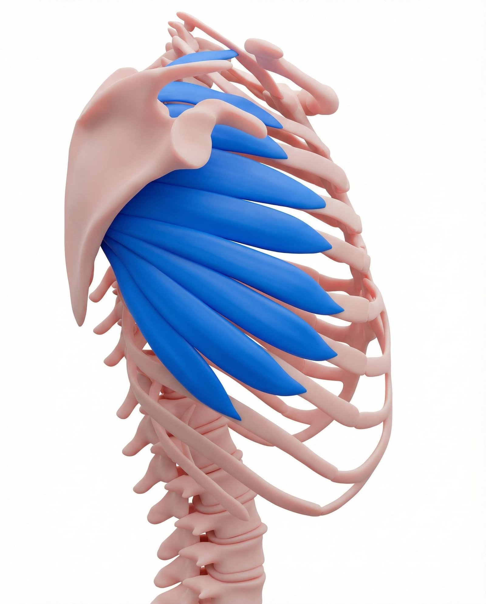
Serratus anterior dysfunction may contribute to lateral chest wall pain, scapular instability, and compensation through the pectoral muscles. It is an important perpetuating factor when chest, shoulder, and scapular pain coexist.
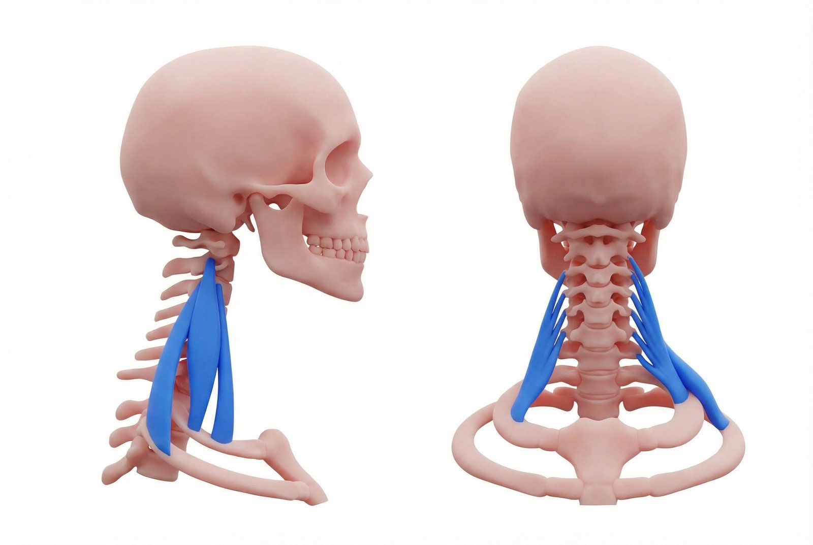
The scalenes can contribute to neck, upper chest, arm, and hand symptoms that overlap with pectoralis minor-related thoracic outlet-type complaints. In many patients, both regions need to be examined together rather than treated as isolated problems.
Referral Patterns: Chest Pain & Arm Symptoms
Pectoralis major may contribute to chest wall pain that feels pressure-like, aching, or tight. Although this can mimic angina subjectively, clinical evaluation must still rule out cardiac causes before a muscular explanation is accepted.
Pectoralis minor may contribute to anterior shoulder pain and thoracic outlet-type symptoms, especially when arm position, chest posture, or prolonged overhead activity influences numbness, tingling, or heaviness.
The pectoral region matters clinically because it sits at the intersection of chest pain, shoulder mechanics, and upper-extremity neurovascular symptoms.
Chest Wall
Arm Symptoms
Posture
Self-Assessment Tests
Self-assessment can help patients notice patterns, but it should stay conservative. Chest pain and neurovascular symptoms require more caution than ordinary muscle self-testing elsewhere.
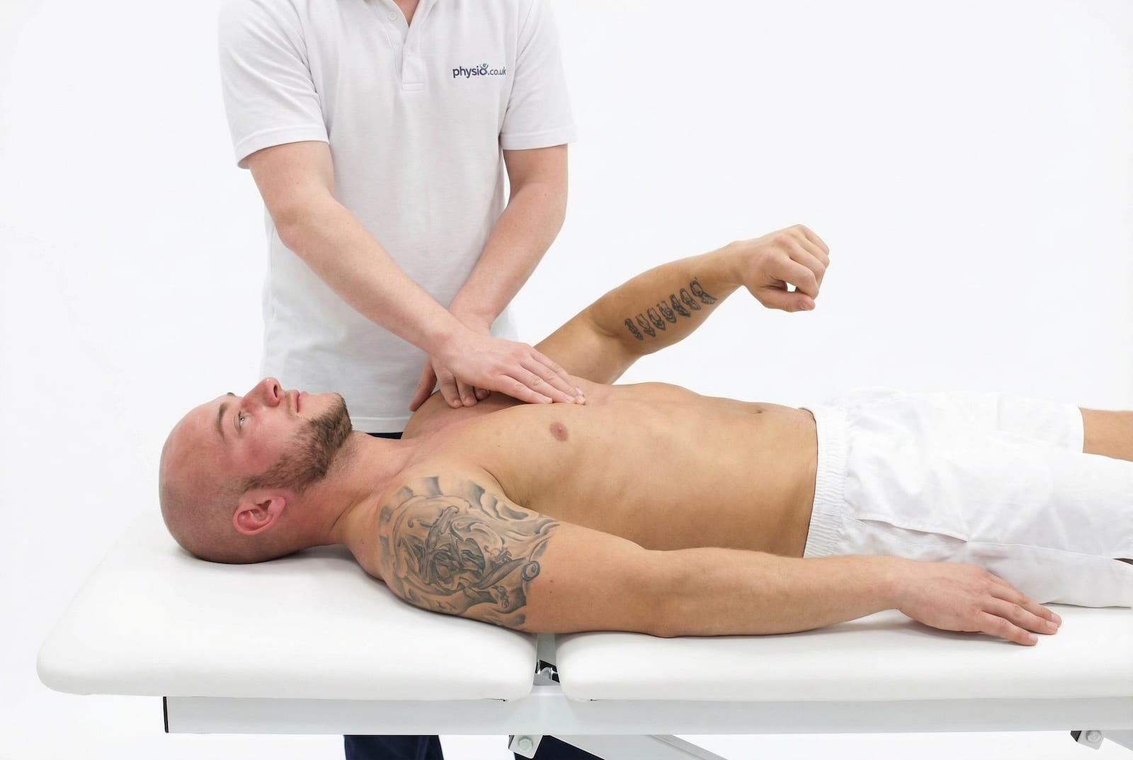
Pectoralis Major Palpation
Pec Minor Compression / Length Assessment
Roos Test (EAST)
Doorway Stretch Response
Treatment Pathways
The most effective pectoral treatment plans usually combine symptom reduction, improved chest-wall length, thoracic mobility, and better scapular and postural control.
Pectoralis Stretching and Chest-Opening Strategies
Stretching & Chest Opening
Gentle chest-opening stretches are often a foundational conservative treatment, especially when forward-shoulder posture and desk-based loading are major perpetuating factors.
- Use tolerable doorway or corner stretches rather than forcing range
- Focus on regular brief practice rather than aggressive intensity
- Pair stretching with breathing and posture reset work
- Stop if stretching reproduces vascular or neurological warning symptoms
- Use stretching as part of a broader plan, not as the only treatment
Chest Wall Self-Management
Simple self-management may help selected patients with pectoral tightness, but chest-wall self-release should stay conservative. The goal is to reduce guarding, not to probe deeply around sensitive structures.
- Use gentle rather than forceful pressure
- Avoid pressing into areas with clear pulsation or strong nerve-like symptoms
- Follow self-treatment with stretching or posture correction
- Stop if you provoke spreading numbness, marked worsening, or chest symptoms that feel medically concerning
- Use clinician guidance if the area feels anatomically uncertain or too sensitive
Needling / Interventional Options
Needling or injection approaches may be considered in selected refractory cases, but the chest wall is an anatomically sensitive region and should be treated only by appropriately trained clinicians.
- Dry needling can be useful in selected cases when performed by a skilled clinician
- Trigger point injections may be considered when clearly indicated
- Procedural technique matters greatly because of nearby lung and neurovascular structures
- Needling should not be presented as first-line care for every pectoral pain pattern
- Conservative management usually remains the starting point
Posture, Scapular Control & Thoracic Mobility
Forward-shoulder posture, thoracic stiffness, and poor scapular control are common perpetuating factors for pectoral pain and thoracic outlet-type symptoms. Addressing these usually matters as much as direct muscle treatment.
- Improve thoracic extension and upper-back mobility
- Work on scapular control and posterior shoulder endurance
- Reduce prolonged forward-rounded positioning during desk work
- Use frequent resets rather than chasing a rigid “perfect posture”
- Build a movement strategy that is sustainable across the workday
Pec Major
May contribute to anterior chest wall pain and shoulder referral.
Pec Minor
May contribute to anterior shoulder pain and thoracic outlet-type symptoms.
Subclavius
Can overlap with clavicular, shoulder, or arm symptoms.
Scalenes + Pecs
Often coexist in upper-quarter neurovascular symptom patterns.
Red Flags — When to See a Doctor
Because this region overlaps with cardiac, vascular, pulmonary, neurological, and breast/chest-wall pathology, red-flag screening is essential before self-management continues.
Pectoralis major trigger points can contribute to chest wall pain, but acute chest pain always requires appropriate medical evaluation first.
Pectoralis minor dysfunction can contribute to anterior shoulder pain and thoracic outlet-type arm symptoms in some patients.
Upper-quarter neurovascular symptoms often involve more than one region, including the scalenes, clavicular region, and pec minor area.
Forward-shoulder posture and prolonged desk-based positioning are common perpetuating factors for pectoral pain patterns.
Stretching, posture correction, thoracic mobility, and scapular control are often core conservative treatments.
Invasive chest-wall treatments should only be performed by clinicians with appropriate anatomical skill and training.