Understanding Meniscal & Myofascial Knee Pain
Meniscal pathology encompasses a spectrum from acute traumatic tears, typically seen in younger athletic populations, to degenerative lesions that frequently present in older adults. The medial and lateral menisci serve critical biomechanical roles in load transmission, shock absorption, and joint stability. However, interpreting meniscal pathology requires significant clinical nuance. Systematic reviews and large-cohort imaging studies consistently demonstrate a high prevalence of asymptomatic meniscal tears in the general population, particularly in individuals over the age of 50. Consequently, the presence of a degenerative tear on magnetic resonance imaging (MRI) does not inherently confirm it as the primary nociceptive source, necessitating a careful differential diagnosis.
Myofascial pain syndrome (MPS) frequently coexists with or mimics meniscal pathology, complicating the clinical picture. The popliteus muscle, which acts to unlock the knee during early flexion, is a primary source of referred pain to the posterior and lateral joint line, often clinically indistinguishable from a lateral meniscus tear. Similarly, trigger points within the semimembranosus and vastus medialis can refer pain directly to the medial joint line, mimicking medial meniscal pathology. Furthermore, patients with true meniscal lesions often develop secondary myofascial trigger points due to altered gait biomechanics, protective muscle guarding, and chronic joint effusion, creating an overlapping pain presentation.
The presence of a meniscal tear on MRI does not guarantee it is the source of pain; popliteus and hamstring trigger points frequently mimic or amplify joint line symptoms.
Differentiating primary meniscal pain from myofascial referred pain relies on a combination of provocative testing, precise palpation, and clinical history. While tests such as the Thessaly and McMurray maneuvers are traditionally utilized to assess meniscal integrity, their diagnostic accuracy is variable, and they may inadvertently provoke myofascial structures. A critical distinguishing factor is the nature of mechanical symptoms; true meniscal locking typically presents as a rigid block to extension, whereas myofascial pseudo-locking—often driven by hamstring or popliteus spasm—may yield to gentle, sustained pressure. Current guidelines generally recommend a trial of conservative management, including targeted myofascial therapy and neuromuscular rehabilitation, for non-obstructive meniscal lesions before considering surgical intervention.
Anatomical Considerations
The knee joint relies on a complex interplay between passive stabilizers (menisci, ligaments) and active dynamic stabilizers (musculature). Understanding their anatomical proximity is crucial for differential diagnosis.
Popliteus
A small, deep muscle located at the posterior knee that functions to internally rotate the tibia and "unlock" the knee during early flexion. Trigger points in the popliteus are frequently misdiagnosed as lateral meniscal tears due to their referral pattern. Hypertonicity here can also create a sensation of stiffness or pseudo-locking that mimics intra-articular mechanical blocks.
Refers pain primarily to the posterior knee and lateral joint line, occasionally extending down the proximal posterior calf.Semimembranosus
The most medial of the hamstring muscles, inserting on the posterior medial condyle of the tibia. It has direct fascial attachments to the medial meniscus. Trigger points in this muscle can refer pain directly to the medial joint line, closely mimicking the presentation of a medial meniscus posterior horn tear.
Refers pain to the medial joint line, posterior thigh, and deep posterior knee.Biceps Femoris
The lateral hamstring muscle that inserts on the fibular head. It plays a significant role in lateral knee stability. Myofascial dysfunction in the short head of the biceps femoris is a common contributor to lateral knee pain and can complicate the clinical picture of lateral meniscal or lateral collateral ligament (LCL) pathology.
Refers pain to the posterolateral knee, fibular head, and occasionally down the lateral calf.Gastrocnemius
The superficial two-headed calf muscle that crosses the posterior knee joint. Trigger points in the medial or lateral heads often develop secondary to altered gait mechanics associated with knee joint effusion or meniscal injury. These trigger points can perpetuate posterior knee pain long after an acute meniscal injury has stabilized.
Refers pain to the posterior knee, posterior calf, and occasionally the plantar aspect of the foot.Vastus Medialis
The medial component of the quadriceps femoris, crucial for dynamic patellar tracking. Trigger points in the distal portion (vastus medialis obliquus) can refer pain to the anteromedial knee. This referral pattern is frequently confused with anterior horn medial meniscus tears or patellofemoral pain syndrome.
Refers pain to the anteromedial knee, medial patellar border, and distal medial thigh.Sartorius
A long, superficial muscle crossing from the anterior superior iliac spine to the medial tibia (pes anserinus). Myofascial pain in the distal sartorius often presents as medial knee pain that exacerbates with activity, requiring differentiation from medial meniscal pathology and pes anserine bursitis.
Refers pain along the medial thigh and directly over the medial joint line and pes anserinus insertion.
Traumatic Meniscal Tear
Acute rotational force on a weight-bearing knee, often resulting in sudden pain, a "pop," and delayed effusion.
Degenerative Meniscal Lesion
Gradual breakdown of meniscal fibrocartilage over time, often coexisting with mild osteoarthritis and presenting with intermittent aching.
Popliteus Overload
Repetitive stress from downhill running or chronic knee instability, leading to trigger points that mimic lateral meniscus pain.
Medial Hamstring Dysfunction
Myofascial tension in the semimembranosus referring pain to the medial joint line, often exacerbated by prolonged sitting or sudden acceleration.
Pain Referral Patterns
Myofascial trigger points in the muscles surrounding the knee can project pain directly into the joint space. Recognizing these specific referral patterns helps differentiate muscular pain from intra-articular pathology.
- Popliteus
- Pain concentrates deep in the posterior knee and along the lateral joint line. This pattern is notoriously difficult to distinguish from a lateral meniscus tear based on patient history alone.
- Semimembranosus
- Pain focuses on the posteromedial joint line and upper calf. Because of its anatomical connection to the medial meniscus, dysfunction here often perfectly mimics posterior horn meniscal pain.
- Vastus Medialis
- Pain refers to the anteromedial aspect of the knee joint. It is frequently described as a deep, aching pain that mimics anterior horn meniscal tears or chondral defects.
- Gastrocnemius
- Pain refers strongly to the posterior knee crease and down the calf. It may present as a feeling of fullness or tension in the back of the knee, similar to a Baker's cyst.
Clinical Assessment & Differentiation
Clinical differentiation requires assessing both structural integrity and muscular function. These assessments help distinguish between intra-articular meniscal lesions and extra-articular myofascial pain.
Joint Line Palpation
With the knee flexed to 90 degrees, systematically palpate the medial and lateral joint lines. Note whether the tenderness is precisely on the joint line (suggestive of meniscal pathology) or slightly above/below it in the muscular attachments.
Positive: Precise joint line tenderness is traditionally associated with meniscal tears, though sensitivity is higher than specificity.
Thessaly Test
Stand on the affected leg with the knee flexed to 20 degrees. While holding a stable surface for balance, internally and externally rotate the body and knee 3 times.
Positive: Medial or lateral joint line discomfort, or a sense of catching/locking, suggests possible meniscal pathology.
Popliteus Resisted Internal Rotation
Sit with the knee flexed to 90 degrees. Attempt to internally rotate the lower leg (point toes inward) while applying manual resistance to prevent the movement.
Positive: Pain at the posterolateral knee during this resisted muscle contraction suggests popliteus involvement rather than an isolated meniscal tear.
Hamstring Length & Provocation
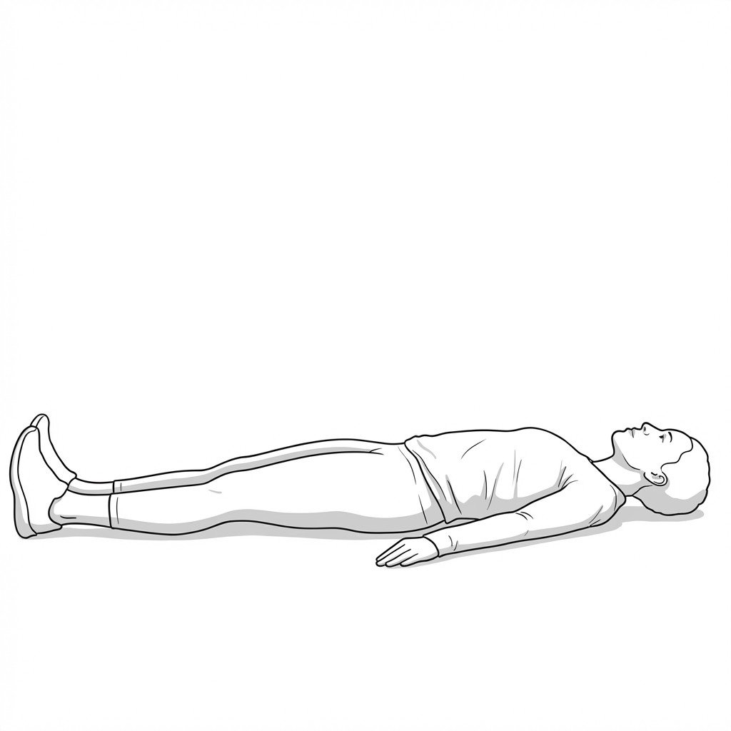
Lie supine and perform a straight leg raise, or passively extend the knee while the hip is flexed to 90 degrees. Palpate the medial and lateral hamstrings during the stretch.
Positive: Reproduction of the patient's familiar knee pain during hamstring stretch, particularly if accompanied by palpable taut bands, suggests a myofascial component.
Evidence-Based Treatment Pathways
Management of knee pain should address both potential intra-articular pathology and secondary myofascial dysfunction. Current evidence strongly supports conservative rehabilitation as a first-line approach for degenerative meniscal tears.
Self-Care
- Activity modification to avoid deep squatting and pivoting
- Application of ice for acute flare-ups or joint effusion
- Gentle, pain-free range of motion exercises (e.g., stationary cycling)
- Self-massage of the quadriceps and calf musculature
- Use of a supportive knee sleeve for proprioceptive feedback
Manual Therapy
- Ischemic compression of popliteus and hamstring trigger points
- Transverse friction massage at muscular insertions
- Patellofemoral and tibiofemoral joint mobilizations
- Myofascial release of the vastus medialis and iliotibial band
- Passive stretching of the posterior chain musculature
Interventional
- Intra-articular corticosteroid injections for acute inflammatory flares
- Hyaluronic acid (viscosupplementation) for concomitant osteoarthritis
- Trigger point injections for refractory popliteus or hamstring MPS
- Diagnostic ultrasound-guided local anesthetic blocks
- Arthroscopic partial meniscectomy (generally reserved for mechanical locking or failed conservative care)
Rehabilitation
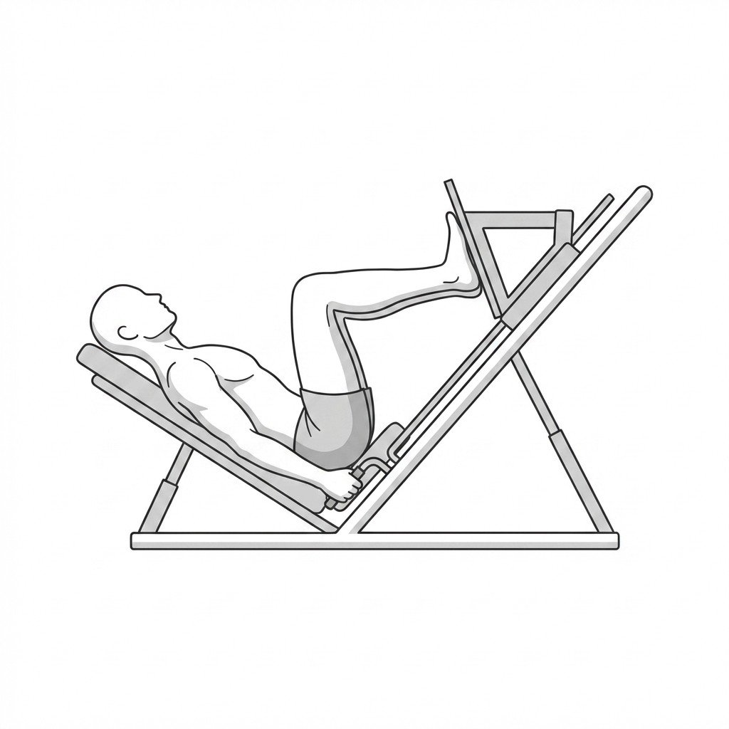
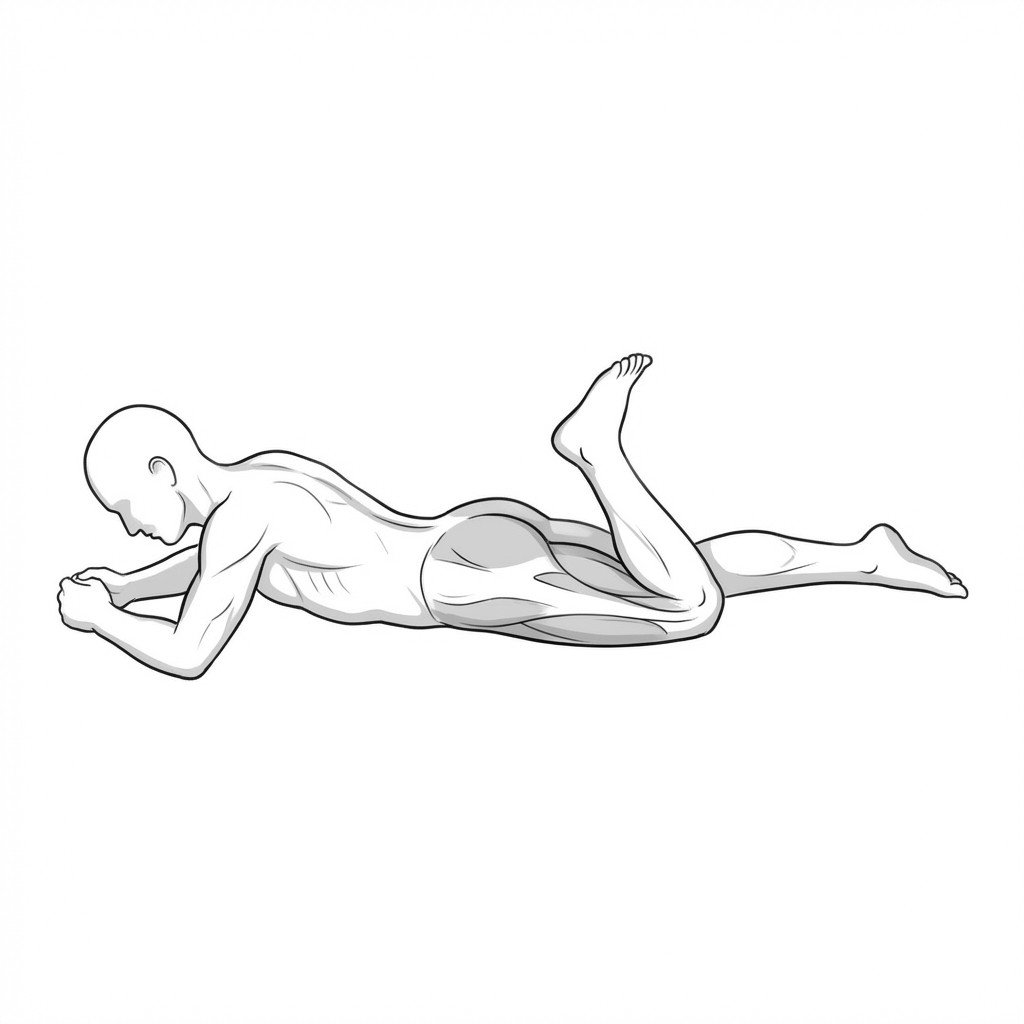
- Progressive quadriceps and hamstring strengthening
- Neuromuscular control and proprioception training
- Hip abductor and external rotator strengthening
- Gait retraining to normalize joint loading
- Graduated return-to-sport or return-to-activity protocols
Red Flags — When to See a Doctor
While many cases of knee pain can be managed conservatively, certain signs indicate severe structural damage or medical emergencies that require prompt physician evaluation.
True Mechanical Locking
A physical inability to fully extend the knee, often accompanied by a hard, mechanical block. This suggests a displaced meniscal fragment (e.g., bucket-handle tear) requiring prompt evaluation.
UrgentAcute Traumatic Hemarthrosis
Rapid swelling of the knee joint within 2 hours of a traumatic twisting injury. This strongly suggests an intra-articular bleed, often associated with ACL or peripheral meniscal tears.
UrgentInability to Bear Weight
Complete inability to put weight on the affected leg following an injury, which may indicate an occult fracture or severe structural instability.
UrgentFever and Erythema
Knee pain accompanied by systemic fever, localized redness, and severe warmth. This presentation requires immediate exclusion of septic arthritis.
EmergencyConcomitant Instability
Frequent episodes of the knee "giving way" or buckling during routine activities, suggesting significant ligamentous compromise alongside potential meniscal injury.
Investigate
Clinical Pearls
Magnetic resonance imaging (MRI) frequently reveals degenerative meniscal tears in asymptomatic individuals; clinical correlation is essential.
Trigger points in the popliteus muscle are an under-recognized mimic of lateral meniscus tears and can be overlooked on clinical examination.
Current evidence suggests that exercise therapy is often non-inferior to arthroscopic surgery for degenerative meniscal tears without true mechanical locking.
Myofascial "pseudo-locking" from hamstring or popliteus spasm can mimic a meniscal block, but typically yields to gentle, sustained pressure.
Patients with true meniscal pathology often develop secondary myofascial pain due to altered gait and protective muscle guarding.
Differentiating joint line tenderness from adjacent muscular tenderness is a critical step in the physical examination of knee pain.
Movement therapy
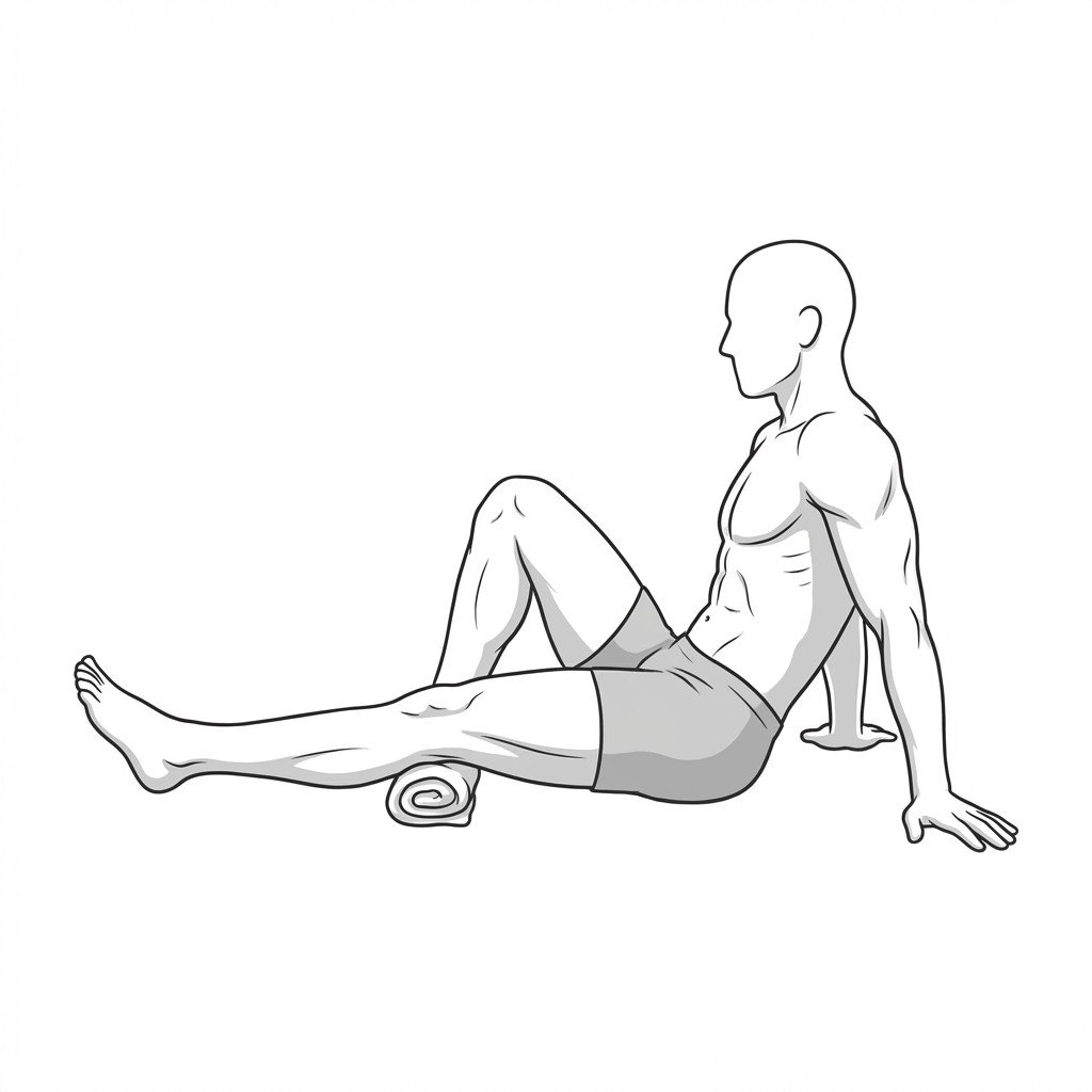
Exercises for Meniscal Rehab
These exercises rebuild the quadriceps and hamstring co-contraction that protects the meniscus under load — beginning with range-of-motion restoration and quad sets, then progressing to single-leg loading as the joint tolerates. Start with 2–3 and progress as tolerated.