Understanding Medial Elbow Pain
Medial epicondylalgia describes load-related pain around the common flexor-pronator origin on the inside of the elbow. Gripping, turning a tool and repeated wrist flexion may reproduce it. A recent increase in force, repetitions or throwing volume is useful context, but pain intensity does not reveal the amount of tendon damage.
Tendon and muscle tenderness can coexist. Pressing a sore forearm may reproduce familiar pain without establishing a separate trigger-point disorder. Symptoms extending from the neck or shoulder, finger tingling, loss of hand strength or pain during the throwing arc broaden the examination beyond the tendon.
A useful examination connects familiar pain with a task and checks competing explanations.
Start with onset, trauma, work or sport demands, symptom distribution and changes in function. Compare active elbow and wrist movement, grip, resisted wrist flexion and pronation, and gentle palpation. Add sensory and motor testing when neural symptoms occur; a throwing history may require a trained ligament examination. Imaging is selected when suspected injury or persistent uncertainty would change care.
Overlapping Findings, Different Next Steps
Pain and provocation
What palpation tells you
Initial direction
When to reconsider
Muscles and Nearby Structures
The flexor-pronator origin lies close to the ulnar collateral ligament and ulnar nerve. Their functions overlap during gripping and throwing, so a painful movement seldom isolates one structure.
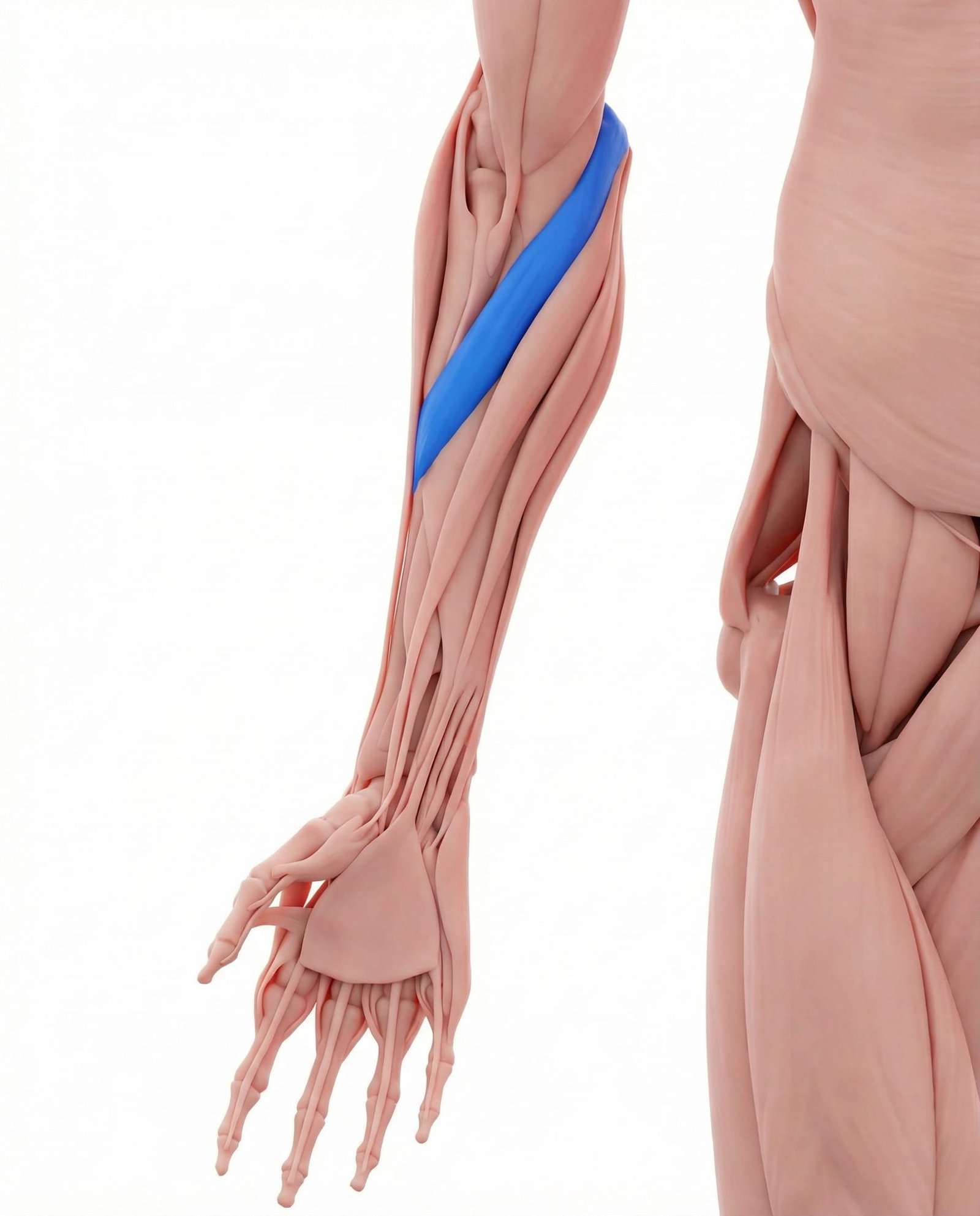
Turns the forearm palm-down and contributes to the flexor-pronator origin. Resisted pronation loads more than this one muscle. Compare local tenderness with wrist-flexion pain, grip tolerance and sensory findings; forearm aching alone does not establish median nerve entrapment.
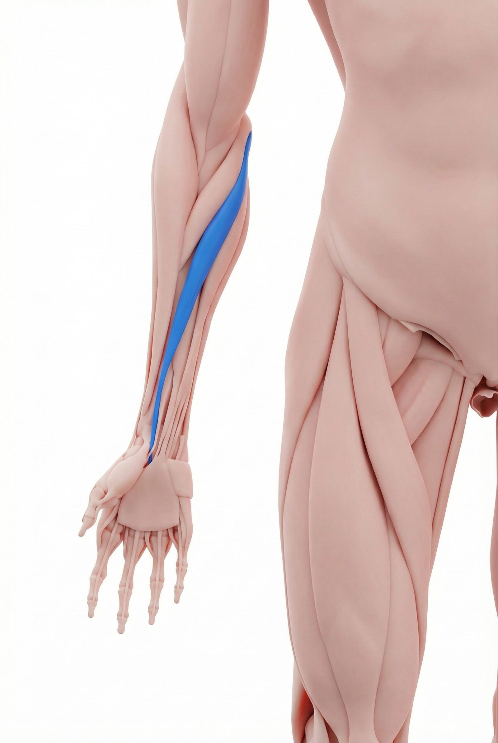
Flexes and radially deviates the wrist from a shared medial origin. Pain with wrist flexion may fit common flexor tendinopathy, but neither resistance testing nor tendon tenderness isolates this muscle or measures tendon damage.
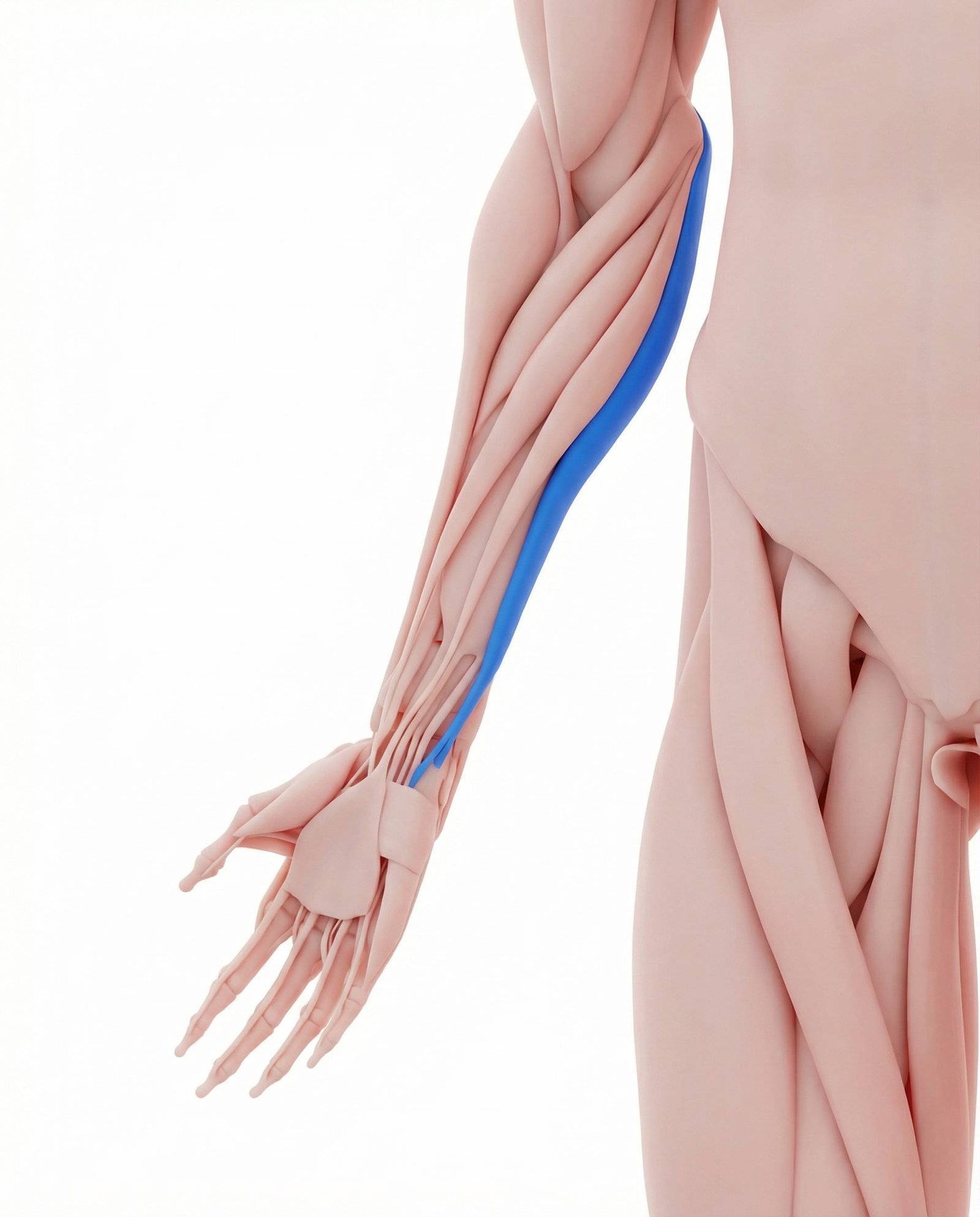
Flexes and ulnarly deviates the wrist. The ulnar nerve passes between its proximal heads; this relationship matters when tingling or hand weakness accompanies pain. Tender muscle does not prove nerve compression or justify nerve gliding.
A variable superficial wrist flexor that may be absent. Its presence, absence or tenderness does not diagnose medial epicondylalgia; assess the whole flexor-pronator task rather than assigning symptoms to a single tender strand.
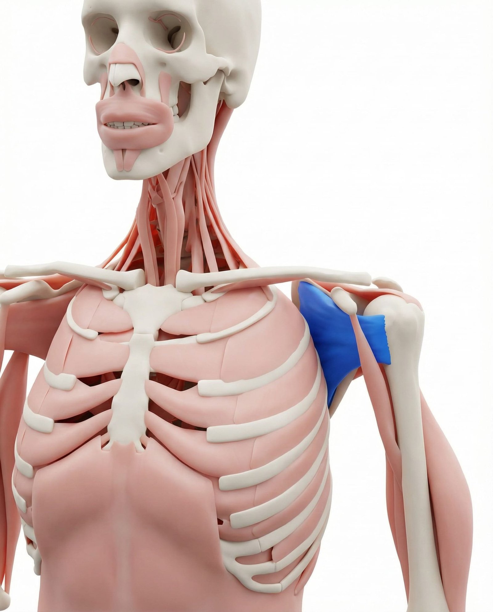
A shoulder internal rotator. Examine shoulder motion and strength when the history includes shoulder pain, restricted rotation or throwing difficulty. Persistent elbow pain is not evidence that this deep muscle is the hidden cause or requires release.
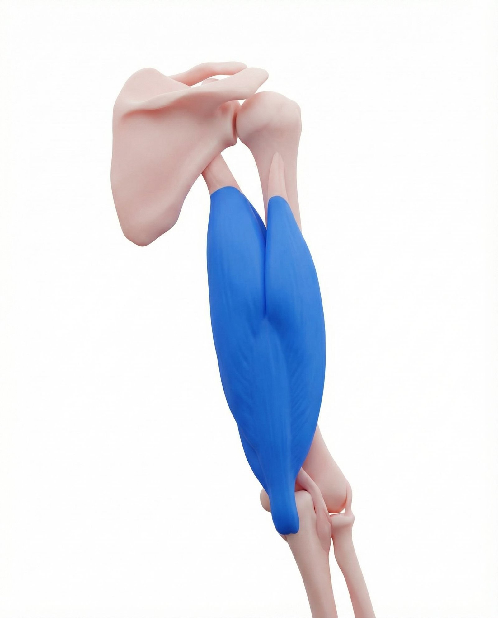
The triceps extends the elbow. Posterior pain, extension weakness or a snapping sensation prompts assessment of the posterior elbow and ulnar nerve. Palpation cannot establish highly specific medial-head referral or exclude flexor-pronator injury.
Flexor-pronator loading
Familiar medial pain during gripping, wrist flexion or pronation may fit a tendon presentation; it does not quantify degeneration.
Regional muscle pain
Muscle tenderness can accompany the same loading problem. Reproduction by pressure does not prove a distinct causal trigger point.
Neural symptoms
Finger tingling or hand weakness needs a sensory and motor examination, with the neck and other nerve sites considered when appropriate.
Valgus loading
Pain during throwing, a pop or instability raises the possibility of ligament injury and changes the rehabilitation decision.
Interpreting Pain Distribution
Use the distribution to choose what to examine. Local tenderness and familiar pain reproduction are observations, not validated maps of a pain generator.
Flexor-pronator region
Shoulder and cervical region
Posterior and medial elbow
Clinical Assessment and Test Limits
These are descriptions of clinical examination, not a home challenge to reproduce maximum pain. Record location, strength and familiar symptom reproduction, then interpret the findings together.
Resisted wrist flexion
Resisted forearm pronation
Ulnar nerve examination
Moving valgus stress test
Palpation and functional baseline
Treatment and Reassessment
After traumatic, neural and ligament problems have been considered, agree on a tolerable loading plan and a functional target such as carrying a bag or completing a work task. The progression below is a clinical framework, not a trial-validated fixed dose. Medial-elbow exercise research is limited; an uncontrolled eccentric-exercise study cannot establish that eccentrics outperform other loading.
Self-care and load adjustment
- Temporarily reduce forceful gripping, wrist flexion or pronation; change tool grip or split long tasks into shorter bouts.
- Keep comfortable elbow and wrist movement instead of stopping all arm use.
- A support or brace is an optional comfort trial, not a tendon-healing treatment; stop if it causes tingling or pressure behind the elbow.
- Avoid hard massage at the bony origin or ulnar nerve. Short-lived comfort does not establish a diagnosis.
- Record one functional task and the next-morning response to work and exercise.
Adjuncts guided by findings
- Manual treatment may be tried for comfort or a measured motion restriction; retain it only if useful function improves.
- Do not prescribe deep subscapularis release as a prerequisite for elbow recovery.
- Nerve glides are not routine tendon care. Neural symptoms first require examination, and any selected movement must not increase tingling or weakness.
- Dry needling is an optional clinician-delivered adjunct with procedure-specific risks, not a substitute for a loading plan or nerve assessment.
- Recheck the same task after an adjunct rather than treating a change in tenderness as proof of tissue repair.
Procedures and further assessment
- Consider ultrasound or MRI when a suspected tear, ligament lesion or unresolved diagnostic question would alter management.
- Discuss steroid injection cautiously: a medial-elbow randomized trial found no sustained advantage over saline at three months or one year.
- That injection study does not demonstrate tendon repair or justify repeated injections.
- PRP, shockwave and tendon procedures should not be presented as proven next steps from these sources; discuss condition-specific evidence, costs and risks with a specialist.
- Persistent disability despite an appropriately adjusted programme calls for diagnostic reassessment before an invasive escalation.
Progressive rehabilitation
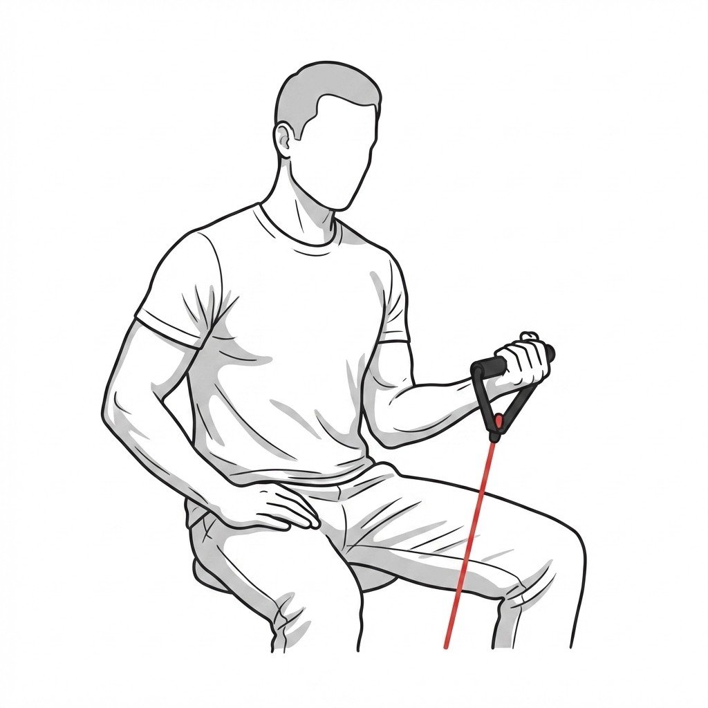
- Start with supported low-resistance wrist flexion and pronation, using a hold or short movement range that is tolerable. Isometrics are an option, not guaranteed pain relief.
- Progress controlled lifting and lowering through a comfortable range; adjust resistance, repetitions or range one variable at a time.
- Add grip endurance and longer task exposure once the current load is stable without a sustained later-day or next-morning flare.
- For sport, rebuild shoulder and whole-arm capacity where deficits are measured, then reintroduce throwing or swing volume before maximal speed and force.
- If the next morning is consistently worse, reduce the last load change. Reassess declining strength, new tingling or a plateau rather than pushing through.
When Further Assessment Matters
Stop provocative exercise after a new injury or neurological deterioration. A hot swollen joint with fever needs urgent care; persistent mechanical symptoms also need examination.
Use history, loading response, strength and sensory findings together; no single palpation or resistance test isolates the painful tissue.
Flexor-pronator tendon and regional muscle symptoms can coexist without requiring separate passive-treatment pathways.
Shoulder examination is relevant when the presentation supports it; subscapularis treatment is not a prerequisite for recovery.
Tingling and hand weakness change the assessment and should not be treated automatically with nerve glides.
Throwing-related instability or an acute pop needs a ligament and injury assessment before progressive loading.
Judge progress by repeatable grip or task tolerance and next-day response. Eccentric-only superiority and fixed-dose protocols are not established by the cited medial-elbow cohort.
Movement options
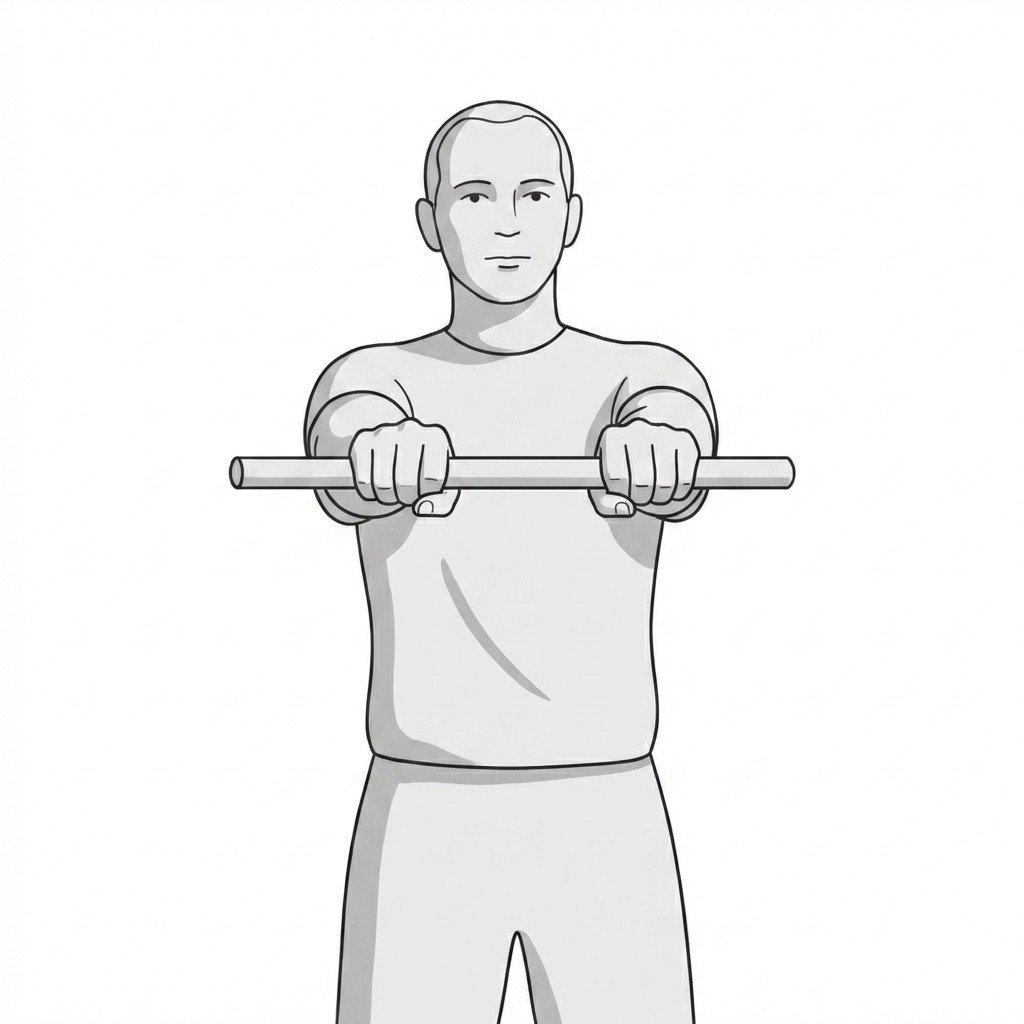
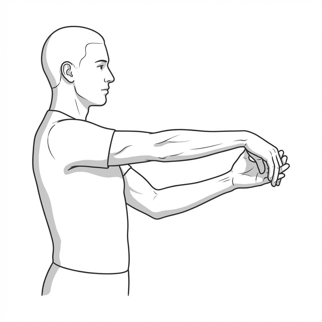
Exercises for Medial Epicondylalgia
These are adaptable examples after injury and nerve concerns have been assessed. Begin with a tolerable resistance and range; compare the next-day response before progressing. The reverse Tyler twist was studied within a combined programme without a comparison group, so it is an option rather than a required or superior method.