Understanding Chronic Ankle Instability & MPS
Chronic ankle instability (CAI) is a complex condition that develops in a notable share of individuals following an acute lateral ankle sprain. It is clinically characterized by recurrent episodes of the ankle "giving way," persistent lateral ankle pain, and subjective feelings of instability. Current literature divides CAI into two primary components: mechanical instability, which involves anatomical laxity of the anterior talofibular (ATFL) and calcaneofibular (CFL) ligaments, and functional instability, which is driven by proprioceptive deficits and neuromuscular control impairments. Differentiating between these components is critical for establishing an effective management plan.
Myofascial pain syndrome frequently coexists with, and complicates the presentation of, chronic ankle instability. During an inversion injury, the peroneal muscles (fibularis longus and brevis) undergo rapid, forceful eccentric loading in an attempt to stabilize the joint. This trauma often leads to the development of myofascial trigger points. Evidence suggests that these trigger points can alter afferent input and inhibit normal efferent motor firing patterns. Consequently, the peroneal reflex response time is delayed, leaving the ankle vulnerable to subsequent inversion stresses and directly contributing to the cycle of functional instability.
Addressing functional instability requires looking beyond the ligaments to the neuromuscular control and myofascial health of the dynamic stabilizers.
Clinical evaluation must carefully distinguish between pain originating from structural damage—such as chronic ligamentous tears, peroneal tendinopathy, or osteochondral lesions of the talus—and referred pain from active myofascial trigger points. While stress radiography or MRI may be utilized to identify mechanical laxity or intra-articular pathology, palpation of the lateral leg compartment is necessary to identify myofascial contributors. In many cases, addressing peroneal trigger points to restore normal muscle length and reactivity is a prerequisite for successful proprioceptive rehabilitation.
Anatomical & Biomechanical Context
The stability of the ankle joint relies on a complex interplay between static restraints (ligaments and joint capsule) and dynamic stabilizers (musculotendinous units). Understanding this relationship is essential for diagnosing the root cause of persistent symptoms.
Peroneus Longus (Fibularis Longus)
The peroneus longus is a primary evertor and dynamic stabilizer of the lateral ankle. Following an inversion sprain, it frequently undergoes eccentric overload, leading to the development of myofascial trigger points. These trigger points may inhibit the muscle's reflex response time, contributing to functional ankle instability and recurrent giving-way episodes.
Pain typically refers to the lateral aspect of the ankle, extending down to the lateral midfoot and occasionally mimicking symptoms of a lateral ligament sprain.Peroneus Brevis (Fibularis Brevis)
Situated deep to the longus, the peroneus brevis inserts at the base of the fifth metatarsal. It is highly active in preventing excessive inversion. Trigger points here often develop secondary to chronic ankle instability as the muscle remains in a state of chronic guarding to protect lax ligaments.
Referred pain concentrates heavily over the lateral malleolus and the lateral border of the foot, often complicating the diagnosis of peroneal tendinopathy.Tibialis Posterior
As a primary invertor and dynamic supporter of the medial longitudinal arch, the tibialis posterior often becomes overworked in patients with chronic ankle instability. Compensatory gait changes to avoid lateral loading can place excessive strain on this muscle, leading to medial-sided myofascial pain.
Pain is generally referred to the medial aspect of the ankle, the Achilles tendon region, and the plantar surface of the foot.Tibialis Anterior
The tibialis anterior provides dorsiflexion and eccentric control during heel strike. In the presence of chronic ankle instability, altered proprioception and gait mechanics can lead to overuse of the anterior compartment. Trigger points here may contribute to a perceived weakness or clumsiness in the foot.
Referred pain typically presents over the anteromedial aspect of the ankle and extends down into the dorsal surface of the great toe.Gastrocnemius
The gastrocnemius is a powerful plantarflexor that crosses both the knee and the ankle. Restricted dorsiflexion, often seen after recurrent ankle sprains, can lead to chronic shortening and trigger point formation in the gastrocnemius, further altering normal gait biomechanics.
Pain often refers to the posterior knee, calf, and down into the posterior aspect of the ankle and heel.Soleus
The soleus acts as a postural muscle, constantly working to maintain upright balance. In patients with compromised lateral ankle ligaments, the soleus works overtime to stabilize the lower leg. Myofascial restrictions here can severely limit ankle dorsiflexion, exacerbating functional instability.
Pain is frequently referred to the posterior heel, Achilles tendon, and occasionally the plantar surface of the heel.
Ligamentous Laxity
Permanent elongation or incomplete healing of the ATFL and CFL, leading to mechanical joint play beyond normal physiological limits.
Neuromuscular Inhibition
Altered proprioceptive feedback and delayed muscle firing times, often exacerbated by myofascial restrictions in the dynamic stabilizers.
Myofascial Referral
Active trigger points in the peroneals or tibialis posterior referring pain to the joint line, mimicking structural tissue damage.
Compensatory Overload
Altered gait mechanics to protect the unstable lateral ankle, leading to secondary overuse of the medial arch and posterior chain musculature.
Myofascial Referral Patterns
Myofascial trigger points in the lower leg can project pain into the ankle and foot, often confounding the diagnosis of structural ankle pathology. Recognizing these patterns aids in accurate differential diagnosis.
- Peroneus Longus and Brevis
- Pain concentrates around the lateral malleolus and extends along the lateral border of the foot. This pattern closely mimics the pain associated with anterior talofibular ligament (ATFL) or calcaneofibular ligament (CFL) pathology.
- Tibialis Posterior
- Pain is typically felt along the medial malleolus, extending into the medial arch of the foot. This may be mistaken for posterior tibial tendon dysfunction or medial compartment overload.
- Soleus
- Referred pain focuses on the posterior heel and Achilles region. It may present as a deep ache that worsens with prolonged standing or walking, complicating the clinical picture of post-sprain recovery.
Clinical Assessment & Differentiation
Clinical assessment of chronic ankle instability involves evaluating both mechanical joint play and functional neuromuscular control. The following tests help differentiate structural laxity from functional deficits.
Single Leg Stance Test
Stand barefoot on the affected leg with the knee slightly bent and eyes open. Attempt to maintain balance for 30 seconds. Note any excessive swaying, corrective movements at the ankle, or feelings of apprehension. Repeat with eyes closed if able.
Positive: Inability to maintain balance for at least 10 seconds, significant reliance on arm movements for balance, or reproduction of lateral ankle pain suggests proprioceptive deficits or functional instability.
Anterior Drawer Test (Clinical Concept)
While this is typically performed by a clinician, patients may notice a sensation of the foot shifting forward relative to the shin during certain movements. The clinician stabilizes the distal tibia and pulls the heel forward to assess the integrity of the anterior talofibular ligament (ATFL).
Positive: Increased anterior translation compared to the uninjured side, often accompanied by a "clunk" or lack of a firm endpoint, indicating mechanical ligamentous laxity.
Peroneal Muscle Palpation
Sit with the affected leg crossed over the other knee. Use the thumb to apply firm, sustained pressure along the lateral aspect of the fibula (the outside of the lower leg), searching for taut bands or exquisitely tender nodules.
Positive: Reproduction of the patient's familiar lateral ankle pain upon pressing the muscle belly, suggesting active myofascial trigger points contributing to the symptom profile.
Knee-to-Wall Test (Weight-bearing Lunge)
Stand facing a wall. Place the foot of the affected ankle perpendicular to the wall. Lunge forward, attempting to touch the knee to the wall without the heel lifting off the floor. Measure the maximum distance the toe can be from the wall while achieving this.
Positive: A significant restriction in dorsiflexion compared to the uninjured side (often >2cm difference) suggests joint capsular restriction or soleus/gastrocnemius myofascial tightness.
Evidence-Based Management
Management of chronic ankle instability generally requires a multimodal approach. Current guidelines emphasize comprehensive rehabilitation to address proprioceptive deficits, combined with targeted therapies to resolve myofascial restrictions and restore normal biomechanics.
Self-Care
- Activity modification to avoid uneven terrain during acute flare-ups
- Use of a semi-rigid ankle brace during high-risk activities
- Gentle ischemic compression of peroneal and calf trigger points
- Daily active range of motion exercises (e.g., ankle alphabet)
- Gradual reintroduction of weight-bearing activities as tolerated
Manual Therapy
- Myofascial release targeting the peroneal compartment
- Joint mobilization to restore talocrural dorsiflexion
- Soft tissue mobilization of the gastrocnemius and soleus complex
- Friction massage over the ATFL and CFL (if tolerated and indicated)
- Manual stretching of the posterior chain to improve biomechanics
Interventional
- Trigger point injections in the peroneals to reduce functional inhibition
- Diagnostic ultrasound to evaluate ligament integrity and tendon pathology
- Corticosteroid injections for localized sinus tarsi inflammation (used judiciously)
- Prolotherapy or PRP injections (evidence is emerging but currently mixed)
- Surgical consultation for lateral ligament reconstruction in refractory cases
Rehabilitation
- Progressive proprioceptive training (e.g., wobble board, balance pads)
- Eccentric strengthening of the peroneal muscles
- Neuromuscular control exercises focusing on reaction time
- Closed kinetic chain strengthening for the entire lower extremity
- Plyometric and sport-specific functional return training
Red Flags & Differential Diagnosis
While many cases of chronic ankle instability can be managed conservatively, certain signs and symptoms may indicate more severe structural damage, intra-articular pathology, or conditions requiring urgent medical intervention.
Inability to Bear Weight
Inability to take four steps immediately after an injury or during clinical evaluation. This aligns with the Ottawa Ankle Rules and requires imaging to rule out a fracture.
EmergencySevere Night Pain or Resting Pain
Pain that is unremitting, wakes the patient from sleep, and does not change with mechanical positioning. This may indicate infection, malignancy, or severe intra-articular pathology.
UrgentCatching, Locking, or Deep Joint Clicking
Mechanical symptoms within the joint space may suggest an osteochondral lesion of the talus or a loose body, which are common sequelae of recurrent ankle sprains.
InvestigateVisible Deformity or Extreme Swelling
Significant alteration in the shape of the ankle or foot, accompanied by rapid, severe swelling, suggesting a fracture or high-grade syndesmotic injury.
EmergencyPersistent Weakness in Eversion
Profound weakness when attempting to turn the foot outward, which may indicate a peroneal tendon tear or subluxation rather than simple myofascial inhibition.
Investigate
Clinical Pearls
Chronic ankle instability (CAI) encompasses both mechanical ligamentous laxity and functional neuromuscular deficits.
Myofascial trigger points in the peroneal muscles frequently develop after inversion injuries due to eccentric overload.
Active peroneal trigger points can inhibit normal muscle firing, delaying the reflex response needed to prevent recurrent sprains.
Referred pain from the peroneals can closely mimic the structural pain of lateral ligament damage or tendinopathy.
Restoring normal ankle dorsiflexion and addressing calf myofascial restrictions are critical components of CAI rehabilitation.
Proprioceptive and balance training are generally recommended by current guidelines as first-line interventions for functional instability.
Surgical intervention is typically reserved for cases demonstrating significant mechanical laxity that have failed comprehensive conservative rehabilitation.
Movement therapy
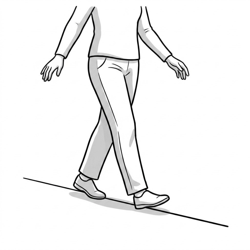
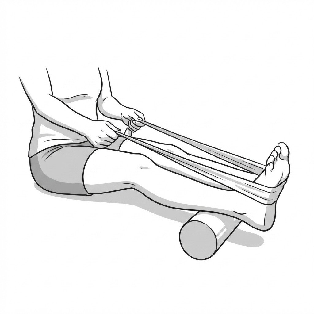
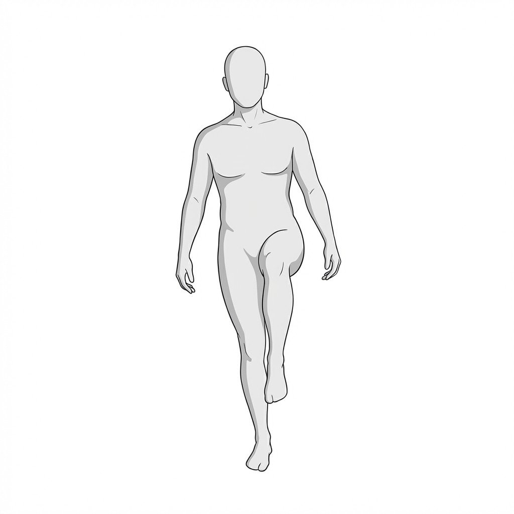
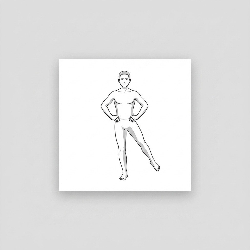
Exercises for Chronic Ankle Instability
Chronic ankle instability is driven as much by proprioceptive deficits and peroneal weakness as by ligament laxity — these exercises retrain neuromuscular ankle control and restore the single-leg stability needed to prevent re-sprain. Start with 2–3 and progress as tolerated.