Understanding the Gastrocnemius & Soleus
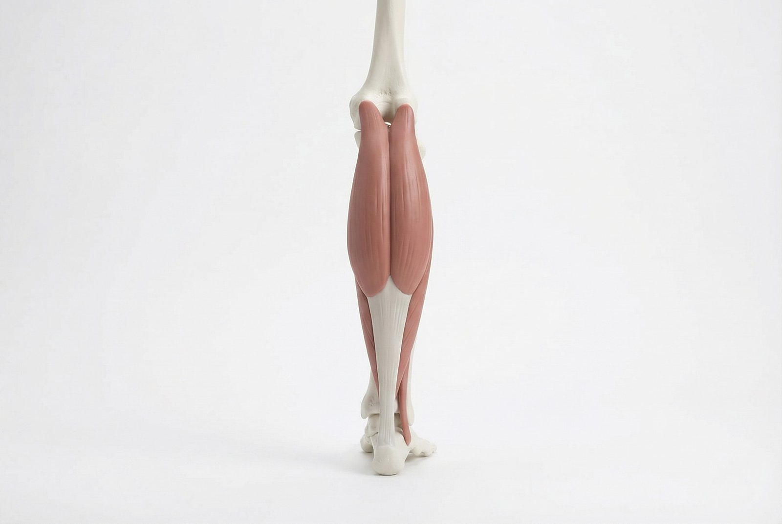
Gastrocnemius and Soleus Anatomy Posterior View Calf Complex
The gastrocnemius is the superficial, two-headed muscle of the calf, originating from the medial and lateral femoral condyles (above the knee) and inserting into the calcaneus via the Achilles tendon. Because it crosses both the knee and ankle joints, it functions as both a knee flexor and an ankle plantarflexor. The soleus lies deep to the gastrocnemius, originating from the posterior tibia and fibula (below the knee) and sharing the Achilles tendon insertion. Together they form the triceps surae — the most powerful muscle group in the lower extremity.
The functional division between these muscles is critical for understanding their trigger point patterns. The gastrocnemius is a fast-twitch, power muscle — designed for explosive plantarflexion (jumping, sprinting, push-off). The soleus is a slow-twitch, endurance muscle — designed for sustained postural support during standing and walking. This means the gastrocnemius develops trigger points from explosive overload (sports, sudden acceleration), while the soleus develops trigger points from sustained static loading (prolonged standing, walking on hard surfaces).
Trigger points in the calf complex develop through several mechanisms: acute overload (sudden sprinting, jumping, or hill running in an unconditioned individual), sustained shortening (habitual high-heel wearing, sleeping with toes pointed), eccentric overload (downhill walking, decelerating during running), and vascular compromise (the soleus has been called the "second heart" because its contraction pumps venous blood back to the heart — prolonged standing or sitting impairs this pump, creating local ischemia that activates trigger points).
The clinical significance of calf trigger points extends far beyond local calf pain. The soleus trigger point — one of the most clinically important in the entire body — refers pain to the heel and plantar surface of the foot, creating a referral pattern that is clinically indistinguishable from plantar fasciitis. Millions of patients are treated for "plantar fasciitis" with orthotics, cortisone injections, and even surgery when the actual pain source is a soleus trigger point that can be resolved with targeted manual therapy in weeks.
Anatomy & Trigger Points
Gastrocnemius trigger points are located in the proximal third of each head — approximately 5-8 cm below the knee crease, in the thickest part of the muscle bellies. The medial head trigger point is the most common and clinically significant. Soleus trigger points sit deeper and more distal — in the proximal half of the muscle, accessed by palpating deeply through or around the gastrocnemius. The soleus trigger point referring to the heel is typically found approximately 8-10 cm below the knee crease on the medial side.
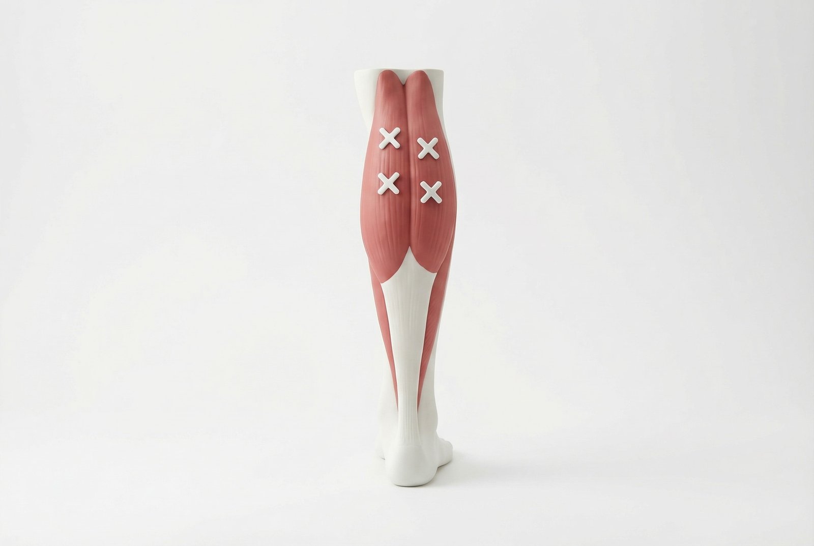
Heel Pain Anatomy
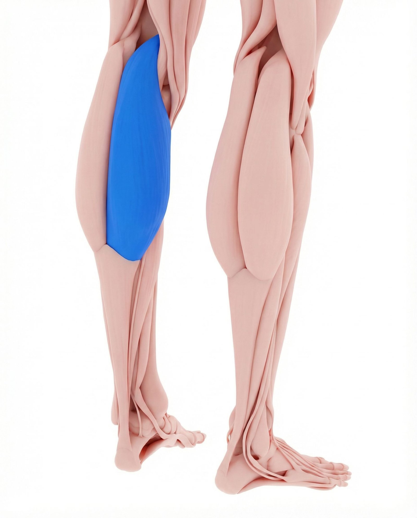
Pain concentrated in the medial calf and extending into the posterior knee crease (popliteal fossa). The medial head trigger points are the primary myofascial cause of nocturnal calf cramps — the agonizing nighttime contractions that wake patients from sleep. They also refer distally to the medial arch of the foot, contributing to "plantar fasciitis" presentations where the actual plantar fascia is not the primary pain generator.
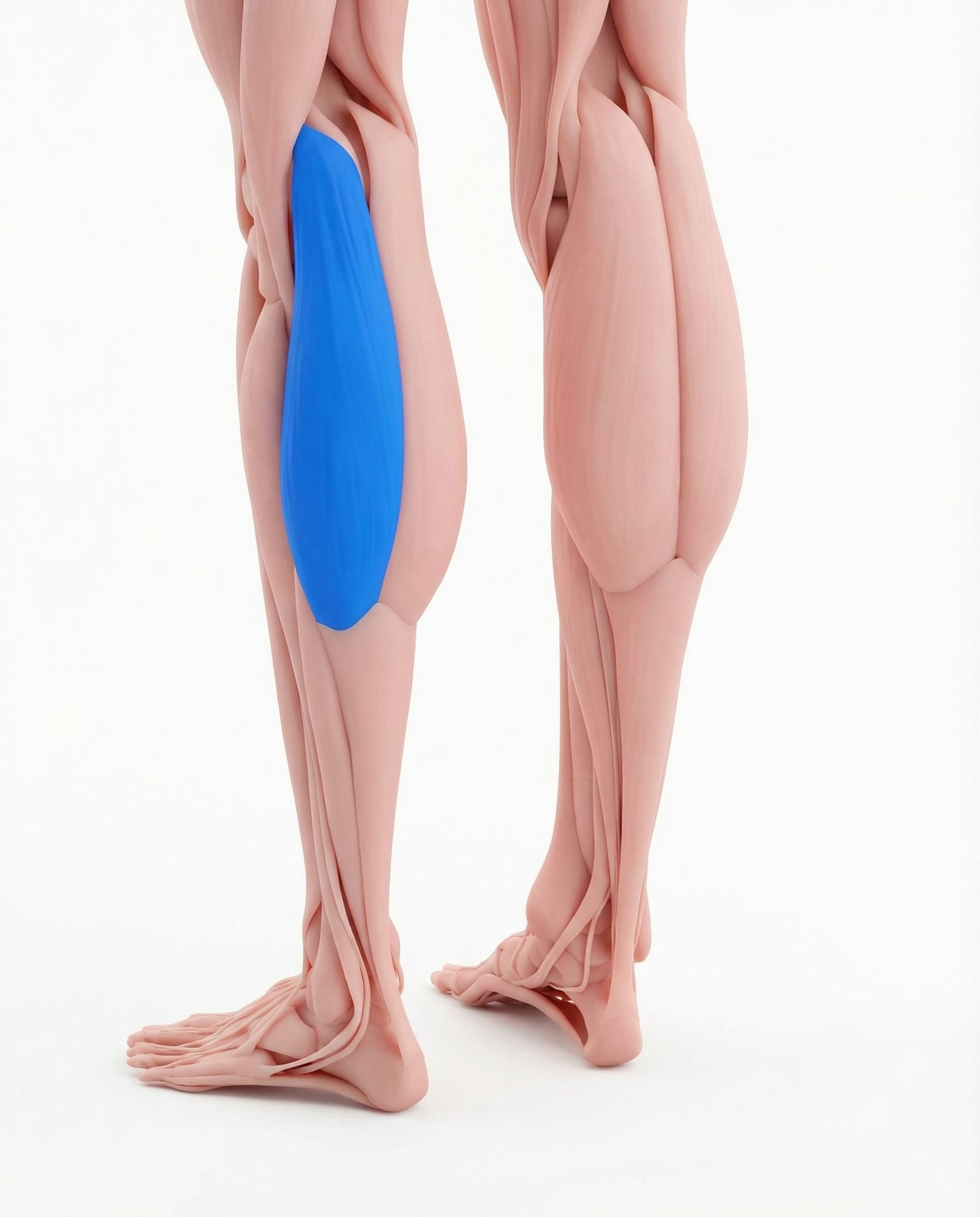
Pain in the lateral calf with referral to the posterolateral knee and occasionally to the lateral ankle. Lateral head trigger points are less common than medial but frequently develop in runners and cyclists. They contribute to posterior knee pain that mimics popliteal cyst (Baker cyst) and can create a sensation of posterior knee "fullness" or tightness.
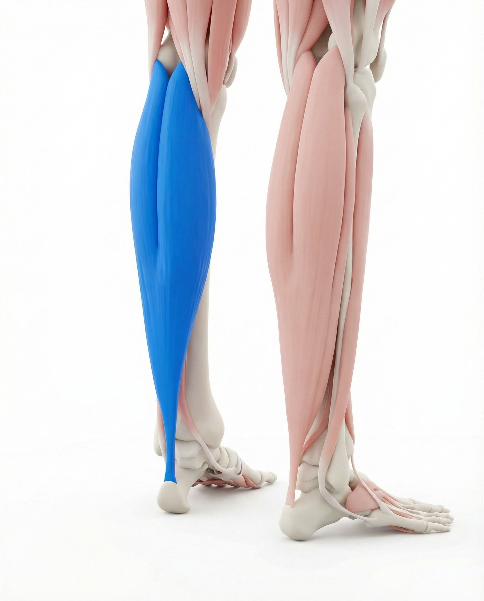
Deep heel and plantar pain — the soleus trigger point is the most important myofascial contributor to "plantar fasciitis" and heel pain. The referral extends from the posterior heel through the plantar surface of the foot. Soleus trigger points also refer to the ipsilateral sacroiliac region — a surprising and frequently overlooked pattern that connects persistent low back pain to calf muscle dysfunction.
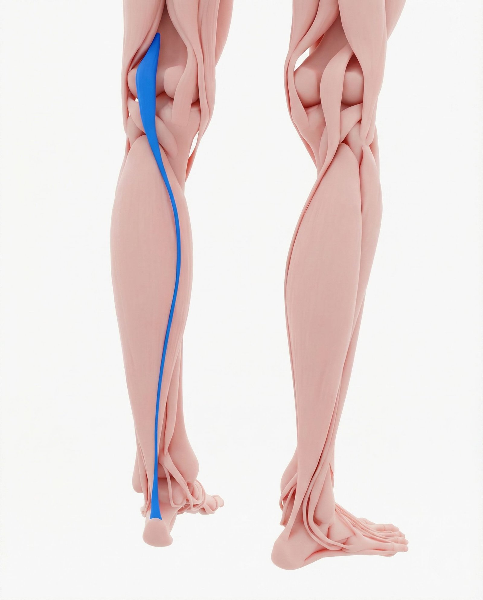
Posterior knee pain deep in the popliteal fossa. The plantaris is a small, thin muscle running between the gastrocnemius heads. While often considered vestigial, its trigger points can produce intense posterior knee pain that mimics meniscal or popliteal pathology. Plantaris rupture (the classic "tennis leg") is frequently confused with gastrocnemius tear.
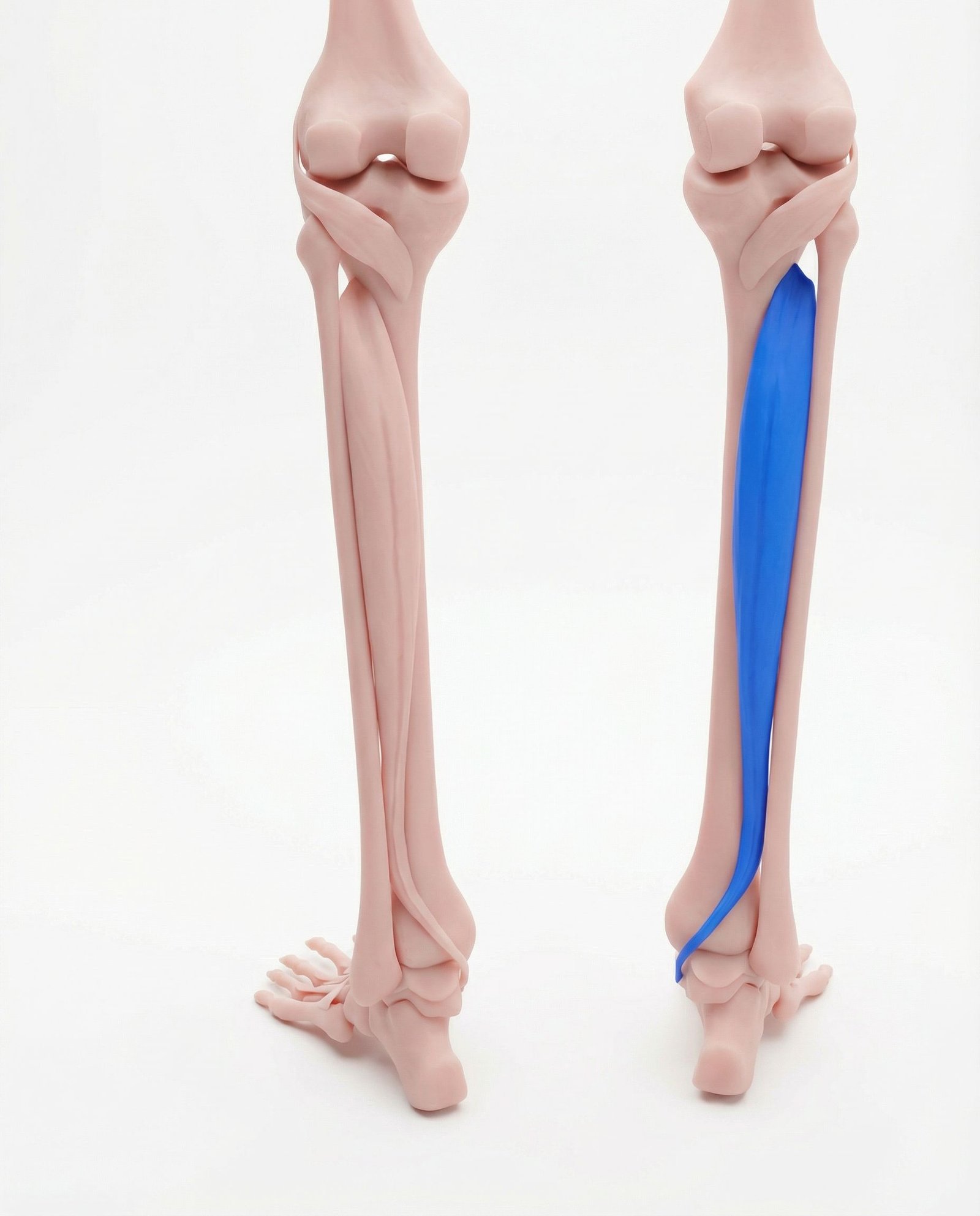
Deep calf pain with referral to the Achilles tendon region and plantar surface of the foot. Tibialis posterior trigger points coexist with soleus dysfunction and contribute to the medial ankle and arch pain pattern. Weakness of this muscle leads to progressive flat foot deformity and increased mechanical demand on the calf group.
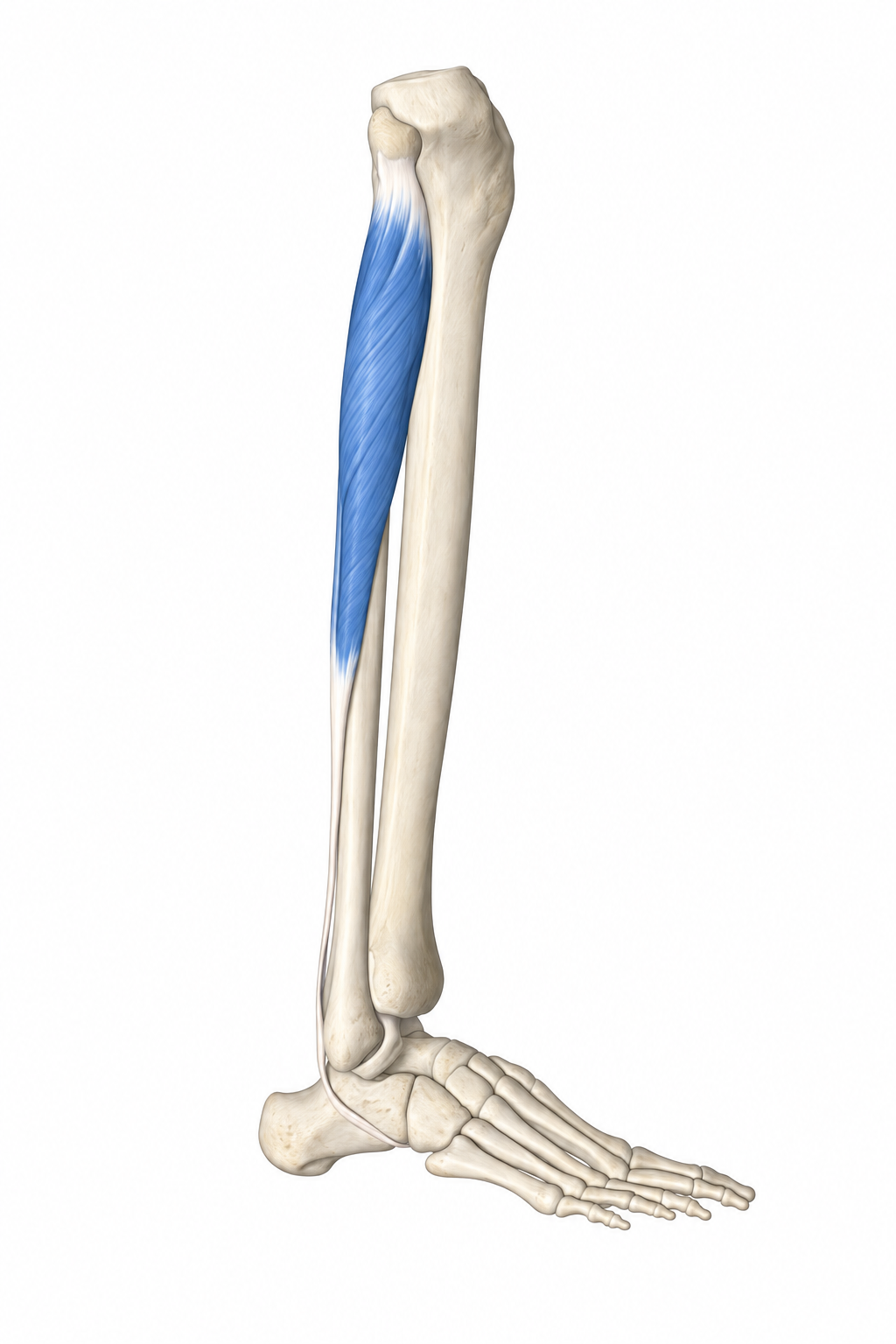
Lateral ankle pain concentrated around and behind the lateral malleolus. Peroneal trigger points develop secondarily to calf tightness through altered ankle biomechanics — when the gastrocnemius and soleus are tight, the ankle loses dorsiflexion range, forcing compensatory pronation that overloads the peroneal muscles.
Referral Patterns: The Calf & Heel Signature
The gastrocnemius trigger points refer in two distinct patterns: the medial head refers to the posterior calf, posterior knee, and importantly to the medial arch of the foot; the lateral head refers to the posterolateral calf and lateral ankle region. The posterior knee referral from the medial head frequently mimics a popliteal cyst (Baker cyst) — patients describe a feeling of "fullness" or "pressure" behind the knee that is actually referred pain from the calf, not fluid accumulation in the joint.
The soleus trigger point generates a uniquely distal referral: deep, persistent heel pain that is concentrated on the plantar surface of the calcaneus and extends along the Achilles tendon. This pattern is clinically identical to plantar fasciitis — the classic "first-step pain" that is worst getting out of bed in the morning. Additionally, the soleus has a surprising proximal referral to the ipsilateral sacroiliac region — connecting calf dysfunction to low back pain in a pattern that is frequently missed by clinicians treating each region in isolation.
If you treat plantar fasciitis for three months without improvement, look higher. The heel pain you are treating may have its source in the soleus muscle, fifteen inches above the foot.
Heel Pain
Night Cramps
Posterior Knee
Self-Assessment Tests
These tests evaluate calf trigger point activity, gastrocnemius-soleus flexibility, functional calf strength, and the differential diagnosis between myofascial calf pain and vascular, neurogenic, or structural causes. The critical test: if deep pressure on the soleus in the proximal calf reproduces the patient's heel pain — the diagnosis is myofascial referred pain, not plantar fasciitis.
Gastrocnemius Palpation
Soleus Squeeze Test
Weight-Bearing Dorsiflexion Test
Single-Leg Heel Raise Test
Treatment Pathways
Effective calf treatment requires separate attention to the gastrocnemius and soleus — they are stretched differently (straight knee vs. bent knee), they respond to different loading protocols, and they have distinct trigger point locations. A "one-stretch-fits-all" approach to the calf leaves the soleus undertreated in virtually every case.
Heel Pain Treatment
Calf Stretching: Wall & Step Protocol
Two distinct stretching positions are required because the gastrocnemius and soleus have different anatomical configurations. The gastrocnemius crosses the knee, so it is stretched with the knee straight. The soleus does not cross the knee, so it is stretched with the knee bent. Both muscles must be addressed separately for complete calf flexibility restoration.
- Wall stretch — gastrocnemius: back leg straight, heel on floor, lean into wall, hold 60 seconds
- Wall stretch — soleus: back leg with knee bent, heel on floor, lean into wall, hold 60 seconds
- Step stretch (eccentric): stand on step edge, slowly lower heels below step level, hold 30 seconds
- Downward dog (yoga) — combines gastrocnemius stretch with hamstring and posterior chain lengthening
- Perform both stretches 3 times daily; increase hold duration as tolerance improves
Calf Self-Release & Foam Rolling
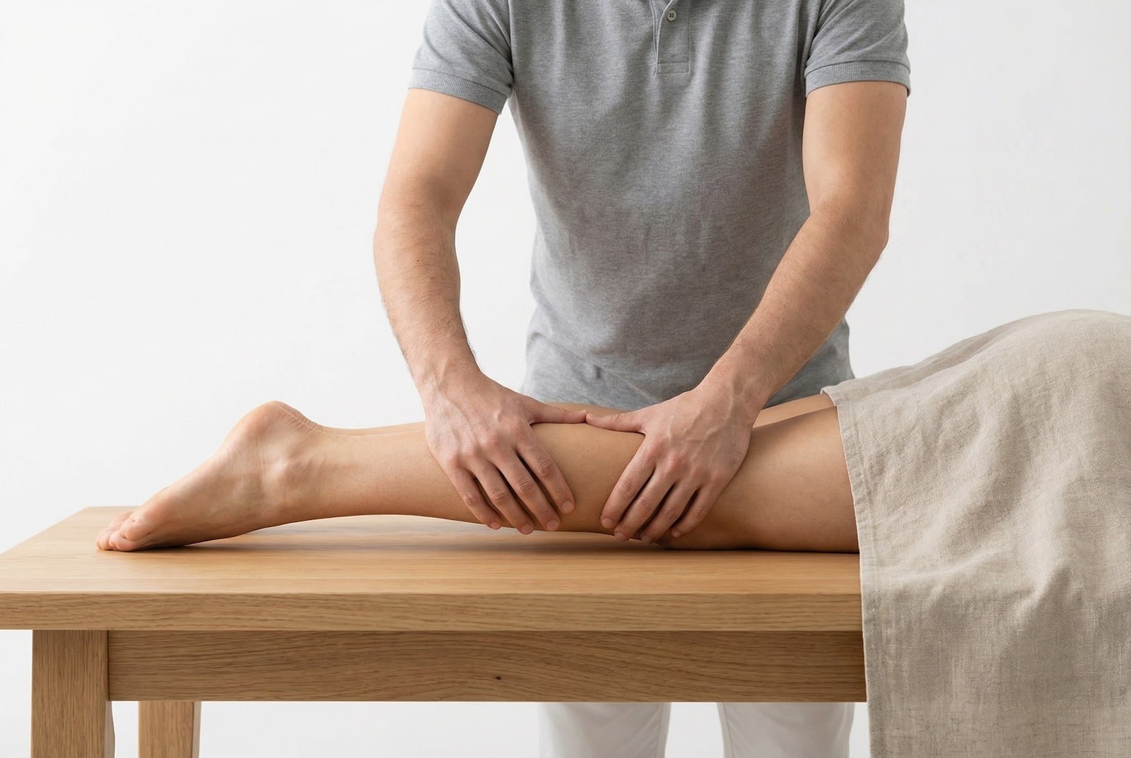
Targeted self-release of the gastrocnemius and soleus using foam roller, lacrosse ball, and cross-friction techniques. The seated position with a lacrosse ball is the most effective approach for the soleus — which is notoriously difficult to reach with a foam roller due to the overlying gastrocnemius.
- Foam roller calf release — seated on floor, calf on roller, cross opposite leg on top for pressure
- Lacrosse ball soleus release — seated on a chair, ball under the calf with knee bent to 90 degrees
- Percussive therapy on gastrocnemius bellies — medium intensity, avoid the Achilles tendon itself
- Cross-fiber massage of the musculotendinous junction — where the muscle meets the Achilles tendon
- Post-release: immediately perform wall stretches to consolidate the range of motion gained
Dry Needling: Calf Complex
Dry needling of the gastrocnemius and soleus is highly effective and produces dramatic local twitch responses that often provide immediate relief of the "cramping" sensation. The soleus requires deeper needling through the gastrocnemius — careful depth control is essential to avoid the posterior tibial artery and tibial nerve in the deep posterior compartment.
- Gastrocnemius medial head — pincer technique with needle directed into the muscle belly
- Gastrocnemius lateral head — flat palpation with needle perpendicular to the skin surface
- Soleus — deeper needling through the gastrocnemius, targeting the proximal muscle belly
- Plantaris — fine needle directed between the gastrocnemius heads in the popliteal region
- Series of 3-4 sessions at weekly intervals; patients often report nocturnal cramp relief after session one
Eccentric Calf Strengthening (Alfredson Protocol)
The Alfredson eccentric calf protocol — originally developed for Achilles tendinopathy — is equally effective for resolving chronic calf trigger points. Eccentric loading stimulates tendon remodeling while progressively loading the muscle through its full range, breaking the cycle of contracture and re-activation.
- Straight-knee eccentric heel drop — on step edge, rise on both feet, lower slowly on one foot
- Bent-knee eccentric heel drop — same protocol with knee flexed 20 degrees to target soleus
- Start with body weight only, 3 sets of 15 repetitions, twice daily
- Progressively add load (weighted backpack) as pain allows — 5% body weight increments
- Full protocol: 12 weeks minimum; expect temporary pain increase in weeks 2-4 before improvement
Gastrocnemius Medial Head
Posterior calf aching with referral to the posterior knee (mimics Baker cyst) and medial arch of the foot.
Gastrocnemius Lateral Head
Posterolateral calf pain extending to the lateral ankle — common in runners and cyclists.
Soleus (Heel Referral)
Deep heel and plantar pain identical to plantar fasciitis — the most important myofascial mimic in the lower extremity.
Soleus (SI Joint Referral)
Surprising proximal referral to the ipsilateral sacroiliac region — connects calf dysfunction to low back pain.
Red Flags — When to See a Doctor
Calf pain has several important differential diagnoses that require urgent exclusion — particularly deep vein thrombosis, Achilles tendon rupture, and compartment syndrome. These conditions present with calf symptoms that can initially resemble trigger point pain but require immediate medical intervention.
The gastrocnemius and soleus form the triceps surae — the most powerful muscle group in the lower extremity, generating the force for push-off during every step. Their trigger points produce three distinct clinical patterns: nocturnal calf cramps (gastrocnemius), posterior knee pain (gastrocnemius), and heel/plantar pain (soleus) — each of which is commonly misdiagnosed as a different condition.
Soleus trigger points are the most important and most overlooked myofascial contributor to "plantar fasciitis." The soleus refers pain directly to the heel and plantar surface of the foot, precisely mimicking the pain pattern attributed to plantar fascia inflammation. When plantar fasciitis fails to respond to stretching, orthotics, and cortisone injections — soleus trigger points should be assessed before considering surgery.
Nocturnal calf cramps — the agonizing nighttime contractions that wake patients from deep sleep — often involve gastrocnemius trigger points and shortened calf posture, rather than electrolyte deficiency alone. Magnesium and potassium supplementation is commonly prescribed but evidence for benefit in idiopathic nocturnal cramps is limited; trials such as Hallegraeff (2012) suggest that pre-sleep calf stretching may reduce cramp frequency and severity in older adults.
The gastrocnemius crosses the knee joint, while the soleus does not — this anatomical distinction has direct treatment implications. Gastrocnemius is stretched with the knee straight (to tension across both joints); soleus is stretched with the knee bent (which slackens the gastrocnemius, allowing the stretch to reach the deeper soleus). Performing only straight-knee calf stretches leaves the soleus untreated.
Calf trigger points are a significant perpetuating factor in Achilles tendinopathy. Chronic gastrocnemius and soleus contracture increases mechanical tension on the Achilles tendon, creating the sustained loading that drives tendon degeneration. Successful Achilles tendinopathy treatment requires addressing calf trigger points concurrently with the eccentric loading protocol.
Prolonged wearing of high heels is the most potent perpetuating factor for calf trigger points in women. The sustained plantarflexion position shortens both the gastrocnemius and soleus, creating chronic contracture that prevents full dorsiflexion during gait. Transition to flat shoes must be gradual (reducing heel height by 1 cm per week) to avoid overloading the already-shortened calf muscles.