Why There Is No Simple Test
Getting Diagnosis
Overview IllustrationThere is no single routine test that identifies myofascial pain syndrome. Diagnosis is based mainly on clinical history and physical examination. Other tests — blood work, imaging — may help exclude overlapping or alternative causes, but they do not detect trigger points directly.
This is because the condition is local and neuromuscular in nature. Trigger points are localized areas of sustained contraction within muscle tissue. They can be identified through hands-on examination, but they do not produce systemic changes visible on routine blood work or standard imaging.
Different clinicians vary in how routinely they assess myofascial findings. Some include trigger point palpation in their standard musculoskeletal exam; others may not assess for it unless specifically asked.
The diagnostic process for myofascial pain syndrome relies on clinical examination skills — knowing what to ask for can help you get an appropriate evaluation.
What tests are for
When your doctor orders blood tests or imaging, they are not looking for myofascial pain syndrome — they are ruling out other conditions that could explain your symptoms. Normal results help narrow the differential diagnosis by eliminating other possibilities.
Several conditions can produce symptoms similar to myofascial pain: hypothyroidism, rheumatoid arthritis, vitamin deficiencies, fibromyalgia, disc herniation, and peripheral neuropathy, among others. Testing helps exclude these, which is an important part of reaching an accurate assessment. The identification of myofascial pain syndrome then rests on what the clinician finds during the physical examination — palpable taut bands, tender nodules, reproduction of the patient's familiar pain, and characteristic referred pain patterns.
Assessment practices vary. If your evaluation has focused primarily on blood work and imaging, it may be worth asking whether a musculoskeletal examination that includes muscle palpation has been performed.
- 01Symptom onset
Pain begins — the cause may not be immediately apparent.
- 02Initial evaluation
Blood tests and imaging are ordered to evaluate for common causes.
- 03Further evaluation
You may see specialists to consider specific conditions before a myofascial assessment is performed.
- 04Trigger point examination
A clinician trained in myofascial palpation identifies taut bands and assesses whether they reproduce your symptoms.
- 05Assessment completed
Myofascial pain syndrome is identified through hands-on assessment, supported by the exclusion of other conditions.
The Diagnostic Process
The Diagnostic Process
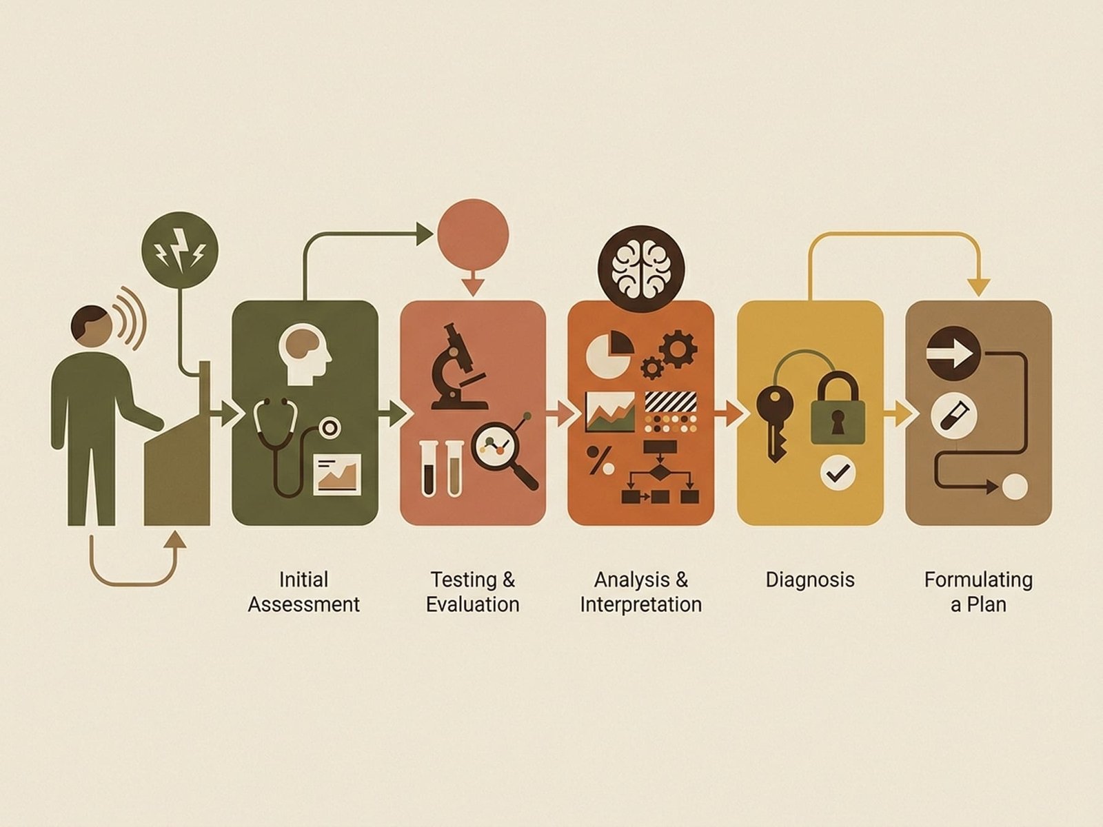
Mechanism DiagramA myofascial pain evaluation typically follows a structured sequence. Here is what each step involves and what you can expect.
Medical History
Your clinician will ask detailed questions about your pain — when it started, what makes it better or worse, where it is, how it feels, and what you have already tried. They will also ask about your medical history, medications, sleep, stress levels, previous injuries, occupational demands, and any other conditions that might contribute to or overlap with myofascial pain.
What to Expect
- Detailed questions about pain location, quality, and duration
- Review of previous treatments and their effectiveness
- Assessment of daily activities and functional limitations
- Questions about sleep quality, stress, and overall wellbeing
- Family history of pain conditions, autoimmune disease, or fibromyalgia
- Review of current medications and supplements
Physical Examination
The examiner will observe your posture, assess range of motion in affected areas, and check for structural asymmetries or biomechanical issues. They will look at how you sit, stand, and move. Muscle strength testing may be performed to identify weakness patterns that could suggest specific trigger point involvement.
What to Expect
- Postural assessment — standing, sitting, and walking
- Active range of motion testing for affected regions
- Passive range of motion to assess end-feel and restrictions
- Muscle strength testing in affected areas
- Evaluation of gait, shoulder height, and spinal alignment
- Assessment of joint mobility or stiffness
Trigger Point Examination
The clinician will systematically palpate muscles in the region of your pain, feeling for the characteristic findings of myofascial trigger points: taut bands, tender nodules, referred pain patterns, local twitch responses, and restricted range of motion. This step is what distinguishes a standard examination from a myofascial-specific evaluation.
What to Expect
- Systematic palpation of muscles in and around your pain area
- Identification of taut bands — tight, rope-like muscle fibers
- Localization of tender points within taut bands
- Testing whether pressure reproduces your familiar pain pattern
- Assessment for local twitch response and jump sign
- Mapping of referred pain patterns
Ruling Out Other Conditions
Your clinician may order blood tests, imaging, or other investigations — not to identify myofascial pain syndrome directly (there is no test for that) but to rule out conditions that could produce similar symptoms or coexist with it. Conditions like rheumatoid arthritis, hypothyroidism, vitamin deficiencies, disc herniation, and fibromyalgia can all present similarly and should be considered.
What to Expect
- Blood work to check inflammation markers, thyroid, and vitamin levels
- Imaging studies if there are specific clinical indications
- Nerve conduction studies if nerve involvement is suspected
- Referral to a rheumatologist if autoimmune disease is possible
- Discussion of findings and what normal test results mean in context
- Explanation of why ruling out other conditions is part of the process
Diagnosis & Treatment Plan
With the information gathered, your clinician will explain their assessment, discuss which trigger points appear to be contributing to your symptoms, outline a proposed treatment approach, and set expectations for what improvement may look like. This is also the time to ask questions and discuss what matters to you.
What to Expect
- Explanation of the assessment findings in plain language
- Identification of primary trigger points and contributing factors
- Proposed treatment approach and timeline
- Discussion of realistic expectations
- Plan for follow-up and treatment adjustments
- Opportunity to ask questions and discuss preferences
What Happens During a Trigger Point Exam
When a clinician evaluates for myofascial trigger points, the examination involves systematic palpation of the muscles in the affected area. Here is what this typically involves.
The exam can be uncomfortable — pressing on tender areas is inherently unpleasant — but it should be tolerable. A careful examiner will communicate throughout and ask for your feedback.
Taut Band Palpation
The examiner runs their fingers across the muscle fibers, feeling for a band of tightness — like a taut string within the softer surrounding muscle tissue. The taut band is a common clinical finding associated with trigger points. Not every taut band contains an active trigger point, but active trigger points are typically found within taut bands.
What you will feel: The examiner pressing across (perpendicular to) the muscle fibers with moderate pressure.
The Tender Nodule
Within the taut band, the examiner locates a small, notably tender spot — the trigger point itself. This is a focal area of contracted tissue, typically a few millimeters in diameter. When pressed, it produces a disproportionate pain response compared to surrounding tissue. The tenderness is sharply localized — moving just a few millimeters away may significantly reduce the pain.
What you will feel: Focused pain when the examiner presses directly on a specific spot.
Pain Recognition
When the examiner compresses a tender area, they will ask: "Does this reproduce your familiar pain?" If pressing on a spot reproduces the pain pattern you have been experiencing, that may help support the assessment. The pain should feel familiar — not just local tenderness, but your recognizable pain.
What you will feel: Your familiar pain being reproduced by pressure on a specific point.
The Jump Sign
When the examiner presses on a particularly active trigger point, you may flinch, pull away, or react involuntarily. This is a reflex response to the intense tenderness of an active trigger point, not a sign of oversensitivity. Clinicians look for this reaction as evidence of a clinically relevant trigger point.
What you will feel: An involuntary flinch or pull-away response.
Local Twitch Response
The local twitch response is a brief, involuntary contraction of the taut band when the area is stimulated by quick pressure or needle insertion. It is a sometimes-observed finding — not present in every case and not necessary for diagnosis. When it does occur, some clinicians consider it a supportive sign. Its role in predicting treatment outcomes is discussed in the literature but not settled.
What you will feel: A quick, visible twitch or jump of the muscle fiber under the examiner's fingers.
Referred Pain Pattern
The examiner maintains sustained pressure on the trigger point for 10-20 seconds to allow referred pain to develop. Referred pain is pain felt at a location distant from where pressure is applied. Each trigger point has a characteristic referral pattern that has been mapped and documented over decades. If pressing a spot in your shoulder reproduces your arm pain, the referral pattern supports the assessment.
What you will feel: Pain or sensation spreading to a location away from where the examiner is pressing.
A Note on Comfort
The trigger point exam involves pressing on tender areas, and it will be uncomfortable at times. This is expected. However, the exam should not be excessively or needlessly painful. A skilled examiner applies graduated pressure, communicates clearly, and respects your tolerance.
You should feel comfortable telling the examiner if the pressure is too much. A thoughtful clinician will adjust their technique. The goal is to identify trigger points accurately, not to test your pain tolerance.
Tests That Rule Out Other Conditions
Your doctor may order various tests as part of the diagnostic workup. These tests are not looking for myofascial pain syndrome — they are looking for conditions that could produce similar symptoms, coexist with it, or perpetuate it.
It can be frustrating when tests come back normal while you are still in pain. It may help to understand that each normal result narrows the field of possibilities. Normal test results do not mean your pain is not real — they mean certain specific causes have been made less likely.
Blood Tests
ESR & CRP
Erythrocyte Sedimentation Rate & C-Reactive Protein
Rules Out
Rheumatoid arthritis, polymyalgia rheumatica, infections, vasculitis
Normal Result Means
Systemic inflammation less likely
CBC
Complete Blood Count
Rules Out
Anemia, infection, blood disorders
Normal Result Means
Hematologic causes of fatigue and pain less likely
TSH & Thyroid Panel
Thyroid Stimulating Hormone, T3, T4
Rules Out
Hypothyroidism — a recognized cause of widespread muscle pain, fatigue, and stiffness
Normal Result Means
Thyroid dysfunction less likely as a contributor
Vitamin D
25-Hydroxyvitamin D
Rules Out
Vitamin D deficiency — associated with chronic musculoskeletal pain and may perpetuate trigger points
Normal Result Means
Adequate vitamin D levels
Vitamin B12
Serum B12 & Methylmalonic Acid
Rules Out
B12 deficiency — can cause neuropathy, muscle weakness, and pain that may overlap with or perpetuate trigger points
Normal Result Means
B12 stores are adequate
Iron Studies
Serum Iron, Ferritin, TIBC
Rules Out
Iron deficiency — can cause fatigue, muscle pain, and restless legs
Normal Result Means
Iron stores are sufficient
ANA
Antinuclear Antibody
Rules Out
Lupus, Sjogren syndrome, scleroderma, mixed connective tissue disease
Normal Result Means
No autoimmune marker detected — though a negative result does not completely exclude autoimmune disease
RF & Anti-CCP
Rheumatoid Factor & Anti-Cyclic Citrullinated Peptide
Rules Out
Rheumatoid arthritis — an autoimmune condition that can coexist with or resemble myofascial pain
Normal Result Means
Rheumatoid arthritis less likely
Imaging Studies
X-Ray
Evaluates bones and joint spaces
Rules Out
Fractures, arthritis, bone tumors, spinal alignment problems
When Ordered
History of trauma, suspected arthritis, chronic back or neck pain, deformity
Important limitation: Cannot visualize muscles, trigger points, or soft tissue — a normal X-ray does not exclude muscular causes of pain
MRI
Detailed imaging of soft tissues, discs, nerves
Rules Out
Disc herniation, spinal stenosis, tumors, labral tears, rotator cuff tears
When Ordered
Neurological symptoms (numbness, weakness), suspected disc pathology, progressive symptoms, treatment not progressing as expected
Important limitation: Standard MRI does not show trigger points — but can identify conditions that may coexist with or be confused for myofascial pain
Nerve Conduction / EMG
Tests nerve and muscle electrical activity
Rules Out
Peripheral neuropathy, nerve entrapment (carpal tunnel), radiculopathy, motor neuron disease
When Ordered
Numbness, tingling, weakness in a nerve distribution pattern, suspected nerve involvement
Important limitation: Standard EMG does not reliably detect trigger points — though specialized techniques can identify spontaneous electrical activity at trigger point sites in research settings
When Imaging Is Indicated
While imaging does not identify myofascial pain syndrome directly, there are specific situations where it is clinically appropriate:
History of significant trauma
Falls, motor vehicle accidents, sports injuries — fractures and structural damage should be excluded.
Progressive neurological symptoms
Increasing weakness, loss of sensation, bowel or bladder changes — these require urgent imaging.
Suspected underlying pathology
Unexplained weight loss, night pain, fever — imaging helps rule out tumors, infections, and serious systemic disease.
Treatment not progressing as expected
If an appropriate treatment course has not produced the expected improvement, imaging may help identify missed structural contributors.
Diagnostic Criteria
Clinicians who assess trigger points often look for a combination of recurring findings. The criteria most commonly referenced were described by Travell and Simons, though their exact application varies in practice.
The findings below are divided into core and supporting categories. No single finding is sufficient in every case — interpretation depends on the overall clinical picture.
Major Criteria (Core Findings)
Palpable taut band in a skeletal muscle
A band of tight muscle fibers that can be felt by an experienced examiner running their fingers across the direction of the muscle fibers.
Notably tender spot within the taut band
A localized area of intense tenderness — pressing on this spot produces significantly more pain than pressing on surrounding tissue just millimeters away.
Patient recognition of the pain complaint
Compression of the trigger point reproduces the patient's familiar pain pattern — the pain they have been experiencing, not just local soreness.
Painful limitation of range of motion
The muscle containing the trigger point cannot stretch to its full length without pain. Range of motion is reduced because the taut band mechanically limits muscle elongation.
Minor Criteria (Supporting Findings)
Visual or tactile identification of a local twitch response
A brief, visible contraction of the taut band when the trigger point is snapped or needled — a spinal cord reflex associated with trigger point presence.
Referred pain or altered sensation with compression
Maintaining pressure on the trigger point produces pain or other sensations at a predictable location away from the compression site.
Spontaneous electrical activity on EMG
Needle electromyography at the trigger point site can detect characteristic low-amplitude, high-frequency electrical discharges not found in normal muscle tissue.
What this means in practice
In practice, most clinicians look for a taut band with a tender area that reproduces the patient's familiar pain. Other findings — twitch response, referred pain patterns — may add support but are not uniformly present. The assessment is clinical rather than formulaic.
Emerging Diagnostic Tools
Emerging Diagnostic Tools
Equipment OverviewTrigger points are currently identified through clinical examination. Researchers are also developing tools that may complement hands-on assessment in the future.
These technologies are at various stages of development and are not yet part of routine clinical practice. They have, however, contributed to understanding the physical characteristics of trigger points.
Ultrasound Elastography
Increasingly availableUses ultrasound technology to measure tissue stiffness in real time. Trigger points and their associated taut bands appear as regions of increased stiffness compared to surrounding muscle tissue. Shear-wave elastography has shown ability to visualize and quantify trigger point stiffness.
Potential clinical role: May become a clinical tool for supporting trigger point identification and monitoring treatment response.
Magnetic Resonance Elastography
Research stageAn MRI-based technique that generates maps of tissue mechanical properties. Can identify regions of increased muscle stiffness consistent with trigger points. Provides good spatial resolution but is currently expensive and impractical for routine clinical use.
Potential clinical role: Primarily a research tool that is contributing to our understanding of the physical characteristics of trigger points.
Microdialysis
Research onlyA technique where a tiny probe is inserted into the trigger point region to sample the local biochemical environment. Research has demonstrated elevated levels of inflammatory mediators and neuropeptides at active trigger point sites compared to normal muscle tissue.
Potential clinical role: Has provided evidence for the biochemical aspects of trigger points — unlikely to become a routine clinical tool due to its invasive nature.
Infrared Thermography
Specialized useUses thermal cameras to detect temperature differences on the skin surface. Active trigger points may create localized temperature variations. Results have been inconsistent across studies, which limits diagnostic reliability at present.
Potential clinical role: May serve as a supplementary screening tool but is not expected to replace manual palpation for clinical diagnosis.
After the Diagnosis
Receiving a diagnosis of myofascial pain syndrome can be clarifying — particularly if you have spent time searching for an explanation for your symptoms. Understanding what is contributing to your pain is a useful step toward finding appropriate treatment.
A diagnosis is a starting point for treatment planning. Here is what typically follows:
Treatment Plan Development
Your clinician will outline an approach — typically starting with the most relevant trigger point treatments and progressively addressing contributing factors, movement patterns, and longer-term prevention.
Finding Appropriate Practitioners
Depending on the approach, you may be referred to a physical therapist trained in myofascial techniques, a physician who performs dry needling or trigger point injections, a pain specialist, or a combination of providers.
Setting Expectations
Recovery timelines vary. Many people see improvement within several weeks of consistent, appropriate treatment, with continued progress over months. Your clinician can help set realistic expectations based on your specific situation.
Learning Self-Management
You will likely learn self-care techniques — stretching, self-massage, ergonomic adjustments, and stress management — that can help you manage your condition on an ongoing basis.
Continue Learning
Treatment Framework
An overview of treatment approaches for myofascial pain syndrome — the options, the evidence, and how they fit together.
Building Your Care Team
How to find clinicians experienced with musculoskeletal pain and myofascial evaluation.
Talking to Your Doctor
Practical suggestions for communicating with healthcare providers about your symptoms.
Trigger Points 101
Background on what trigger points are, how they develop, and why they produce pain.