Therapeutic Exercise Library
Your comprehensive resource for myofascial pain and trigger point therapy exercises.
187 of 187 exercises
Shoulder
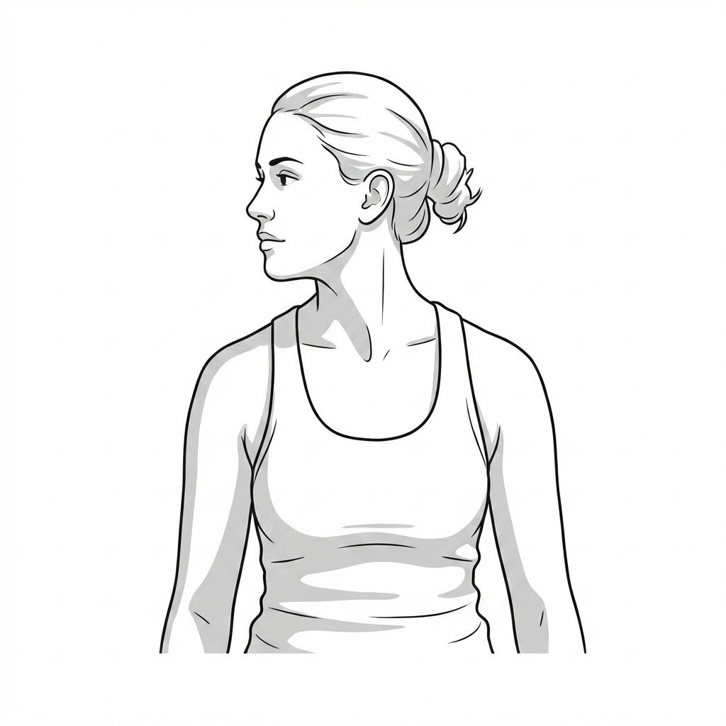
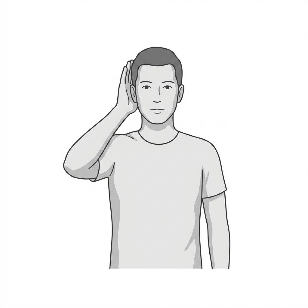
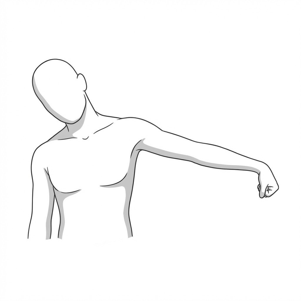
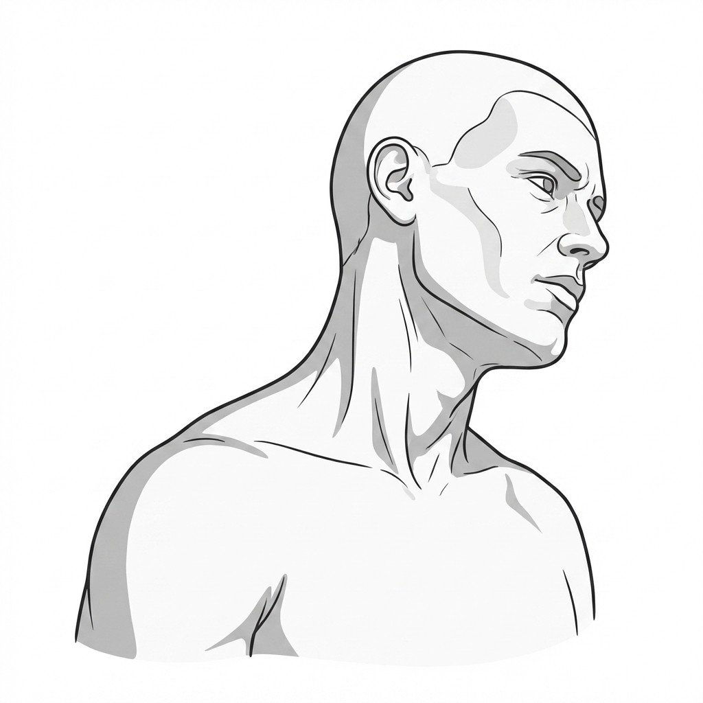
Active cervical rotation
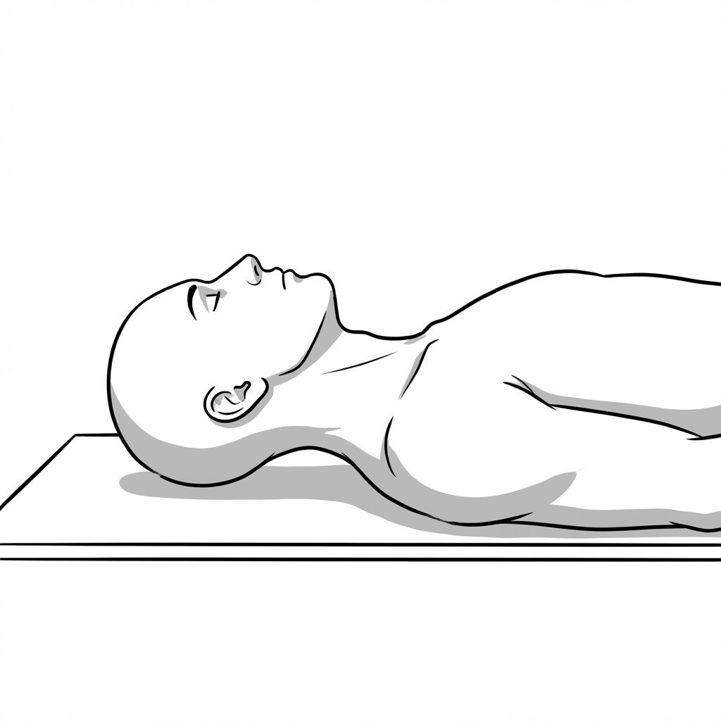
This exercise involves actively turning the head from side to side to improve cervical spine mobility. For Levator Scapulae Syndrome, it helps restore normal joint mechanics and may ease protective muscle guarding, which can reduce pain and stiffness by gently moving the muscle through its functional range.
Start Exercise →Lower Leg
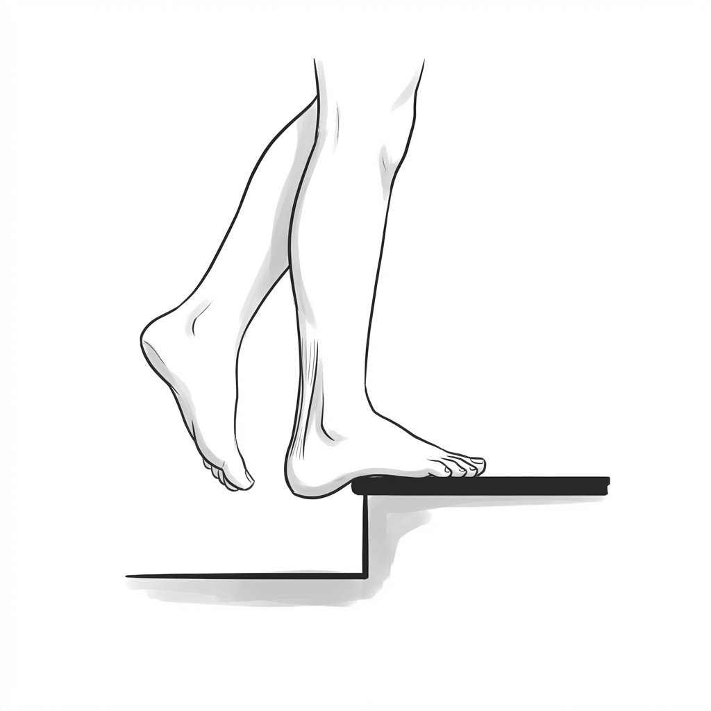
Alfredson eccentric calf drops
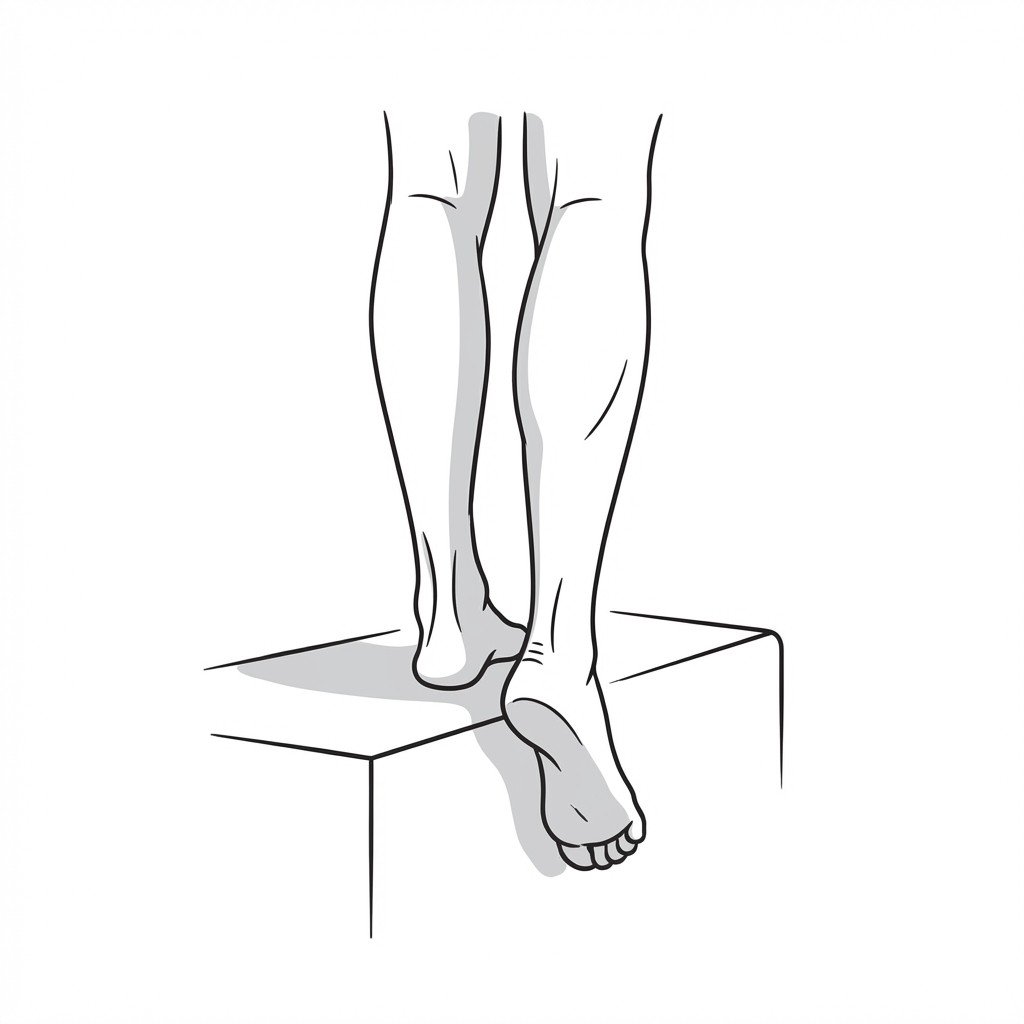
This exercise applies a controlled, high-load eccentric (lengthening) force to the Achilles tendon and calf muscles. This specific loading is thought to stimulate tendon remodeling, increase collagen production, and reduce pain associated with tendinopathy.
Start Exercise →Hip
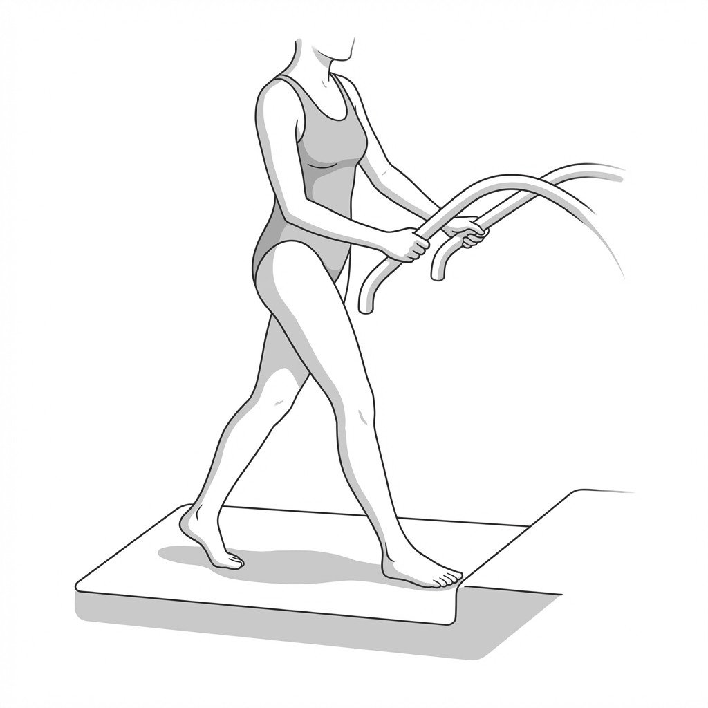
Aquatic walking
Aquatic walking utilizes the buoyancy of water to reduce weight-bearing stress on the arthritic hip joint, allowing movement with less pain. The water's viscosity provides gentle, multi-directional resistance to strengthen the muscles surrounding the hip and improve cardiovascular fitness.
Start Exercise →Knee
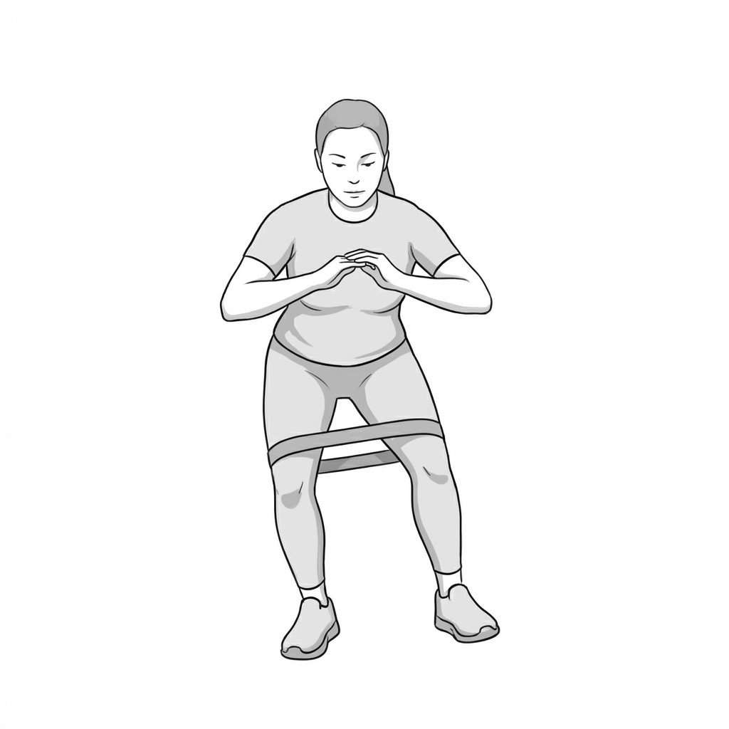
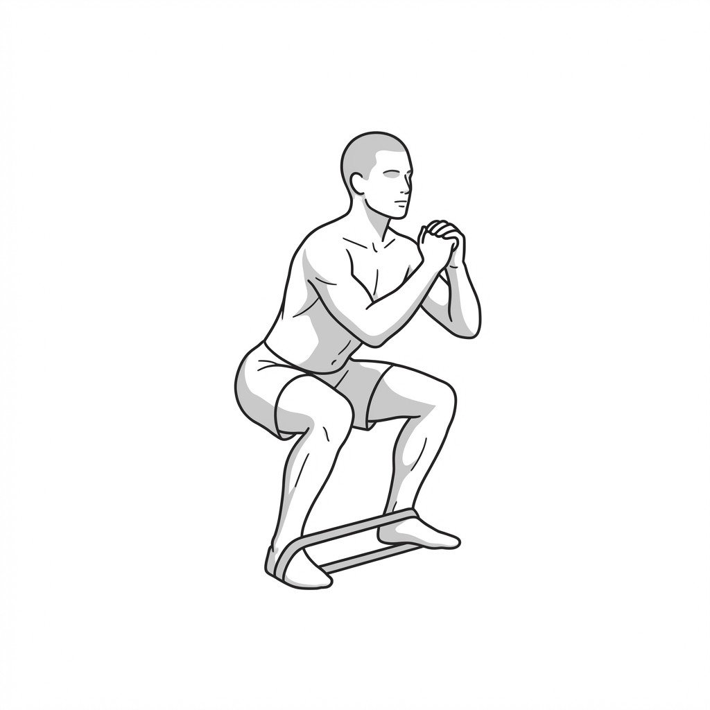
Banded crab walks
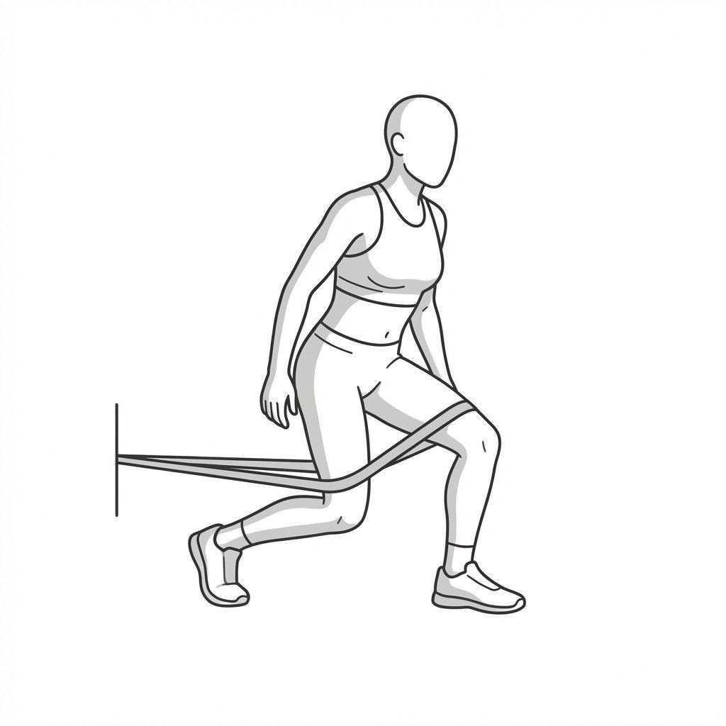
Banded crab walks target and strengthen the hip abductors, specifically the gluteus medius, in a functional weight-bearing position. By improving lateral hip stability, this exercise helps control excessive hip adduction and internal rotation, which may reduce mechanical compression on the iliotibial (IT) band.
Start Exercise →Shoulder
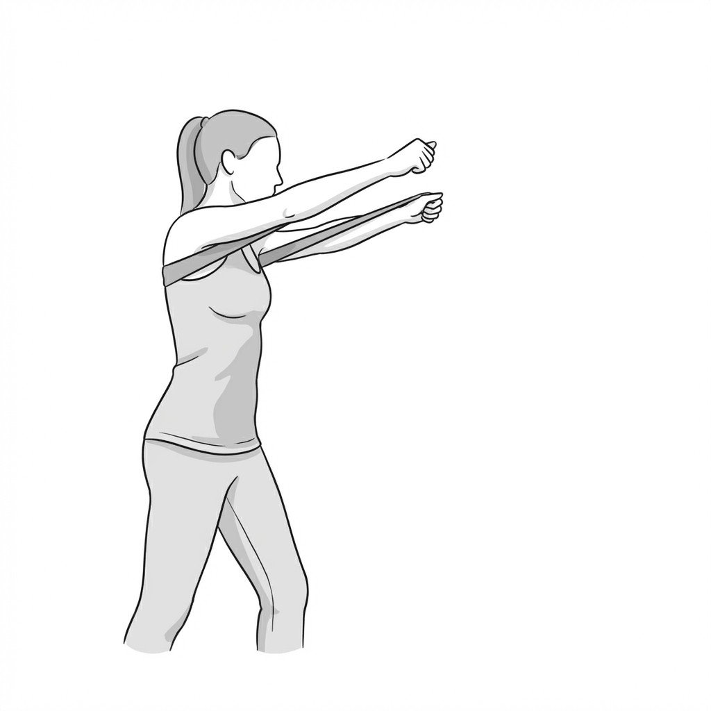
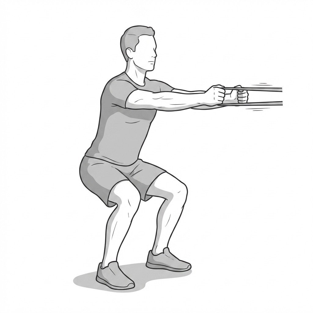
Banded dynamic hugs
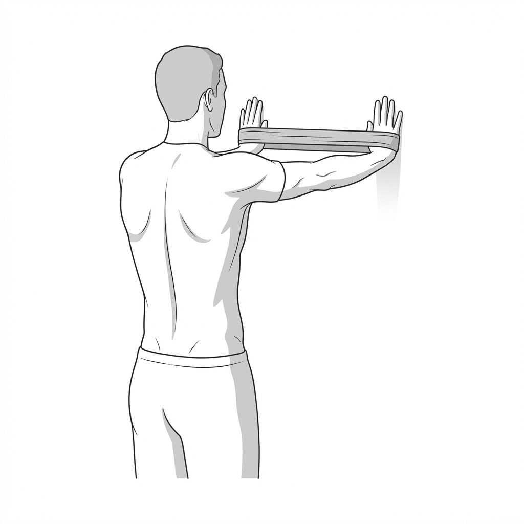
The banded dynamic hug facilitates scapular protraction and upward rotation against resistance to preferentially activate the serratus anterior. This helps correct abnormal scapular kinematics characteristic of scapular dyskinesis by improving scapulothoracic muscle balance and dynamic stability.
Start Exercise →Shoulder
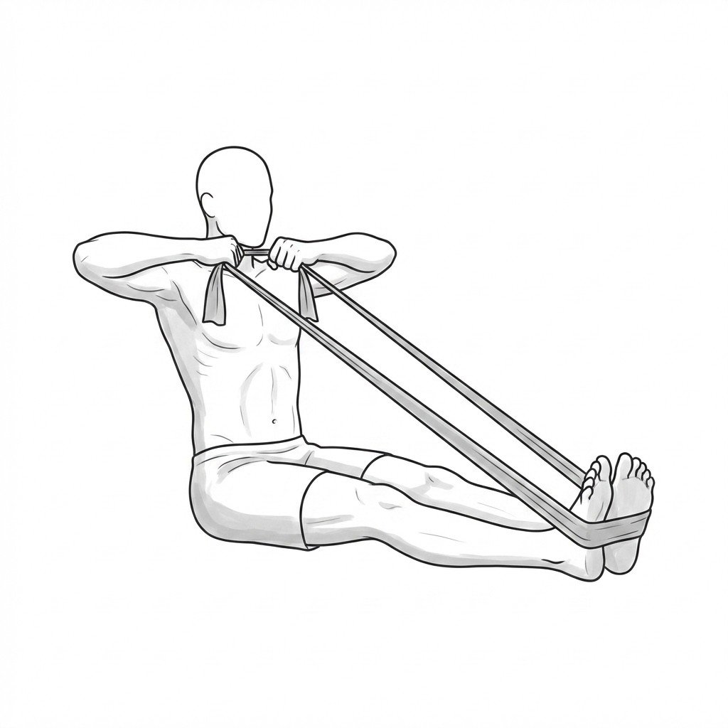
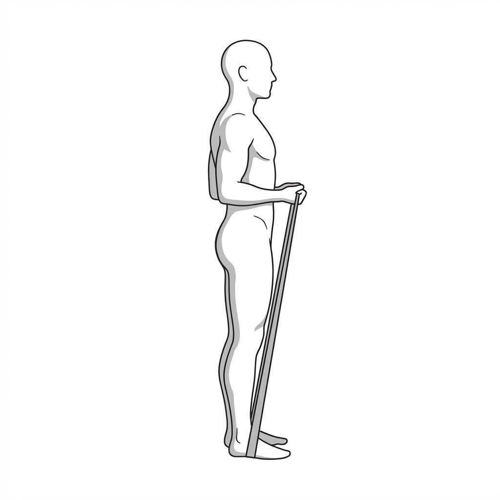
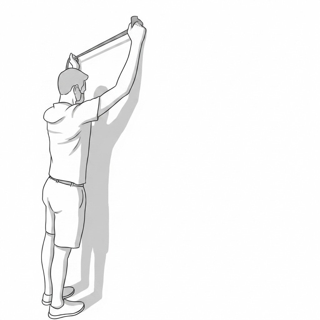
Banded face pulls
Banded face pulls target the scapular retractors and posterior shoulder musculature to combat rounded shoulder posture. By actively engaging the rhomboids and trapezius, this exercise improves interscapular muscular endurance and may help reduce pain related to prolonged postural overstretch of the tissues.
Start Exercise →Lumbar & Pelvis
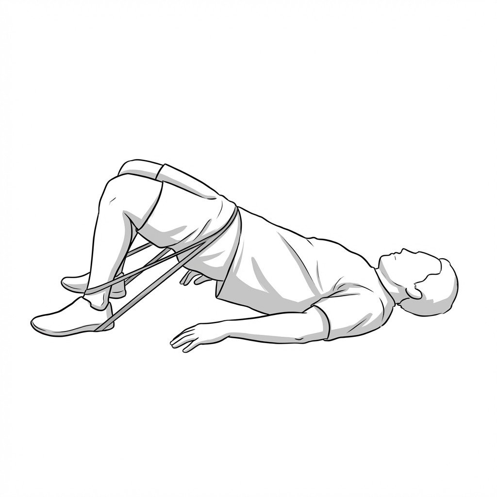
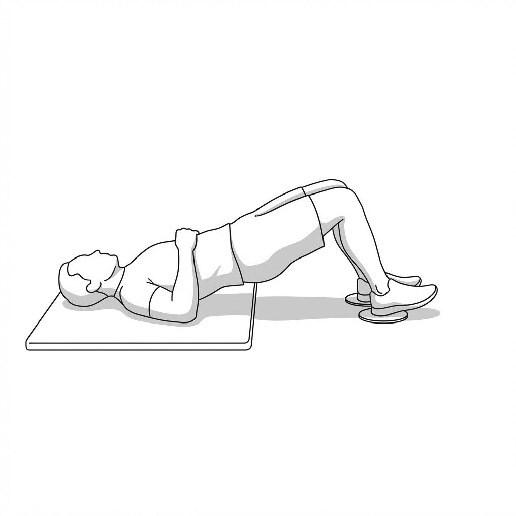
Banded glute bridge
The banded glute bridge activates the gluteus maximus while simultaneously engaging the gluteus medius to work against the band's resistance. This co-contraction is thought to improve force closure across the sacroiliac joint, which may help stabilize the pelvis and reduce shear forces associated with SIJ pain.
Start Exercise →Hip
Banded monster walks
Banded monster walks target the hip abductors and external rotators against progressive resistance. By strengthening the gluteal complex in a functional, weight-bearing position, this exercise may help improve pelvic stability and reduce compressive load on the gluteal tendons at the greater trochanter.
Start Exercise →Shoulder
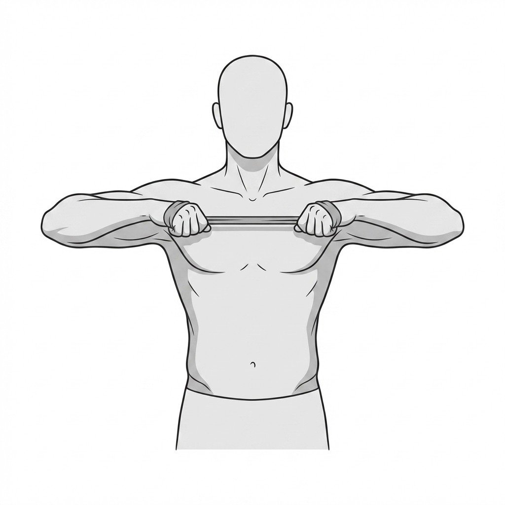
Banded rows
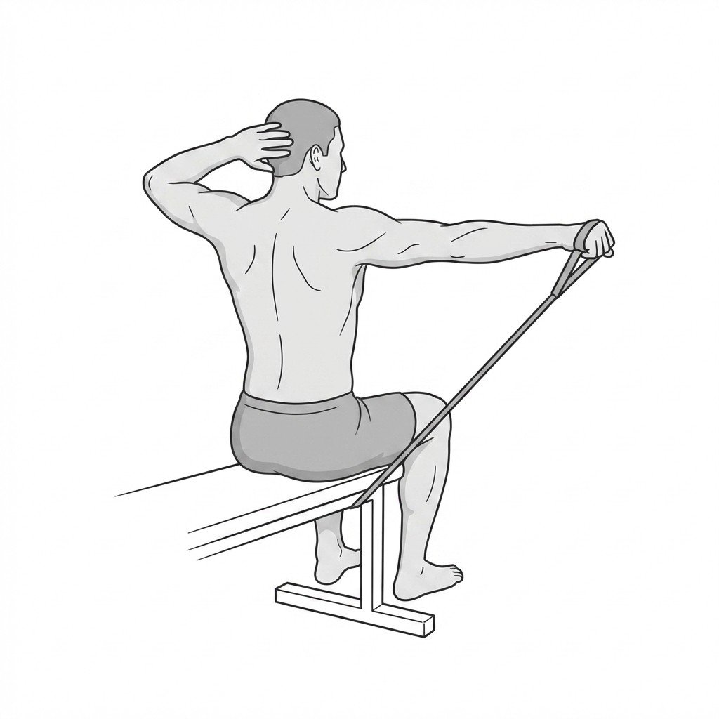
Banded rows target the scapular retractors to improve neuromuscular control and strength of the posterior shoulder complex. By dynamically engaging the rhomboids and middle trapezius, this exercise helps restore normal scapulohumeral rhythm. This helps counter the protracted and anteriorly tilted scapular positioning frequently observed in scapular dyskinesis.
Start Exercise →Shoulder
Banded W-raises
Banded W-raises target the posterior rotator cuff and scapular retractors through resisted external rotation and scapular upward rotation. This exercise helps restore the force couple balance in the shoulder complex, helping to reduce mechanical impingement and potentially promoting adaptive remodeling in the affected tendons.
Start Exercise →Thoracic & Core
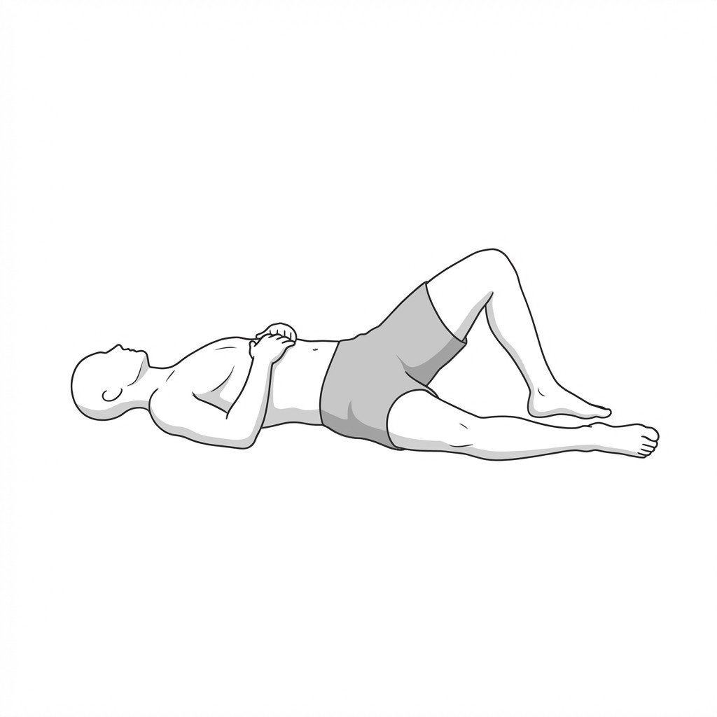
Bent knee fall-outs
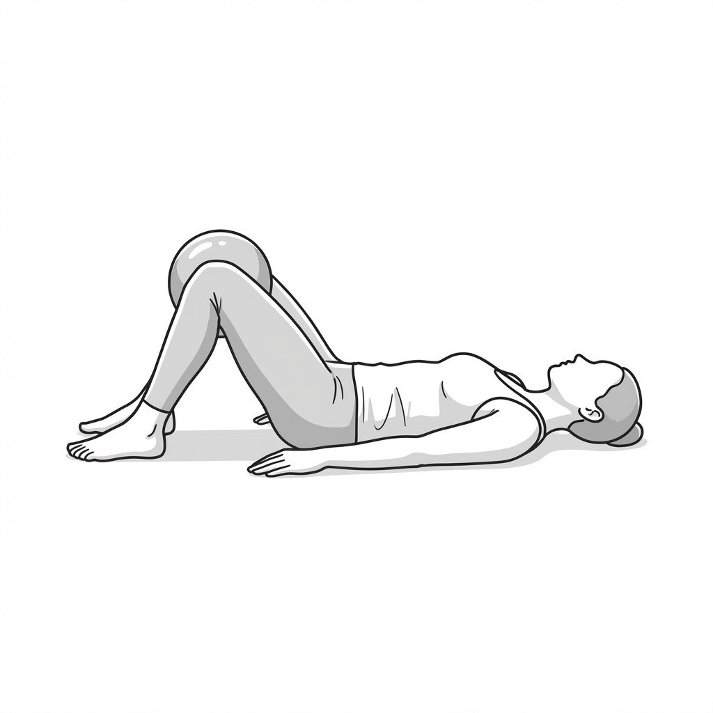
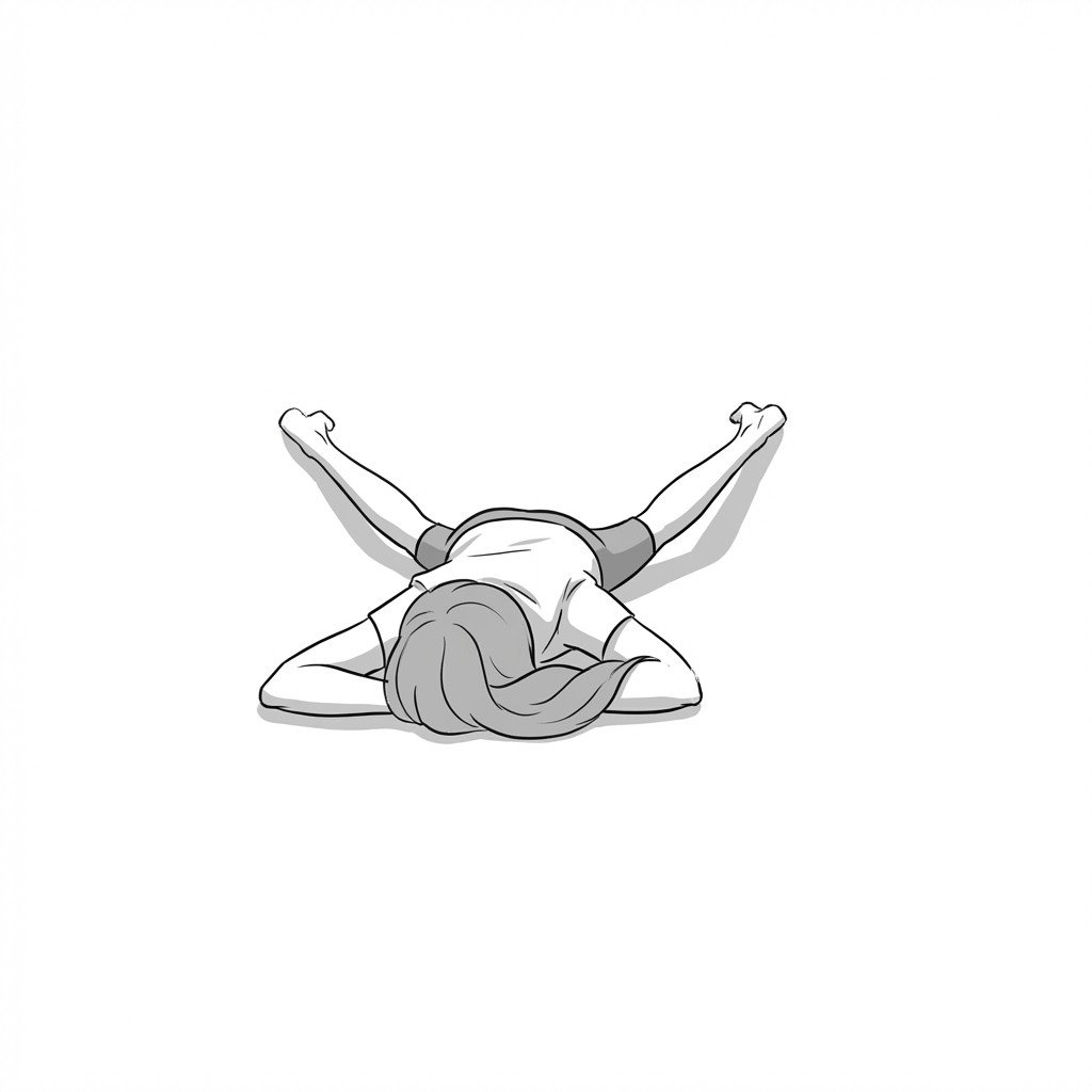
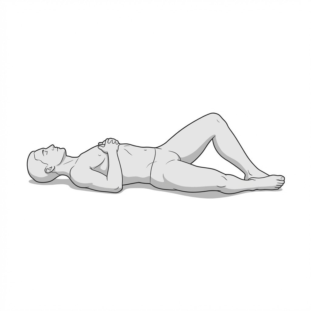
Bent knee fall-outs target the deep core stabilizers while promoting lumbopelvic dissociation. By requiring the transversus abdominis to stabilize the pelvis against the rotational torque of the moving leg, this exercise helps safely build fascial tension across the linea alba without causing outward bulging.
Start Exercise →Thoracic & Core
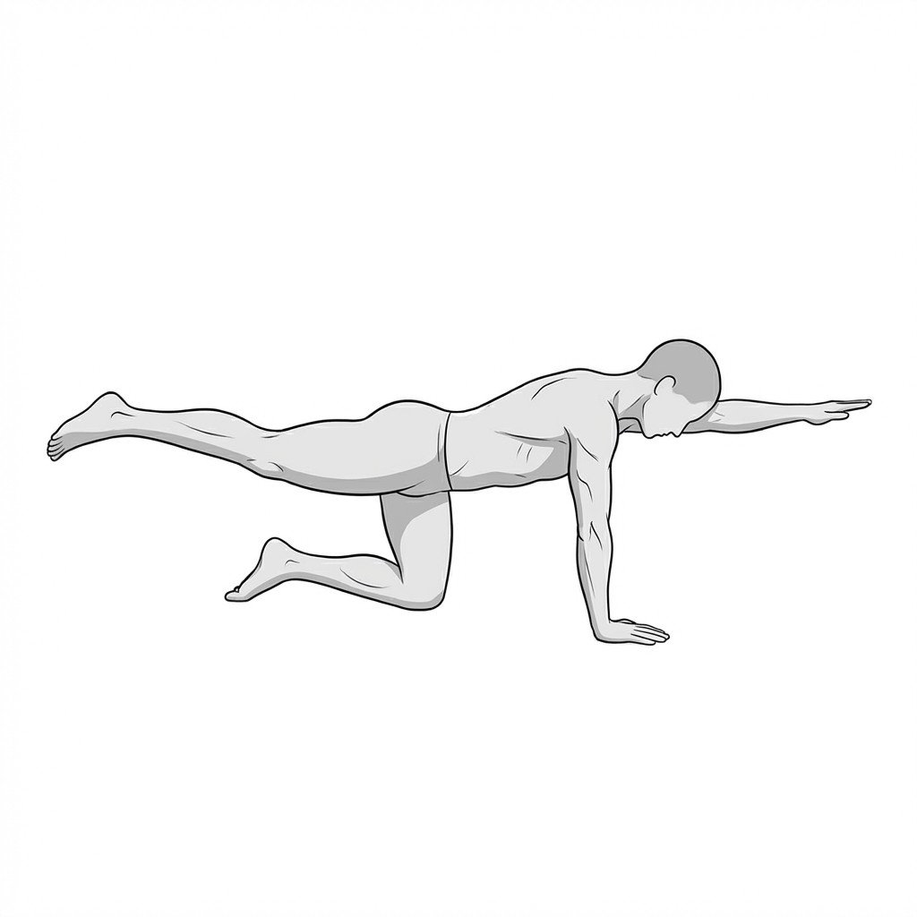
Bird-dog
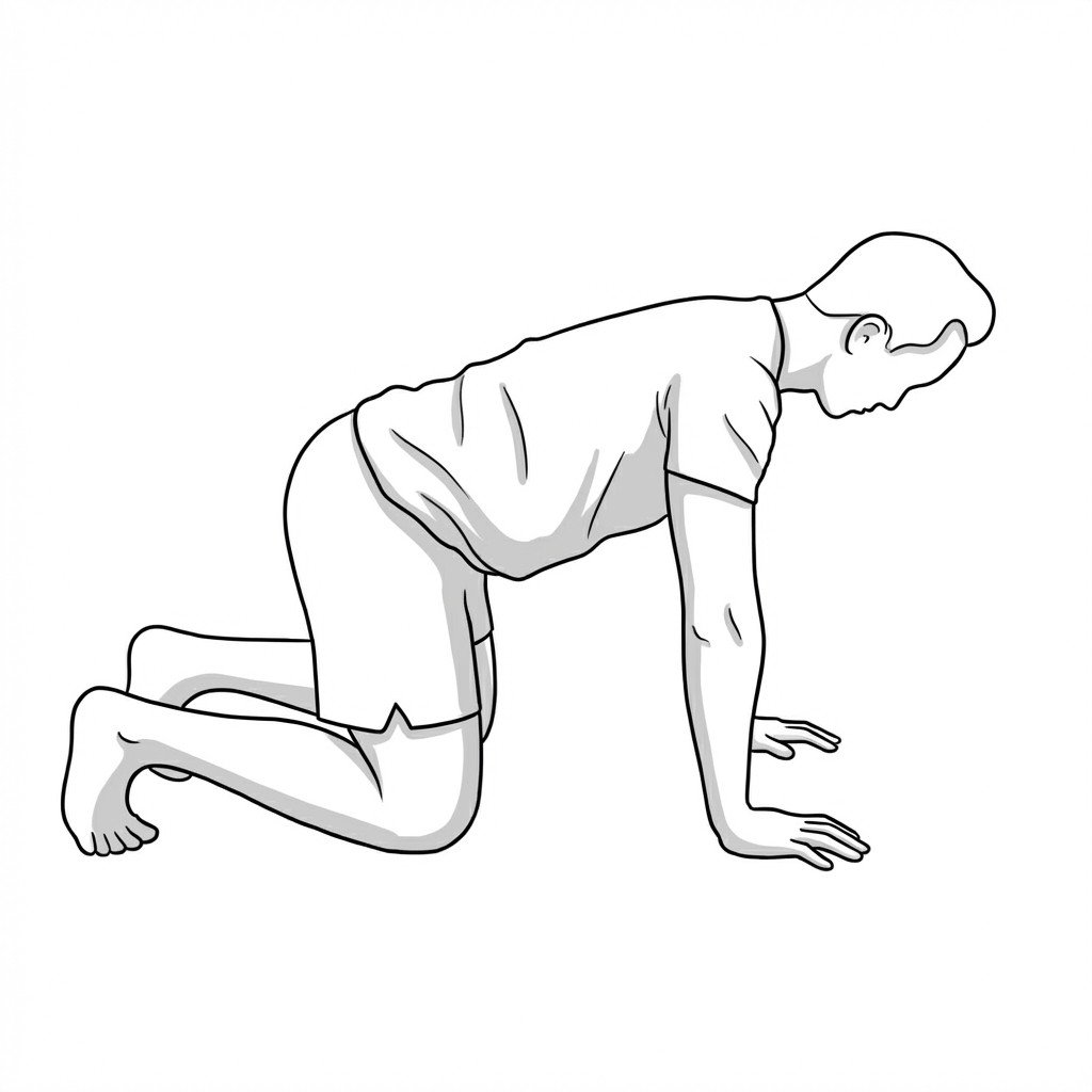
The Bird-dog exercise enhances core stability by requiring anti-rotation and anti-extension control while the extremities are in motion. It selectively activates the lumbar multifidus and transverse abdominis with minimal compressive load on the spinal discs. By training the lumbopelvic stabilizers to maintain neutral alignment during limb movement, it may help reduce pain related to lumbar instability.
Start Exercise →Thoracic & Core
Brachial plexus nerve glides
Brachial plexus nerve glides mechanically mobilize the nerve bundle through tight anatomical spaces like the scalene triangle and subpectoral tunnel. This continuous sliding motion is thought to support neural mobility and may help reduce the nerve mechanosensitivity commonly seen in Thoracic Outlet Syndrome.
Start Exercise →Hip
Bridge with hamstring walkouts
This exercise emphasizes eccentric and isometric loading of the hamstrings while maintaining gluteal and core engagement. By gradually lengthening the hamstrings under tension, it is thought to support tissue remodeling and fascicle-length adaptation, an important part of hamstring rehabilitation that may help reduce reinjury risk.
Start Exercise →Hip
Butterfly stretch
The butterfly stretch applies gentle, controlled elongation to the adductor muscle group and medial hip fascial structures. By promoting tissue remodeling and improving flexibility in the inner thigh, it helps alleviate tension and may help improve range of motion following an adductor strain.
Start Exercise →Knee
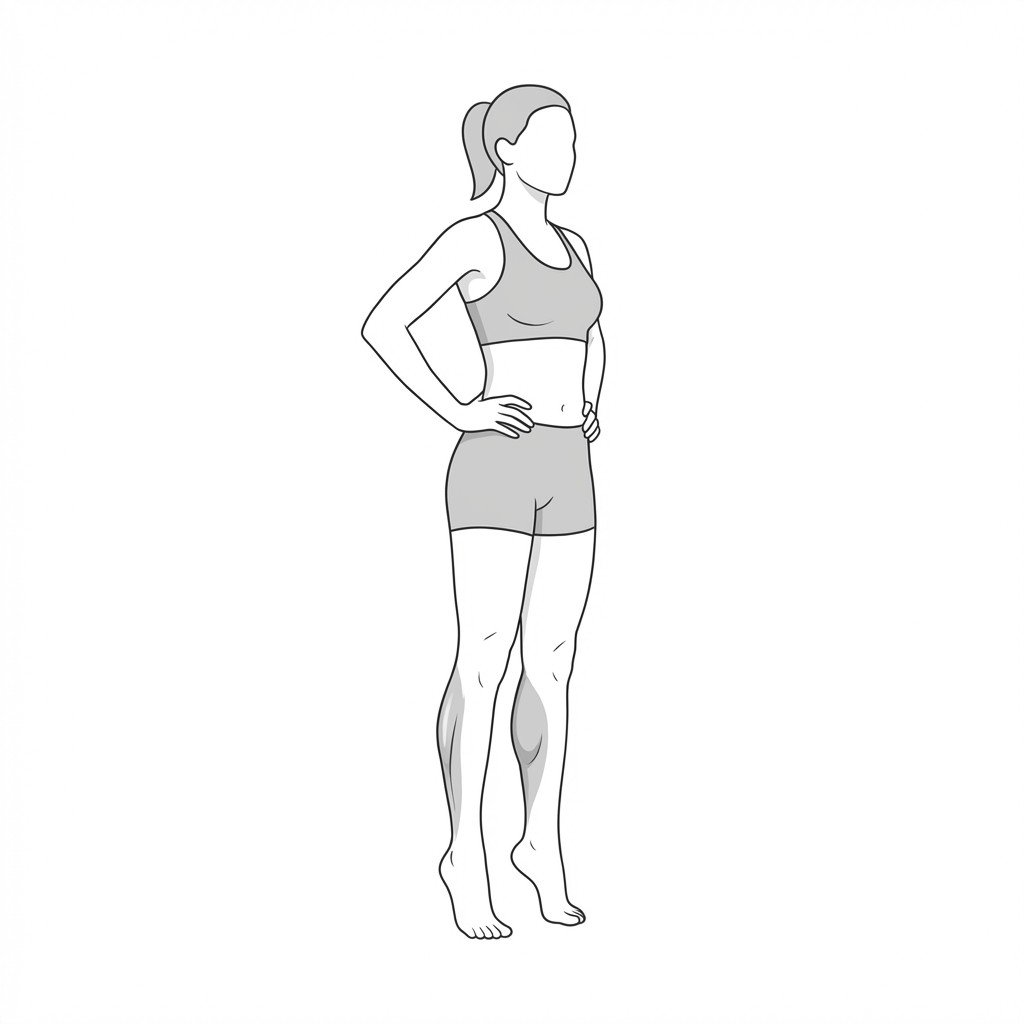
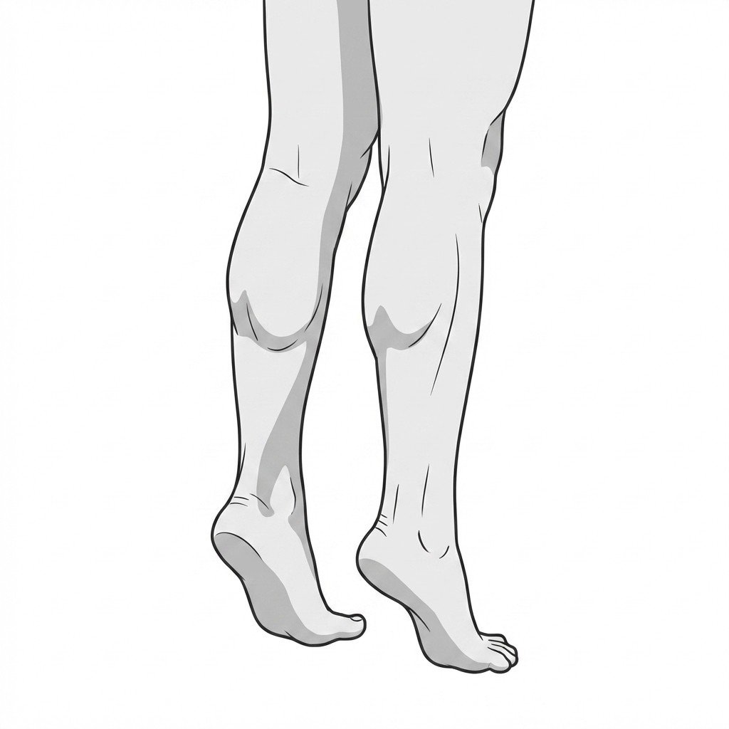
Calf raises
Calf raises strengthen the gastrocnemius and soleus muscles, which are vital for lower extremity force attenuation and forward propulsion. In knee osteoarthritis, improving plantarflexor capacity may help improve walking mechanics and reduce compensatory loading on the affected tibiofemoral joint.
Start Exercise →Lower Leg
Calf stretching
Calf stretching reduces tension in the gastrocnemius-soleus complex, addressing the equinus contracture frequently seen in patients with Posterior Tibial Tendon Dysfunction. By improving ankle dorsiflexion, this stretch decreases compensatory overpronation at the midfoot, which may help reduce mechanical strain on the posterior tibial tendon during stance.
Start Exercise →Shoulder
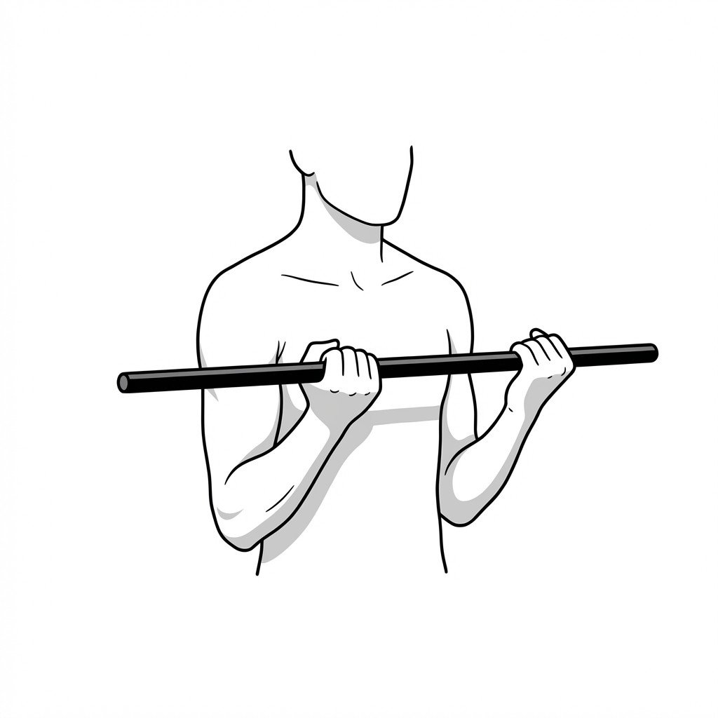
Cane-assisted external rotation
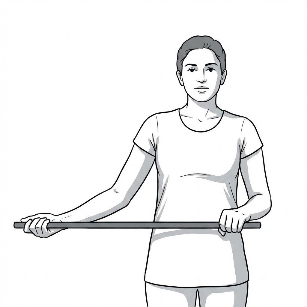
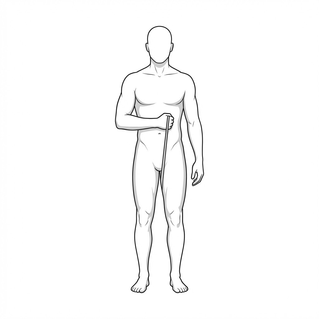
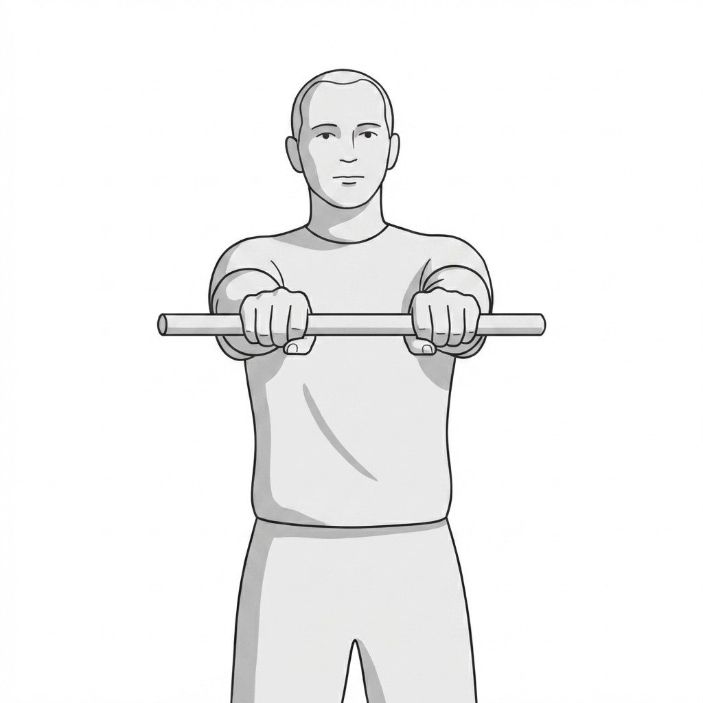
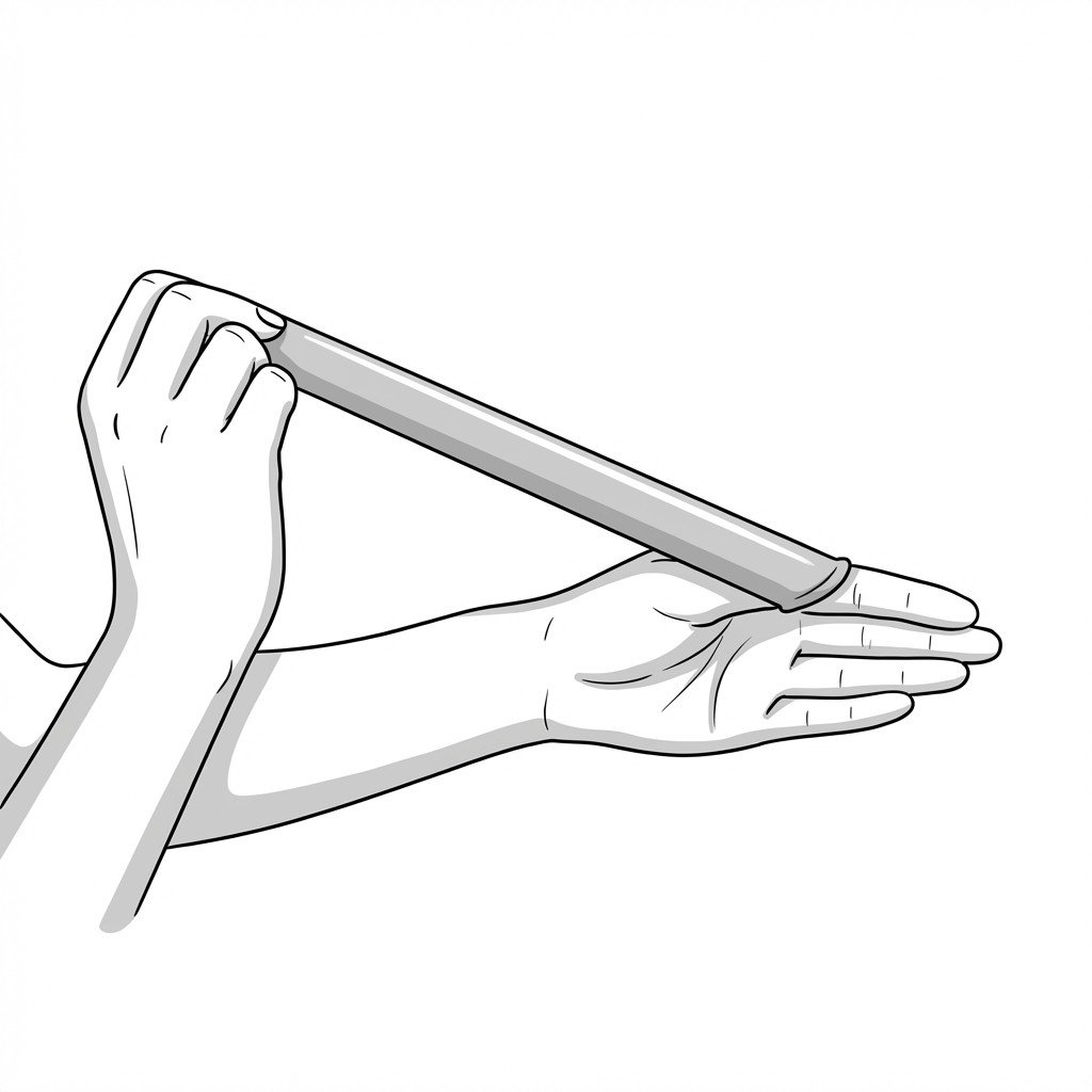
This exercise utilizes a rigid lever to apply an active-assisted stretch to the anterior glenohumeral joint capsule and internal rotators. By passively driving the affected shoulder into external rotation, it targets the capsular restriction characteristic of adhesive capsulitis. This controlled mobilization is intended to help restore functional reaching mechanics and ease tissue stiffness.
Start Exercise →Lumbar & Pelvis
Cat-cow
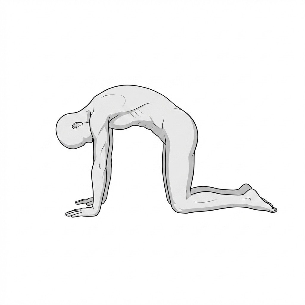
The cat-cow exercise is a gentle active mobility movement that promotes segmental spinal flexion and extension. By oscillating between these positions, it is thought to ease neural tension, promote synovial fluid movement in facet joints, and help modulate nociceptive input. This helps alleviate mechanical low back pain by reducing muscle guarding and restoring normal lumbar kinematics.
Start Exercise →Cervical
Cervical extension in sitting
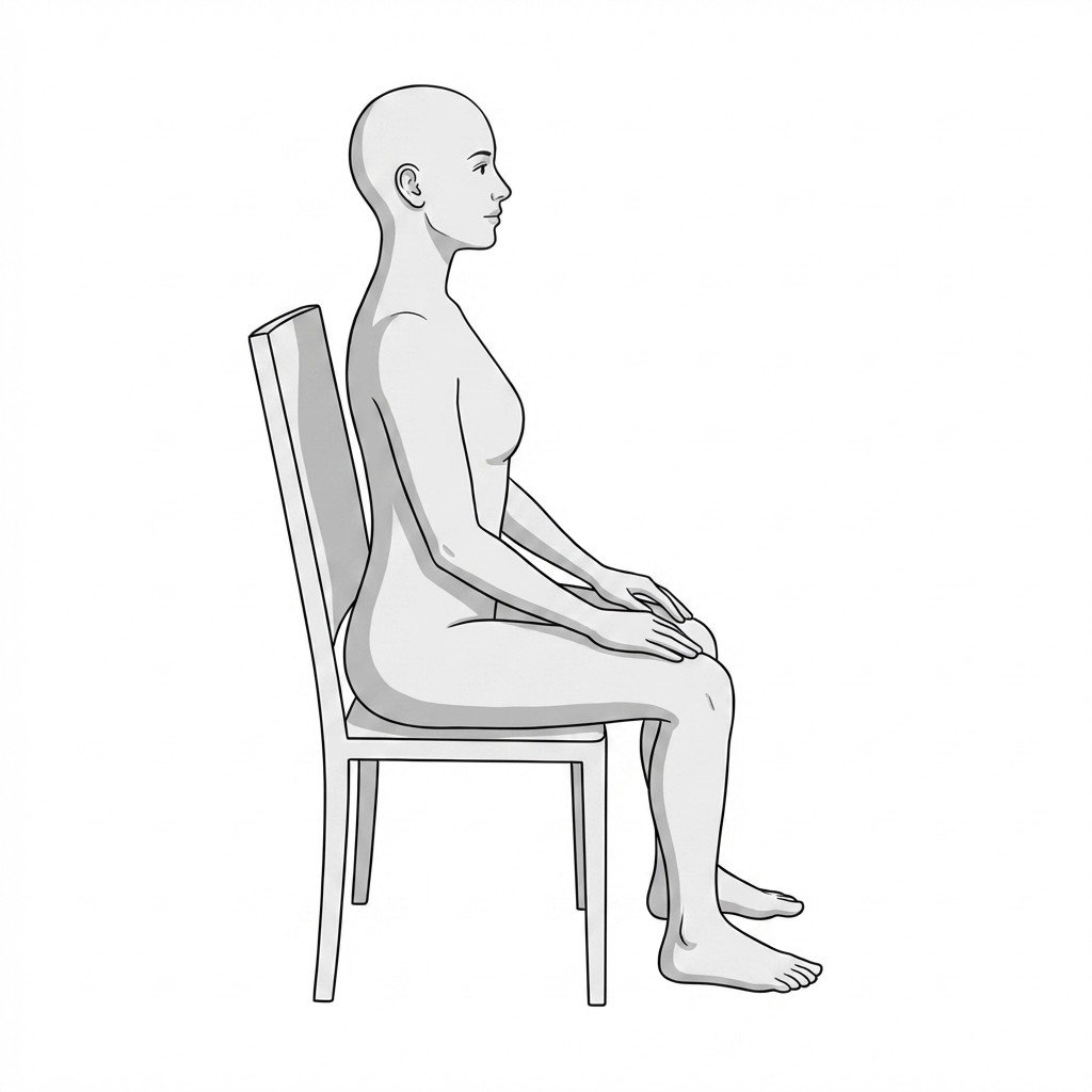
Cervical extension may promote centralization of radiating symptoms, an effect commonly attributed to changes in mechanical load on the intervertebral disc and opening of the anterior disc space. This model proposes that the movement encourages anterior migration of the nucleus pulposus, which is thought to reduce nerve root compression and ease radicular pain traveling down the arm.
Start Exercise →Cervical
Cervical lateral glides
Cervical lateral glides involve the horizontal translation of the head and neck to mobilize the cervical nerve roots as they exit the intervertebral foramen. This rhythmic technique acts as a neurodynamic slider that is thought to support neural mobility and may help reduce intraneural pressure. Mechanistically, it decreases nerve mechanosensitivity and alleviates the radiating pain or numbness associated with cervical radiculopathy.
Start Exercise →Cervical
Cervical retractions
Cervical retractions target the deep neck flexors while actively lengthening the suboccipital muscles and upper cervical extensors. This movement helps restore a more neutral cervical alignment, reducing stress on the intervertebral discs and facet joints caused by forward head posture.
Start Exercise →Cervical
Cervical rotation mobilization
Cervical rotation mobilization may help improve segmental motion in the upper cervical spine, particularly at the atlantoaxial joint. Because the trigeminocervical nucleus links cervical nociceptive afferents with the trigeminal nerve, reducing cervical spine dysfunction may help reduce referred pain and tension in the masticatory muscles, thereby improving TMJ mechanics.
Start Exercise →Thoracic & Core
Chest wall mobility
Chest wall mobility exercises mechanically stretch the myofascial structures of the rib cage and mobilize the costovertebral and costochondral joints. By improving thoracic compliance and breathing mechanics, this exercise may help reduce repetitive mechanical stress on the anterior costal cartilage during respiration.
Start Exercise →Lumbar & Pelvis
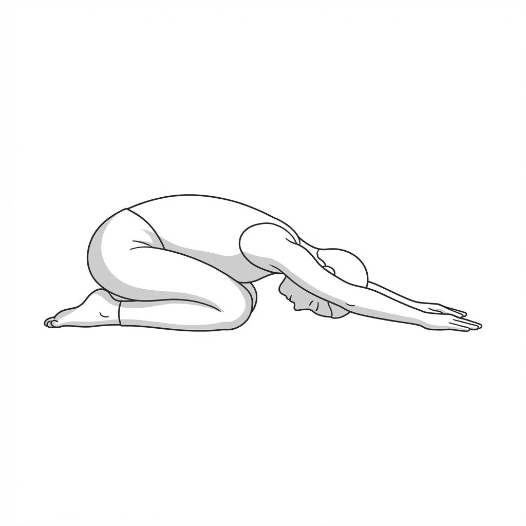
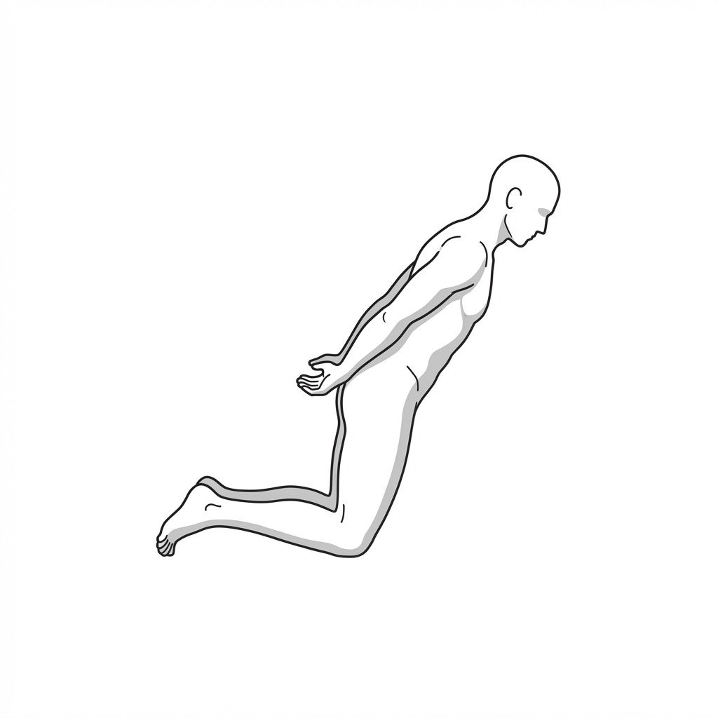
Child's pose
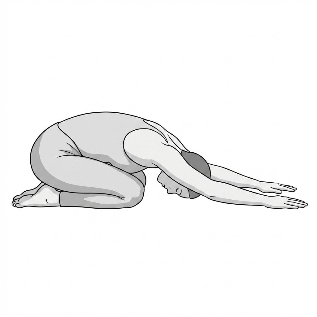
Child's pose gently introduces lumbar flexion, which may increase the neuroforaminal space and is thought to reduce load on structures associated with lumbar radiculopathy. It also provides a prolonged, low-load stretch to the paraspinal musculature, reducing reactive muscle spasm and improving overall lumbar mobility.
Start Exercise →Hip
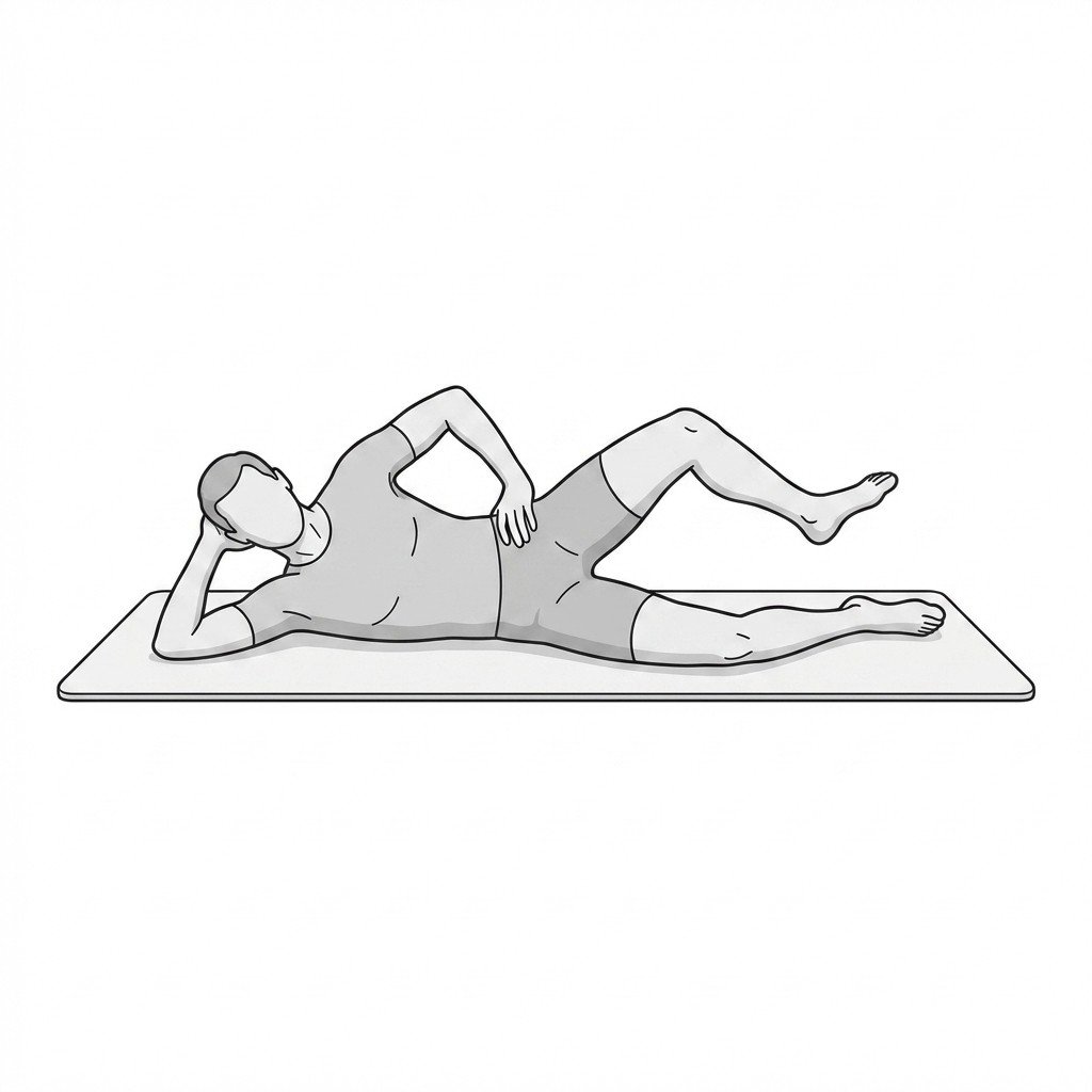
Clamshells
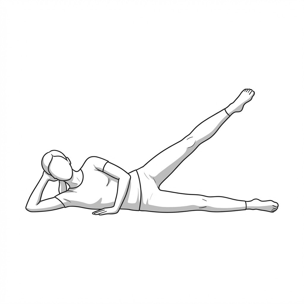
The clamshell exercise isolates and strengthens the hip abductors and external rotators, particularly the gluteus medius. By improving lateral hip stability and neuromuscular control, it may help reduce excessive pelvic drop and femoral internal rotation, which may in turn reduce stress on the greater trochanter and sacroiliac joint.
Start Exercise →Shoulder
Controlled eccentric shrugs
Controlled eccentric shrugs emphasize the lengthening phase of the upper trapezius muscle under a controlled load. This eccentric overload is thought to help desensitize myofascial trigger points, support tissue remodeling, and may improve local circulation in muscle fibers associated with Trapezius Myofascial Pain Syndrome.
Start Exercise →Cervical
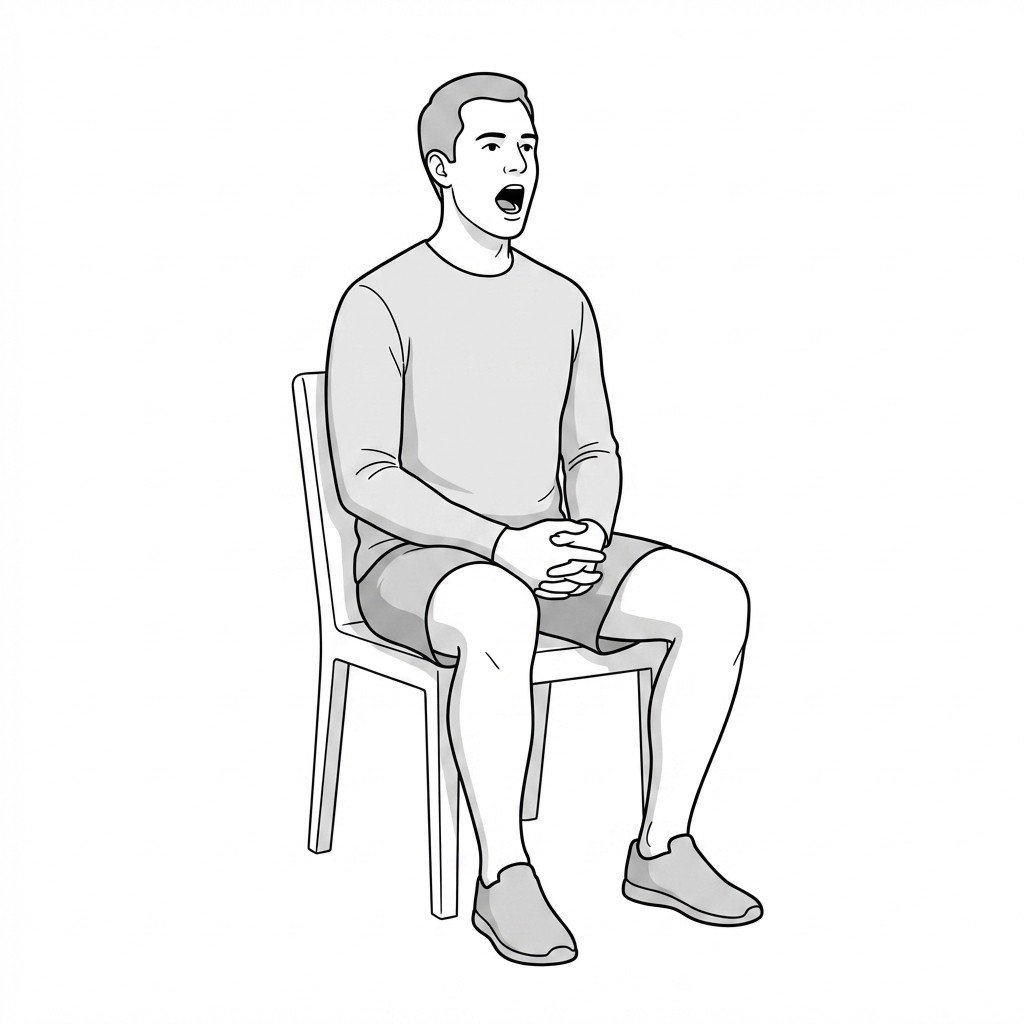
Controlled mouth opening
Controlled mouth opening aims to support proper biomechanics of the temporomandibular joint by maintaining anterior tongue placement, which encourages initial condylar rotation rather than excessive anterior translation. This may help reduce mechanical stress on the articular disc, ease joint clicking, and support more coordinated activation patterns of the masticatory muscles.
Start Exercise →Hip
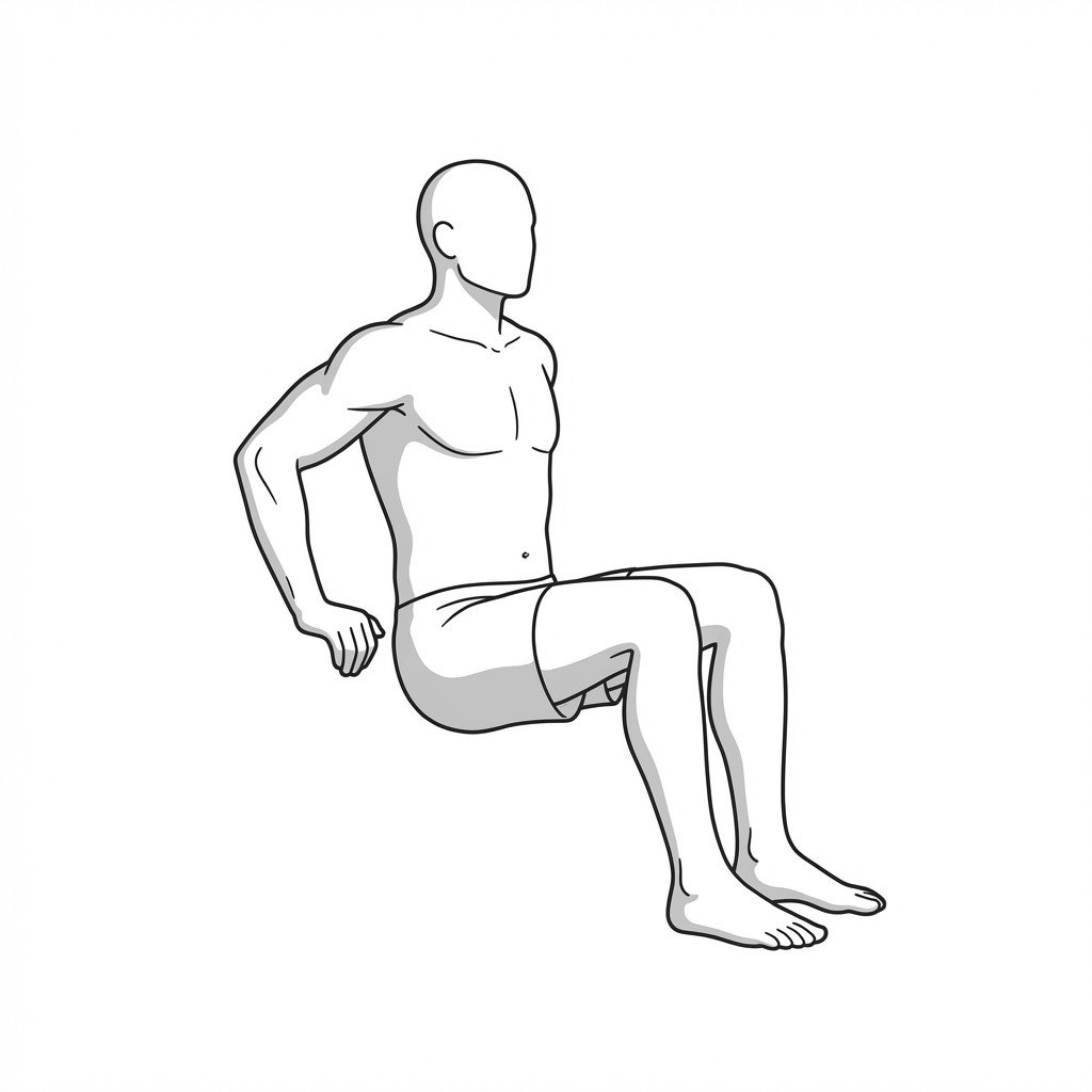
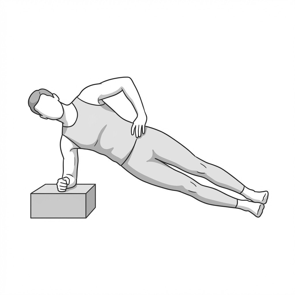
Copenhagen planks
The Copenhagen plank is a high-intensity eccentric and isometric strengthening exercise targeting the adductor complex and lateral core musculature. It helps rehabilitate adductor strains by increasing the tissues' load tolerance and eccentric strength, which may help reduce the risk of recurrent groin injury.
Start Exercise →Shoulder
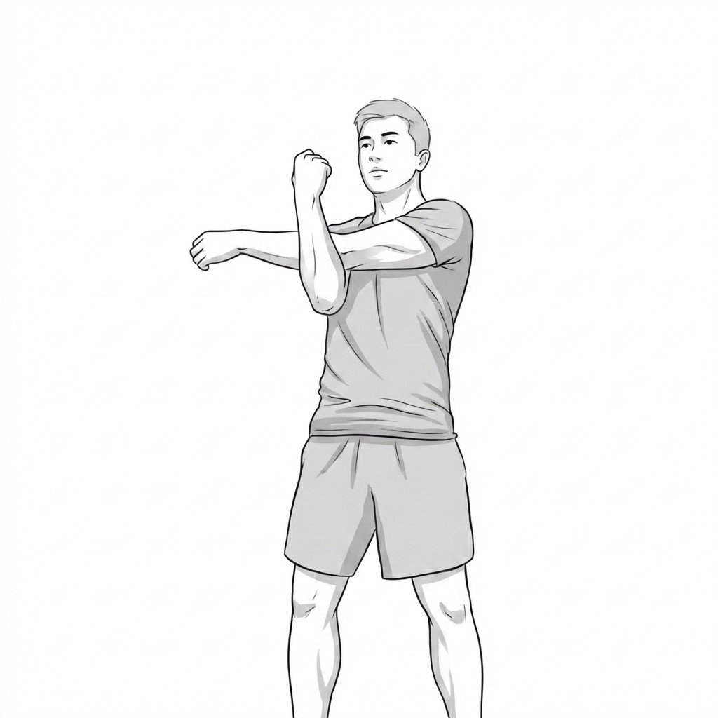
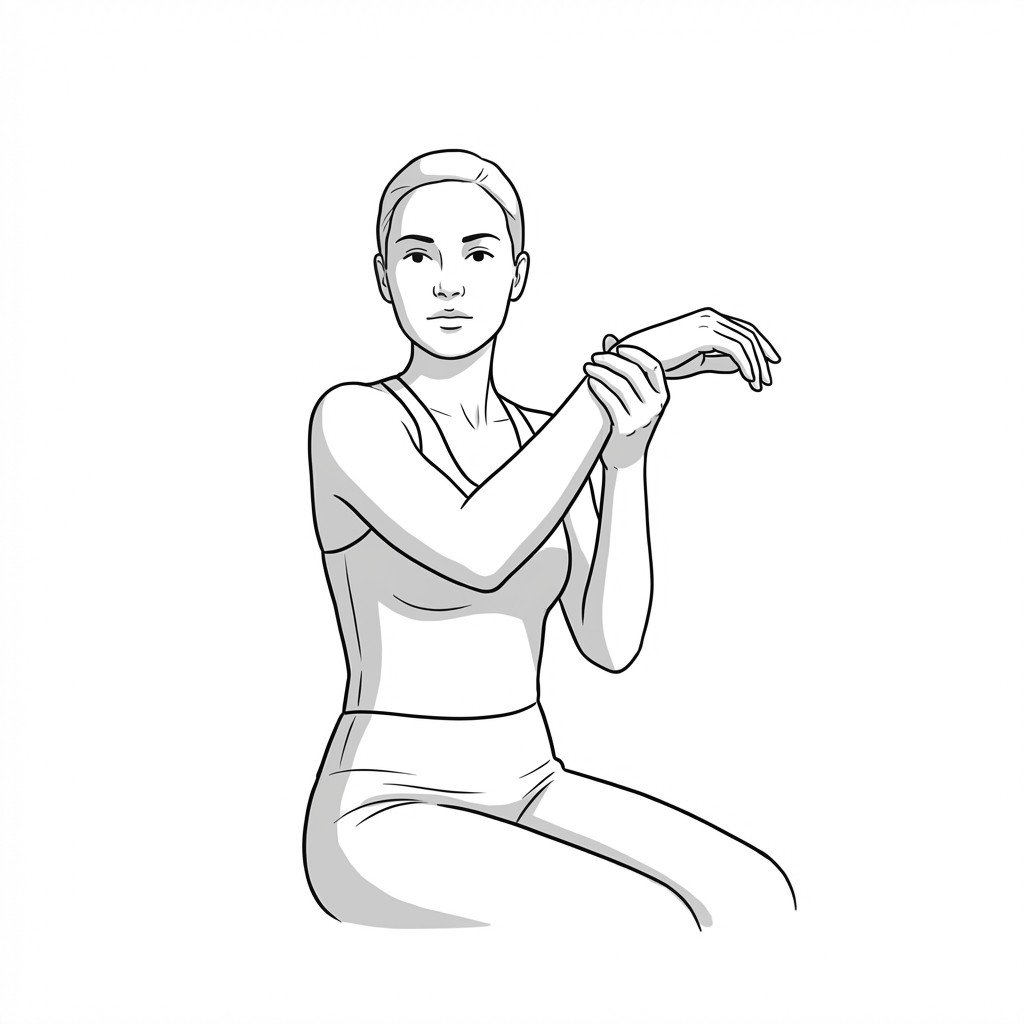
Cross-body stretch
The cross-body stretch targets the posterior capsule and posterior rotator cuff musculature to improve internal rotation and horizontal adduction. By applying prolonged, low-load stress to the contracted capsular tissues, it helps restore glenohumeral joint mobility and may reduce movement restrictions characteristic of adhesive capsulitis.
Start Exercise →Thoracic & Core
Dead bugs
The dead bug exercise targets the deep abdominal musculature to improve lumbopelvic motor control and dissociation of the extremities from the core. By challenging the core with contralateral limb movements, it actively trains the abdominals to resist unwanted lumbar extension and anterior pelvic tilt.
Start Exercise →Knee
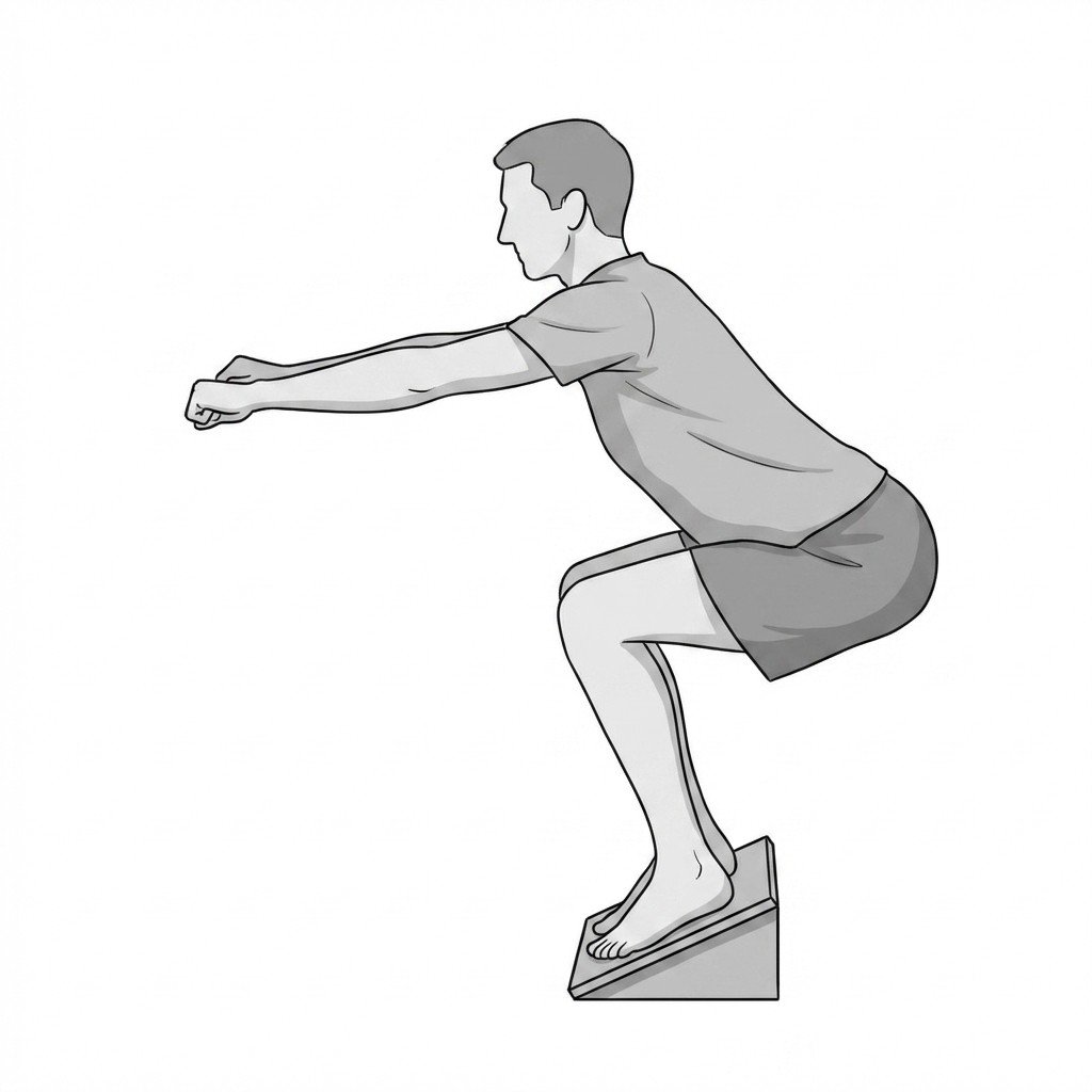
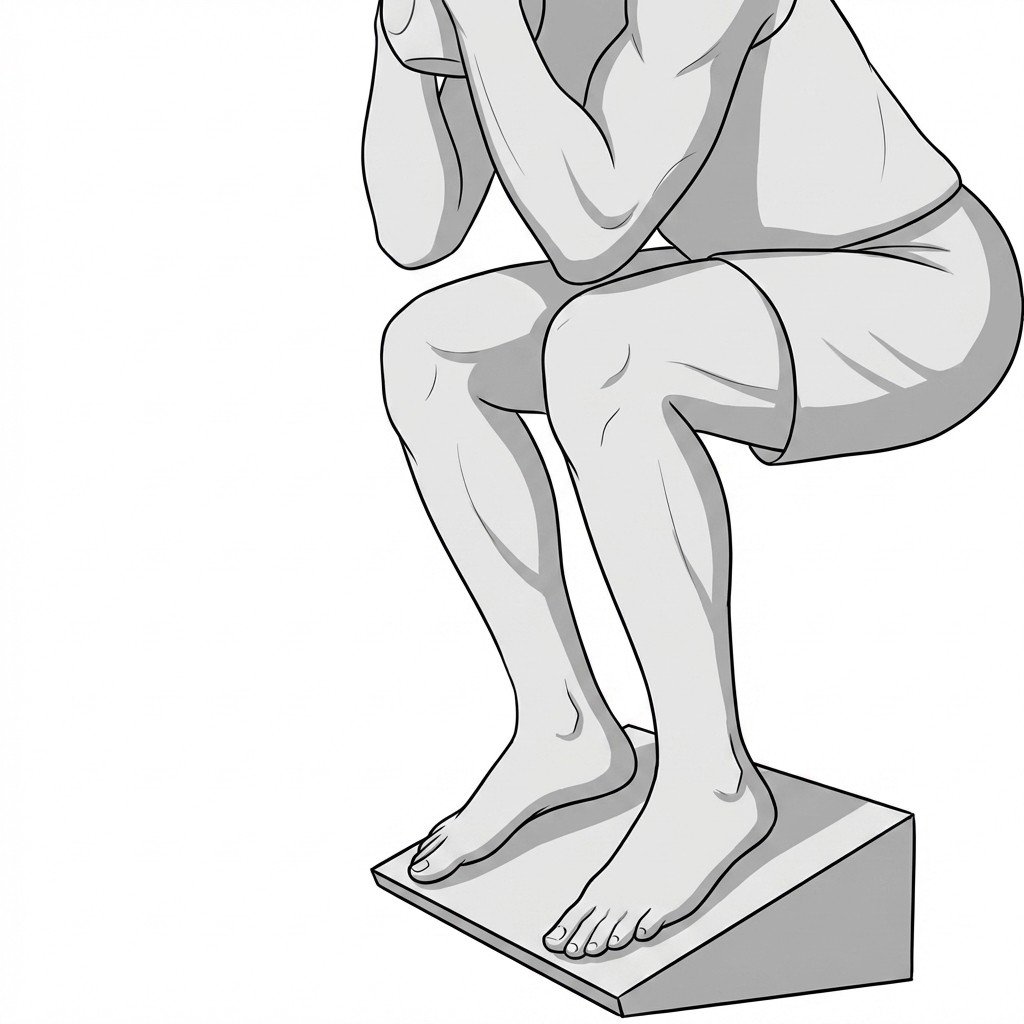
Decline eccentric squats
Decline eccentric squats specifically target the patellar tendon by shifting the mechanical load away from the hips and ankles directly onto the knee extensors. The controlled eccentric (lengthening) phase induces mechanical tension that is thought to stimulate collagen synthesis and tendon remodeling. This targeted structural loading may help improve the tendon's load tolerance and reduce pain over time.
Start Exercise →Cervical
Deep neck flexor activation
Deep neck flexor activation targets the longus colli and longus capitis muscles, which are often inhibited and weakened in patients with axial neck pain. By restoring deep segmental motor control, this exercise may help reduce the compensatory overactivity of superficial muscles and improve dynamic cervical spine stability.
Start Exercise →Lower Leg
Eccentric calf raises
Eccentric calf raises selectively load the gastroc-soleus complex and deep posterior compartment while the muscles are lengthening to improve tissue capacity. This may help Medial Tibial Stress Syndrome by enhancing the ability of the plantar flexors to absorb ground reaction forces and reducing traction stress along the posteromedial border of the tibia.
Start Exercise →Shoulder
Eccentric external rotation
Eccentric external rotation focuses on lengthening the posterior rotator cuff under tension to promote tissue remodeling. This controlled eccentric loading is thought to stimulate collagen synthesis and realignment within the tendon, which may help reduce pain and improve function in rotator cuff tendinopathy.
Start Exercise →Shoulder
Eccentric shoulder flexion lowering
This exercise focuses on eccentric loading of the long head of the biceps tendon by slowly controlling shoulder extension from a fully flexed position. Mechanistically, eccentric muscle contractions are thought to stimulate collagen synthesis and structural tissue remodeling, which may help improve tendon load tolerance and reduce pain in tendinopathic tissues.
Start Exercise →Elbow & Hand
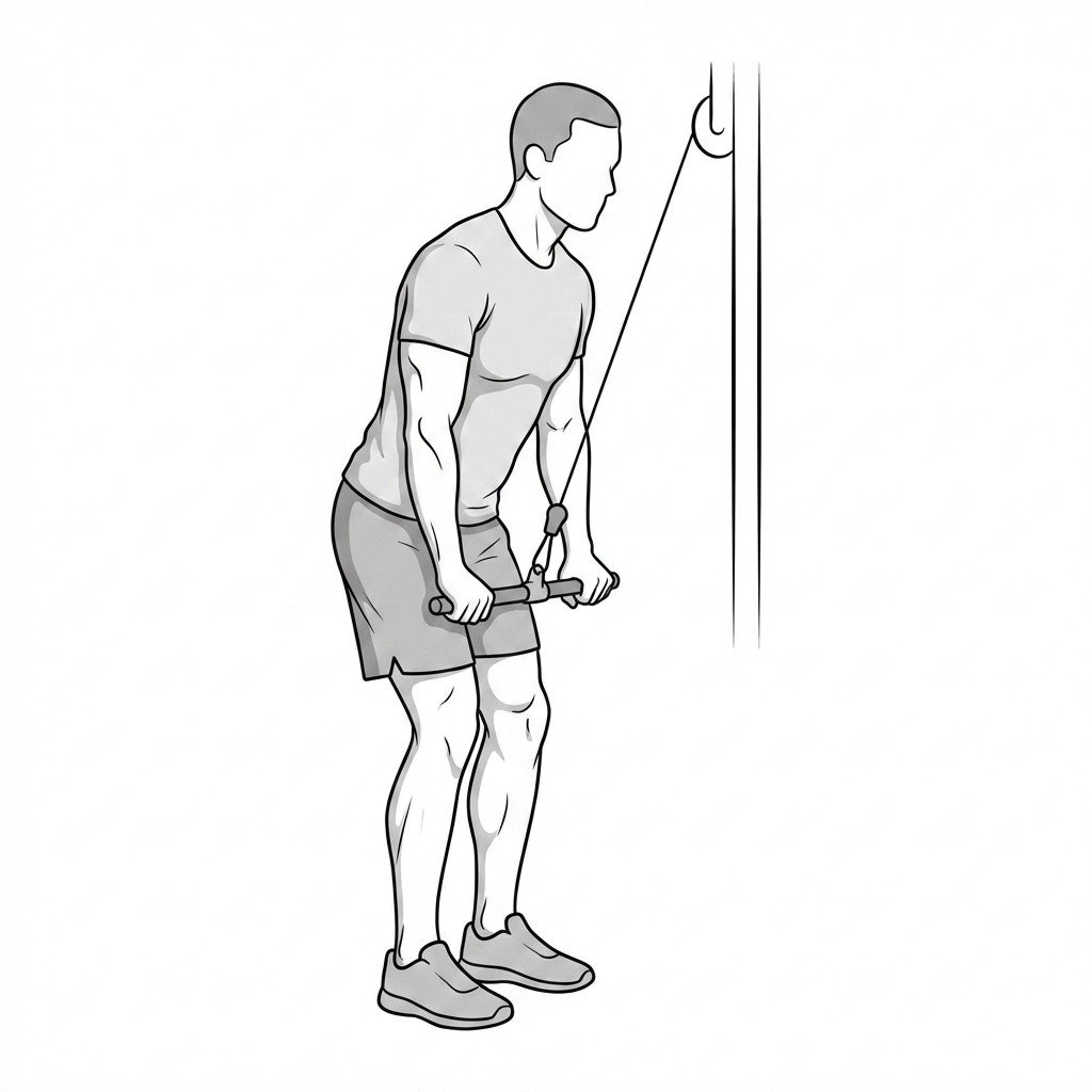
Eccentric triceps push-downs
Eccentric triceps push-downs focus on the lengthening phase of the muscle contraction, applying controlled mechanical load to the triceps tendon. This process stimulates collagen synthesis and promotes the remodeling of disorganized, degenerative tendon tissue. By emphasizing the eccentric phase, it safely builds tensile strength and reduces pain in patients with triceps tendinopathy without overstressing the tissue.
Start Exercise →Elbow & Hand
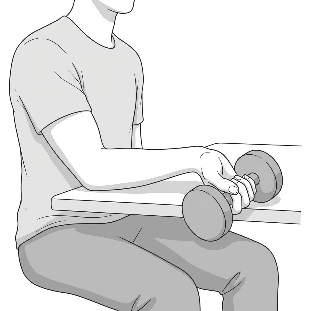
Eccentric wrist radial deviation
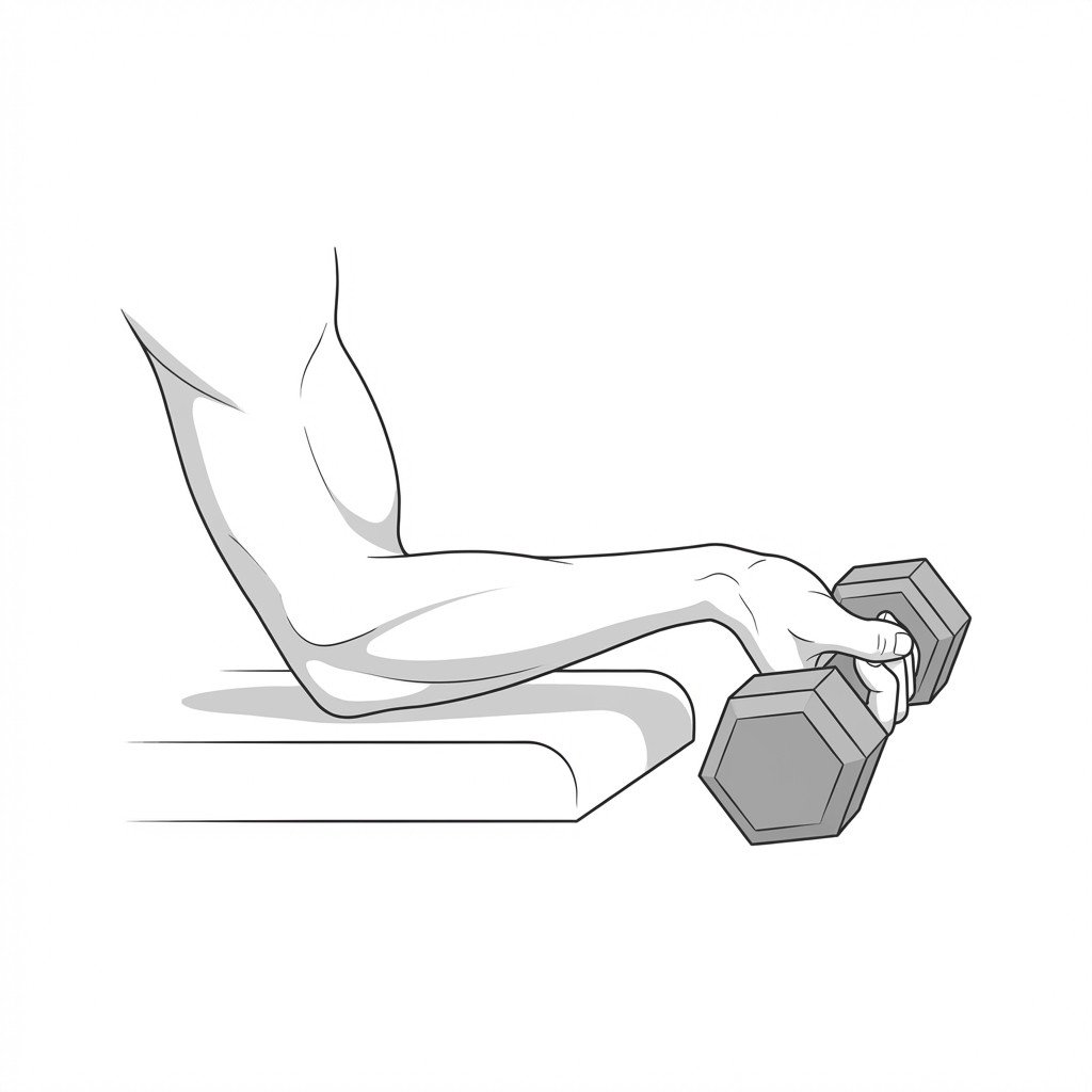
This exercise focuses on lengthening the abductor pollicis longus and extensor pollicis brevis under tension. Eccentric loading stimulates collagen synthesis and tendon remodeling within the first dorsal compartment of the wrist. This gradually improves the tendon's load-bearing capacity and reduces pain associated with De Quervain's tenosynovitis.
Start Exercise →Elbow & Hand
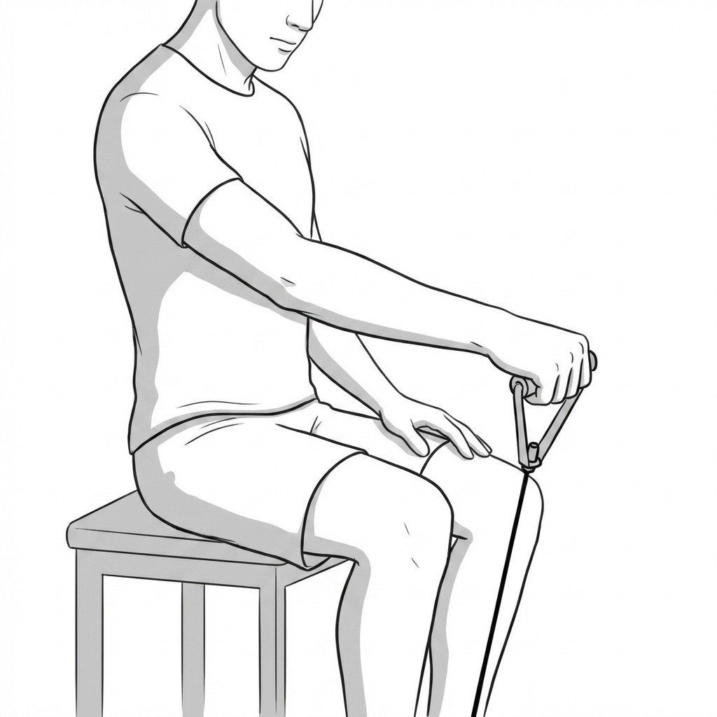
Eccentric wrist supination
Eccentric wrist supination loads the supinator muscle while it is lengthening, promoting tissue remodeling and improving load tolerance. Since radial tunnel syndrome involves compression of the posterior interosseous nerve by the supinator muscle, controlled eccentric loading helps stretch and condition this tissue without exacerbating compressive irritation.
Start Exercise →Elbow & Hand
Elbow extension stretch
This exercise lengthens the soft tissues of the anterior elbow and relieves mechanical tension on the ulnar nerve within the cubital tunnel. By promoting full elbow extension, it counteracts the sustained flexion postures that typically aggravate ulnar neuropathy, thereby reducing nerve ischemia and irritation.
Start Exercise →Shoulder
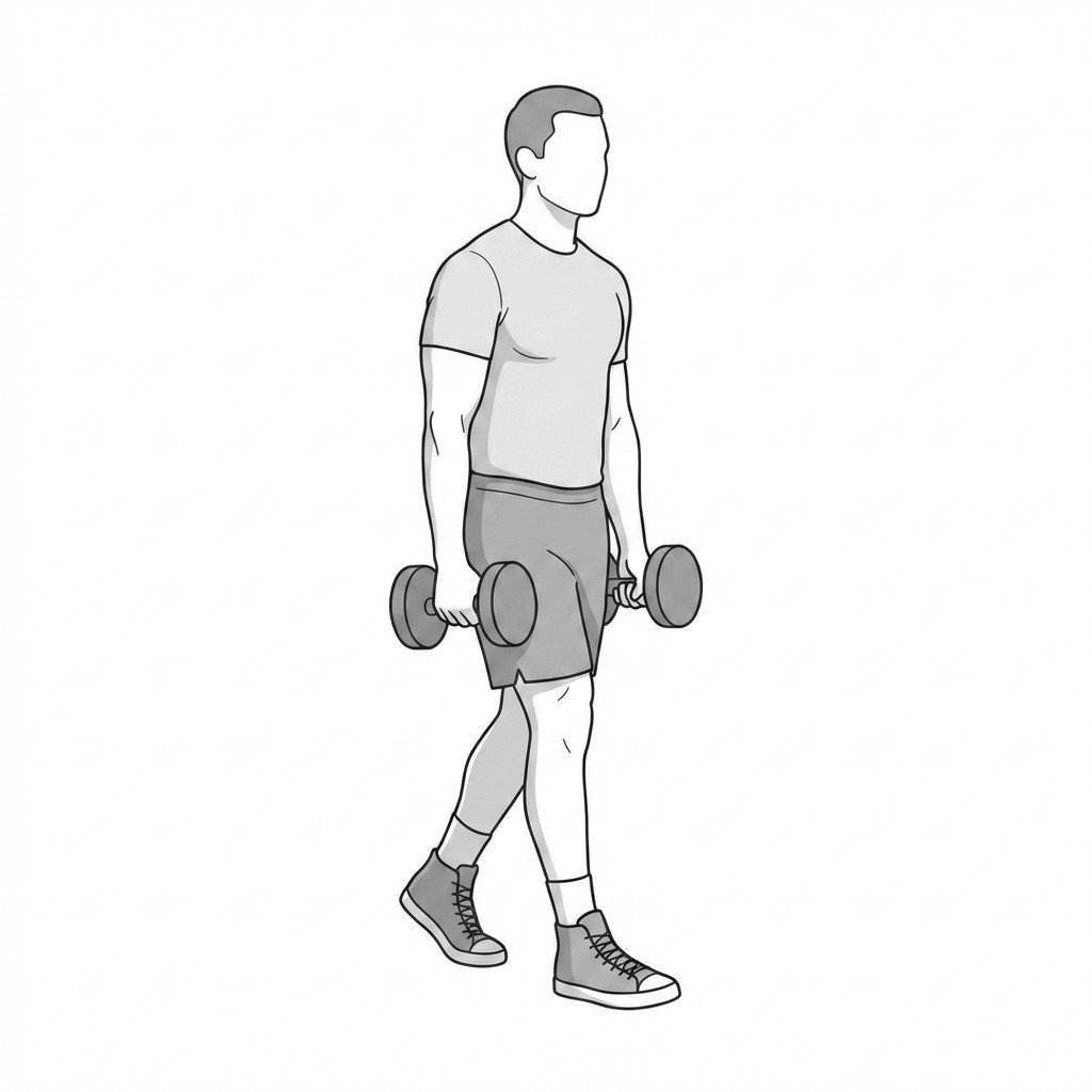
Farmer's carries
Farmer's carries provide heavy isometric loading to the shoulder girdle, requiring the trapezius muscles to stabilize the scapula against a downward distractive force. This sustained isometric contraction helps desensitize hyperirritable trigger points in the upper trapezius and builds local tissue capacity. It also improves overall scapulothoracic endurance and postural awareness.
Start Exercise →Elbow & Hand
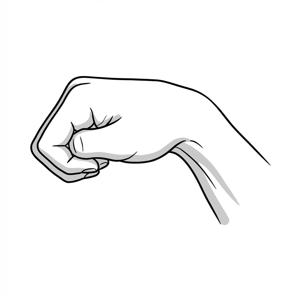
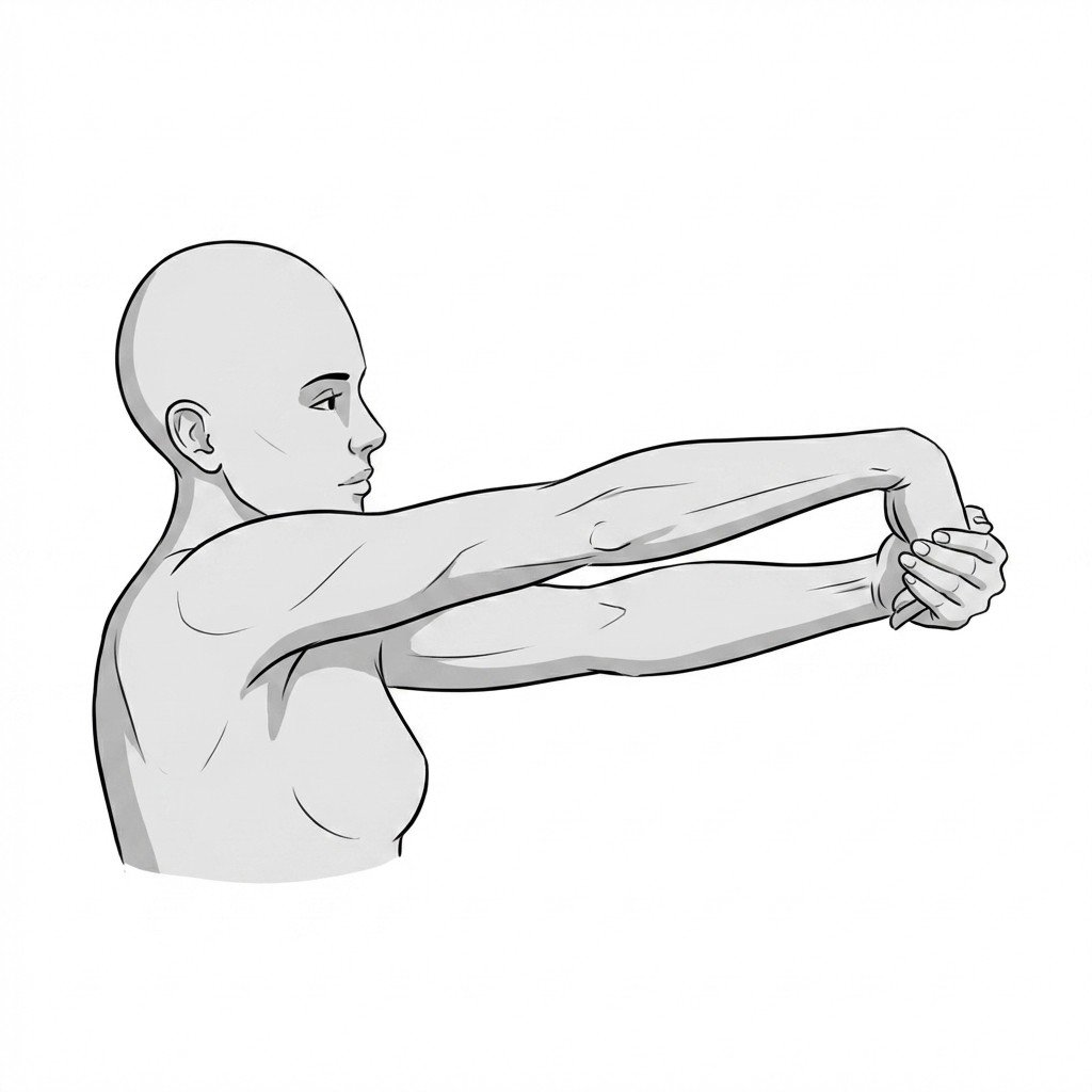
Finkelstein stretch
The Finkelstein stretch applies a targeted tensile load to the tendons of the first dorsal compartment of the wrist, specifically the abductor pollicis longus and extensor pollicis brevis. This controlled stretching may help improve tissue extensibility, reduce tendon gliding resistance, and ease pain associated with De Quervain's tenosynovitis.
Start Exercise →Thoracic & Core
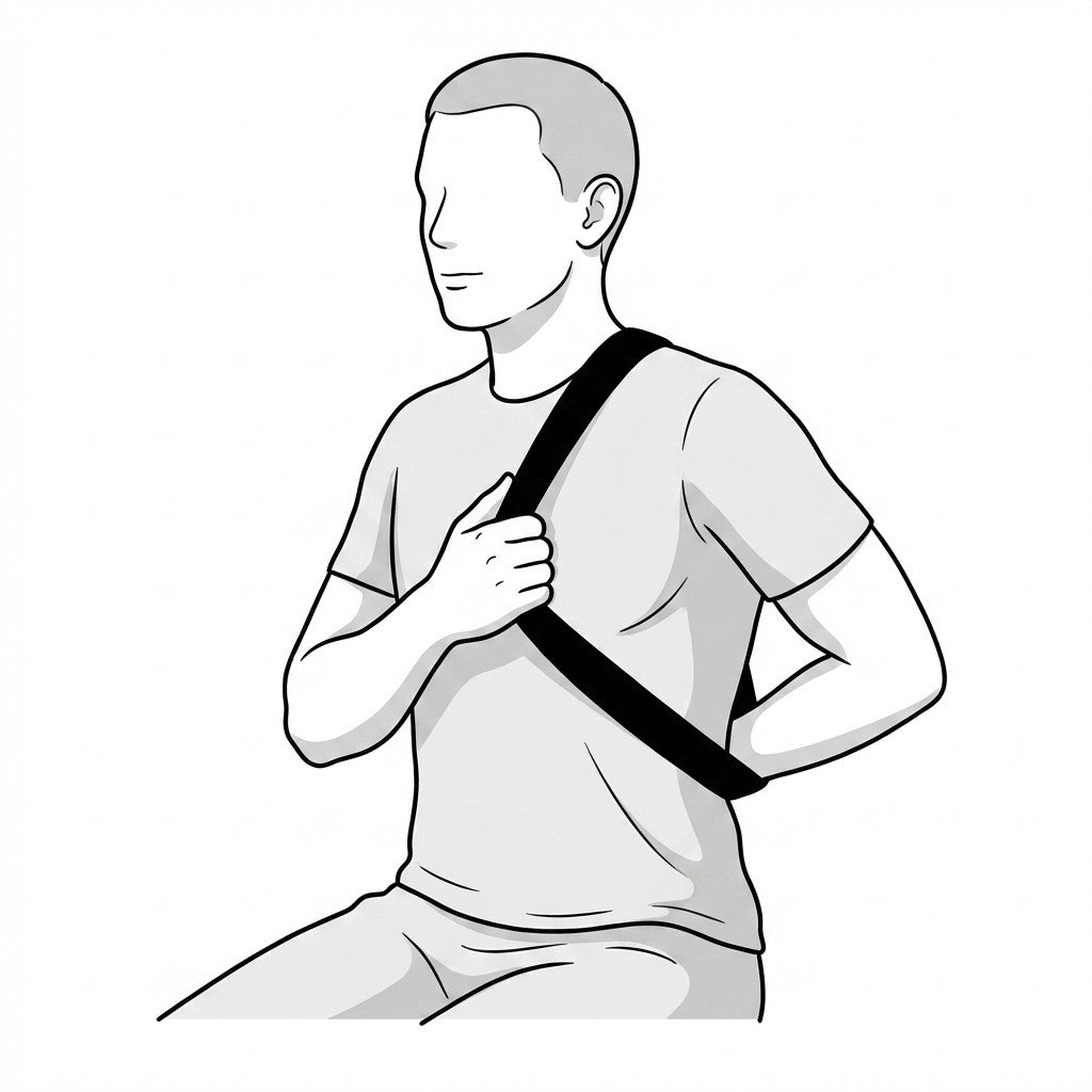
First rib mobilization with strap
This self-mobilization technique applies a localized inferior glide to an elevated first rib using a rigid strap. By mechanically depressing the first rib, it helps open the costoclavicular space, thereby reducing compression on the brachial plexus and subclavian vessels associated with Thoracic Outlet Syndrome.
Start Exercise →Shoulder
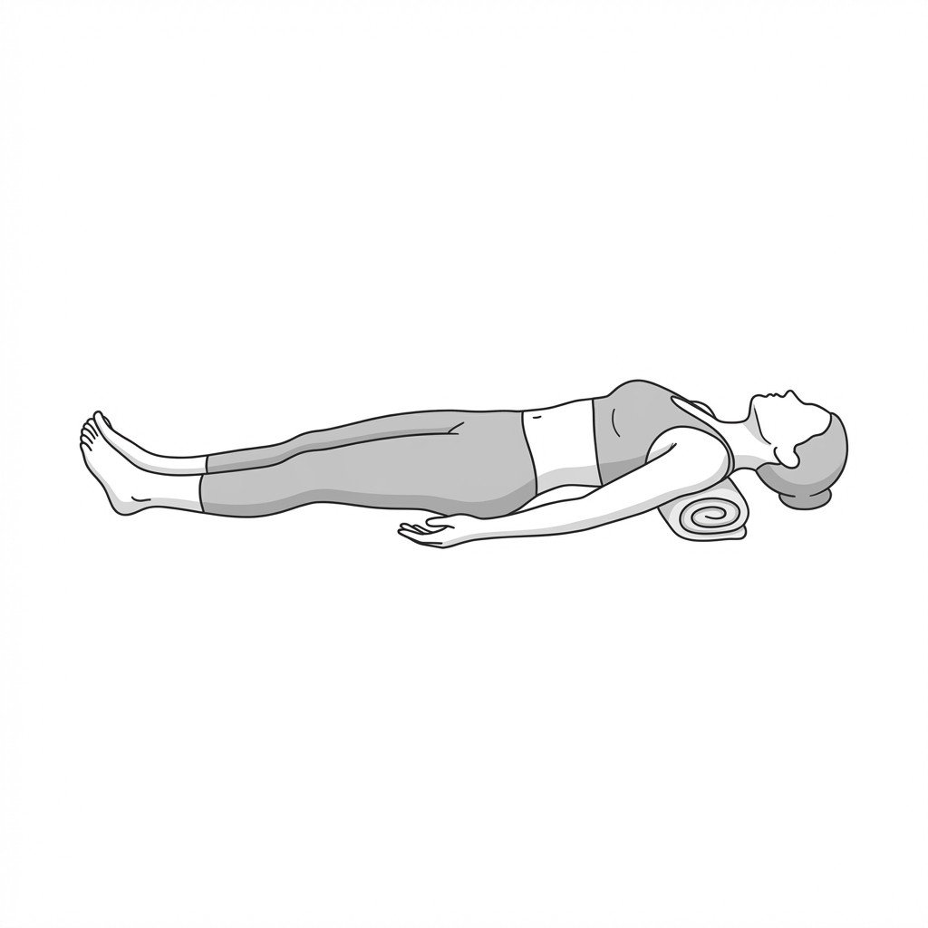
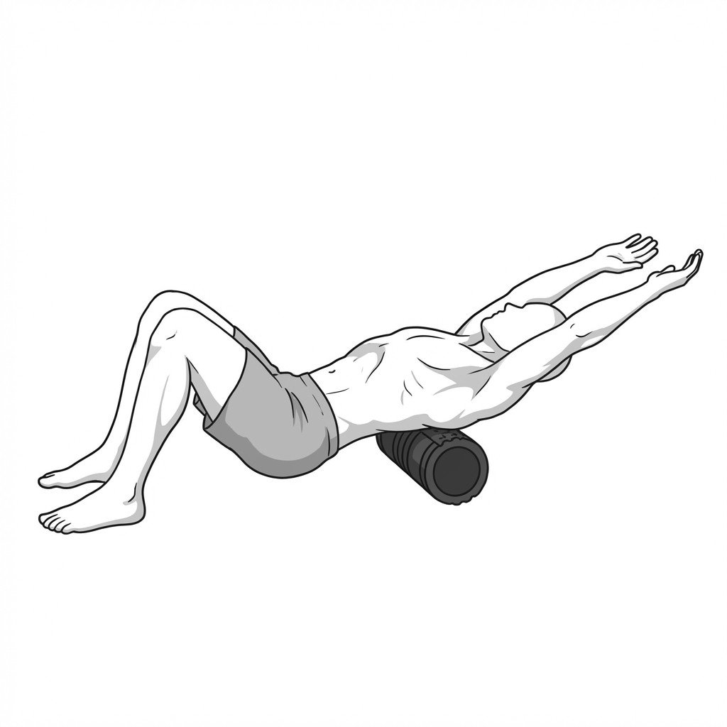
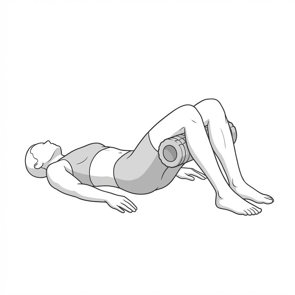
Foam roller thoracic extensions
Foam roller thoracic extensions mechanically mobilize the thoracic facet joints into extension, stretching the anterior chest wall and reversing chronic flexion posture. By improving thoracic segmental mobility, this exercise reduces compensatory hypertonicity in the trapezius and alleviates the biomechanical stress associated with thoracic kyphosis.
Start Exercise →Elbow & Hand
Forearm pronation stretch
This exercise improves flexibility in the forearm by stretching the supinator and biceps muscles as the arm moves into end-range pronation. Restoring full rotational mobility helps reduce mechanical tension and fascial restriction around the elbow joint, which may help reduce secondary load on the ulnar nerve in cubital tunnel syndrome.
Start Exercise →Shoulder
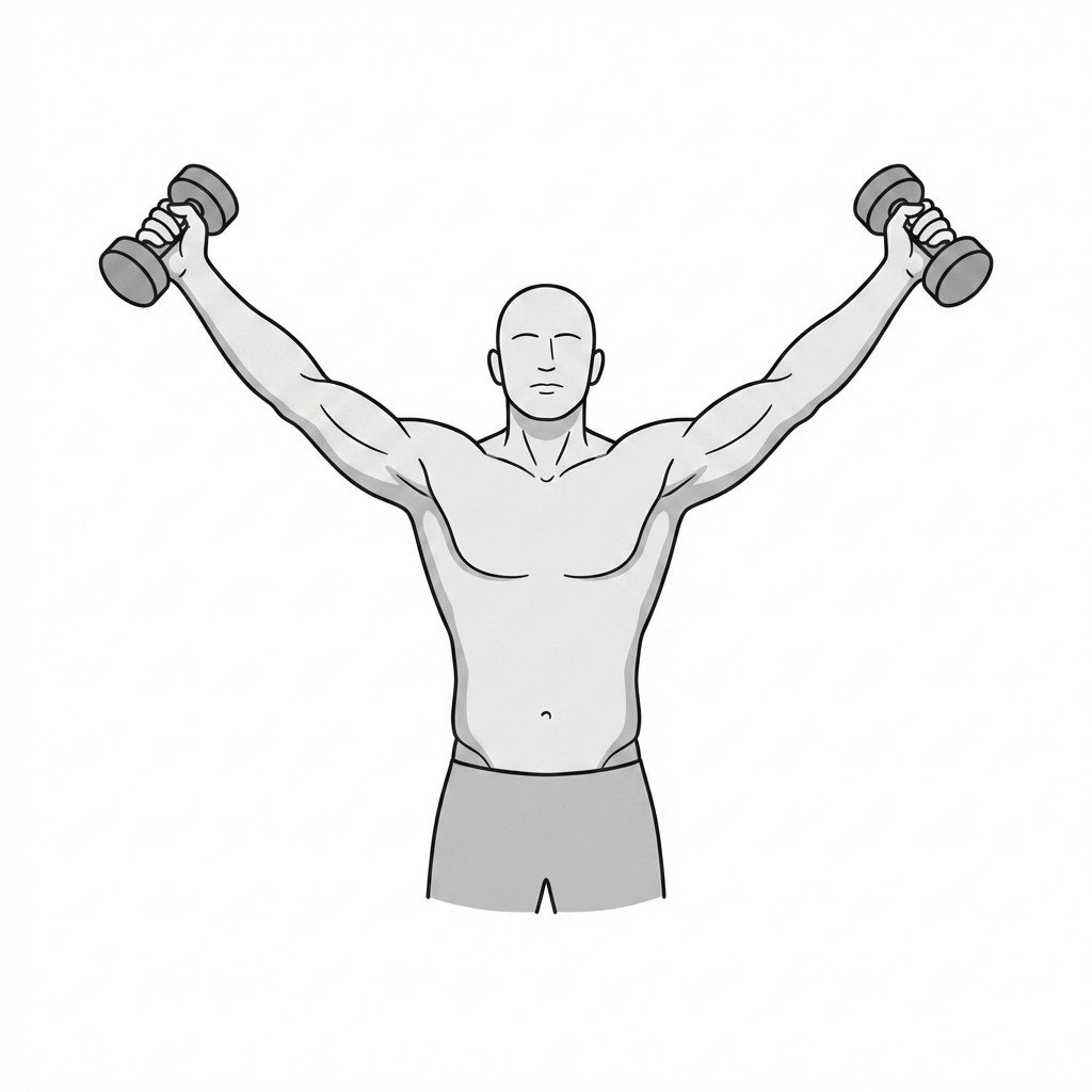
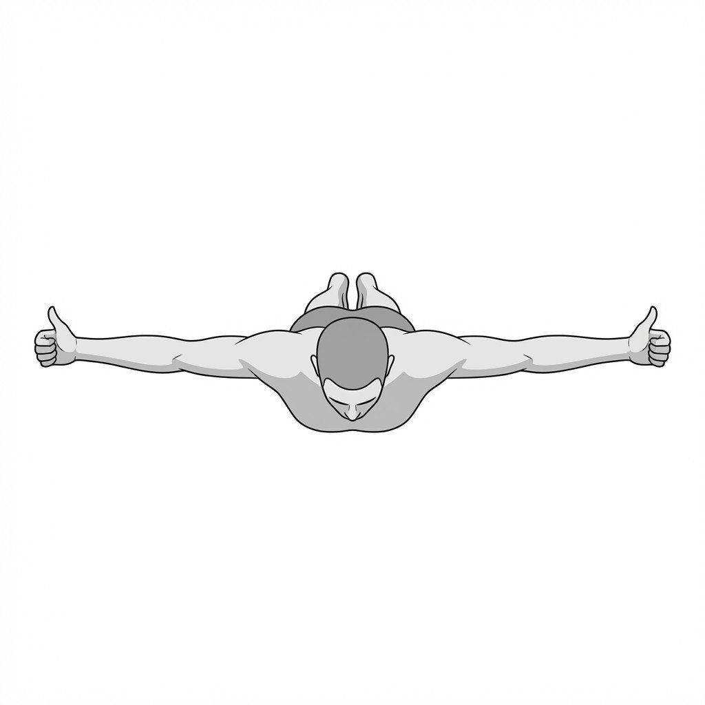
Full can scaption
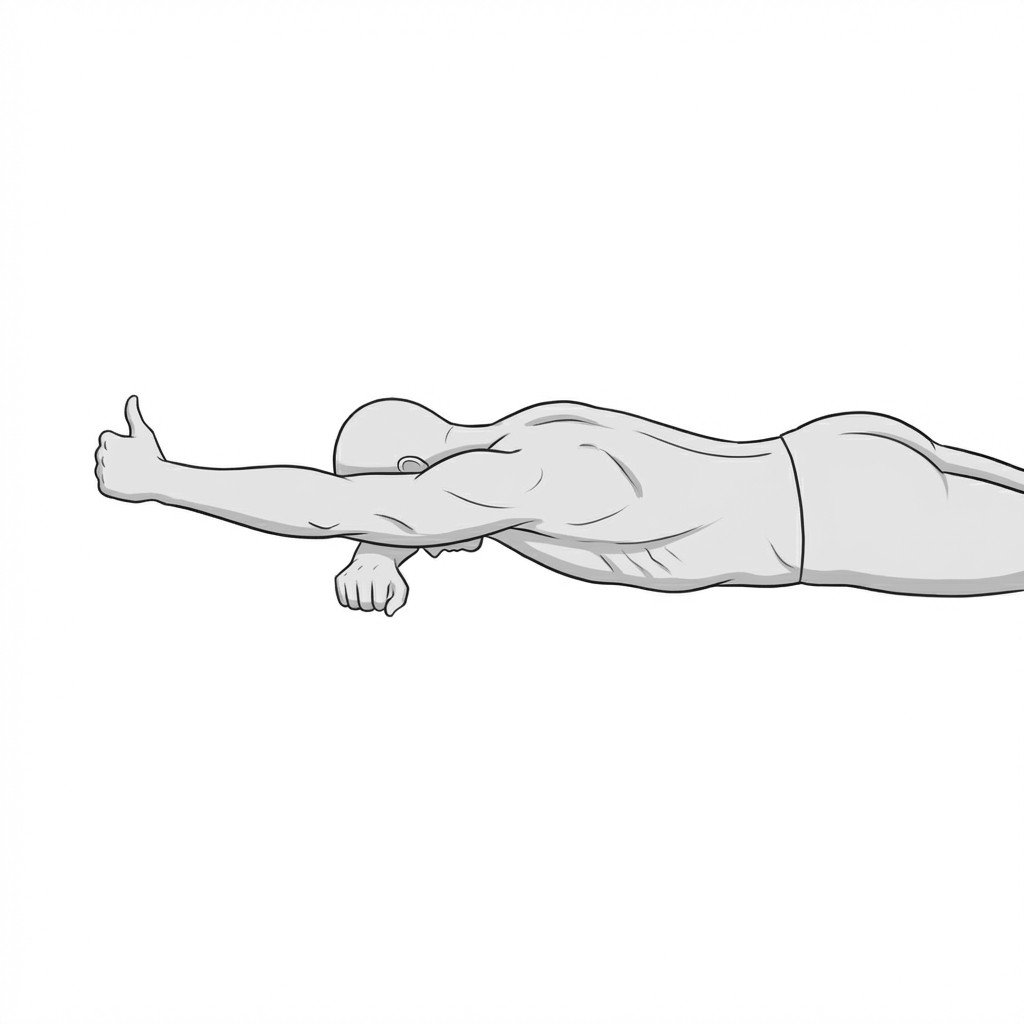
The full can scaption exercise isolates the supraspinatus muscle by lifting the arm in the scapular plane with external rotation (thumbs up). This alignment optimally recruits the rotator cuff while maximizing the subacromial space, which may help reduce impingement and support tendon healing.
Start Exercise →Lower Leg
Gastrocnemius stretch
The gastrocnemius stretch lengthens the superficial posterior lower leg musculature, reducing passive tension on the Achilles tendon. By restoring normal ankle dorsiflexion range of motion, it helps decrease abnormal mechanical loads on the tendon during the gait cycle.
Start Exercise →Cervical
Gentle AROM away from pull
This exercise utilizes active antagonist muscle contraction to induce reciprocal inhibition of the overactive, dystonic cervical muscles. By gently moving the head away from the direction of the dystonic pull, it helps restore sensorimotor control and maintain cervical range of motion while minimizing the risk of triggering a spastic stretch reflex.
Start Exercise →Elbow & Hand
Gentle thumb opposition
Gentle thumb opposition promotes controlled gliding of the abductor pollicis longus and extensor pollicis brevis tendons through the first dorsal compartment. This active movement improves local synovial fluid circulation and may help limit tendon sheath adhesions without over-stretching inflamed tissues.
Start Exercise →Hip
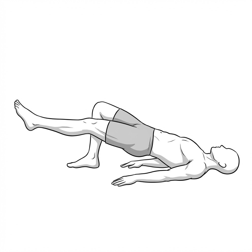
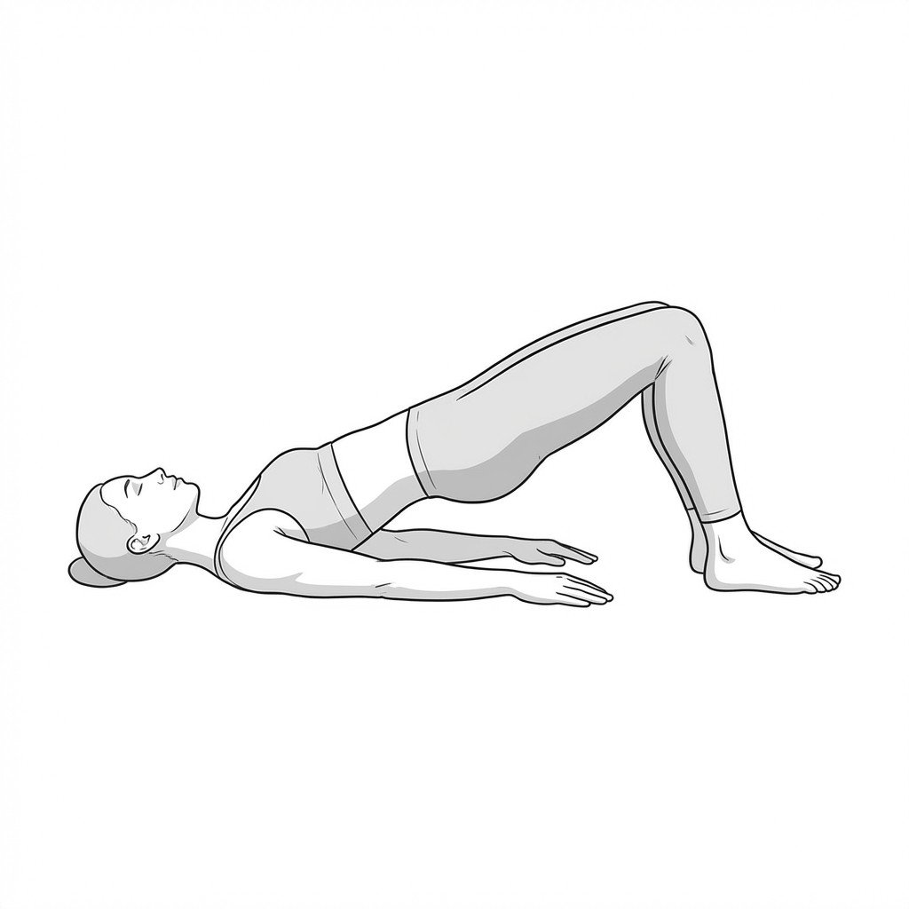
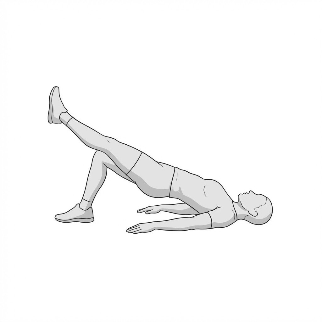
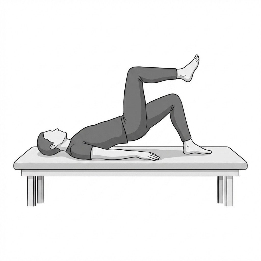
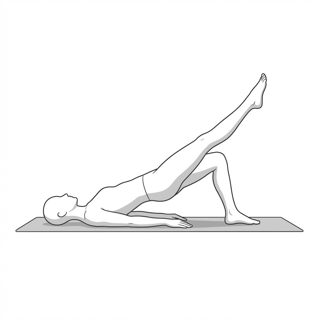
Glute bridge
The glute bridge promotes active hip extension and gluteal muscle recruitment without placing excessive compressive loads on the lateral hip. For Greater Trochanteric Pain Syndrome, it helps restore load tolerance in the gluteal complex while maintaining a neutral, non-adducted hip position to avoid compressing the gluteal tendons against the greater trochanter.
Start Exercise →Knee
Gluteus medius strengthening
This exercise isolates and strengthens the hip abductors to improve frontal plane pelvic stability. By enhancing gluteus medius strength, it helps limit excessive femoral adduction and internal rotation during weight-bearing, which may help reduce strain on the iliotibial (IT) band.
Start Exercise →Knee
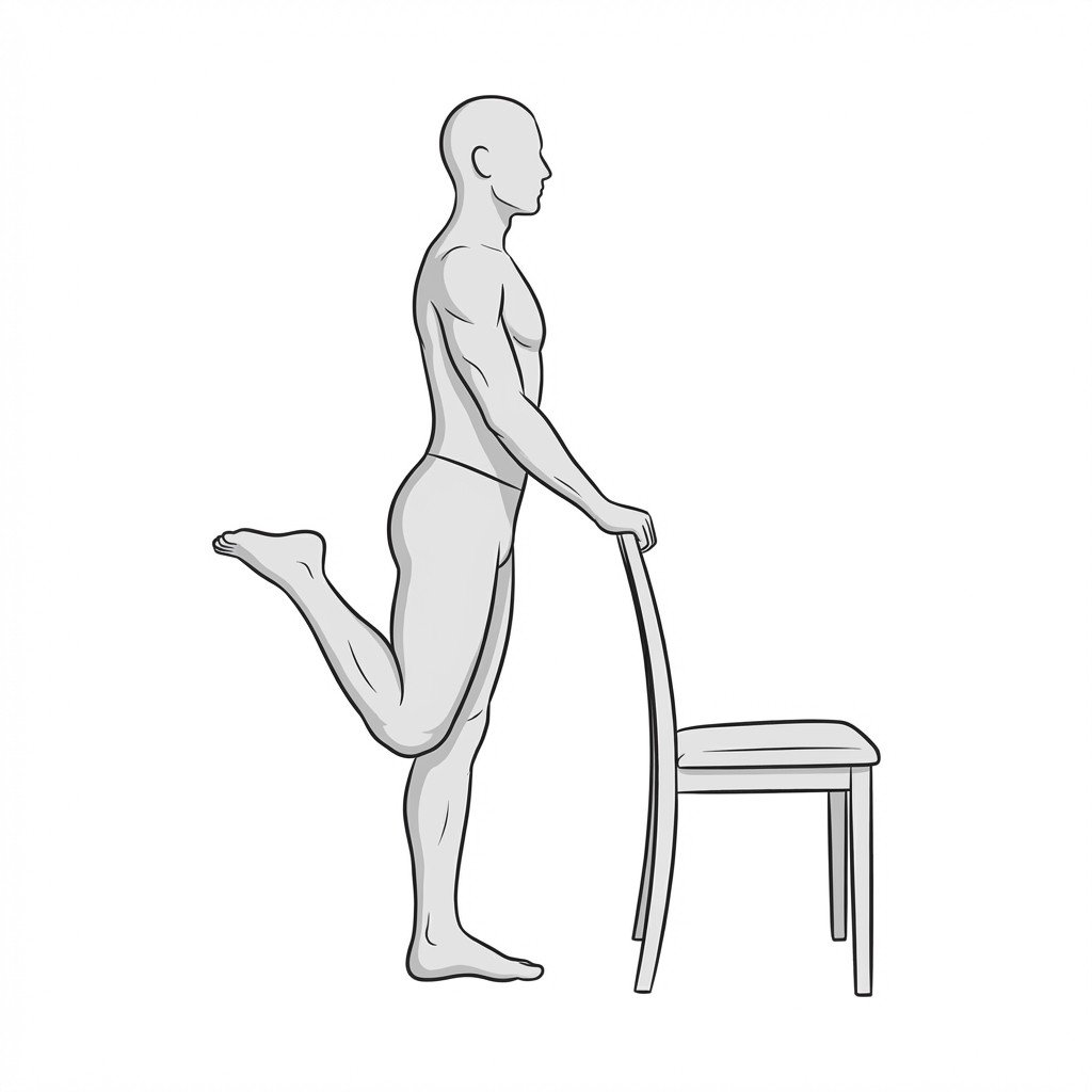
Hamstring curls
Hamstring curls actively recruit the posterior thigh musculature to promote dynamic stability and active shock absorption at the knee joint. Strengthening the hamstrings helps balance co-contraction forces, which may help reduce anterior tibial shear and load on the osteoarthritic joint.
Start Exercise →Knee
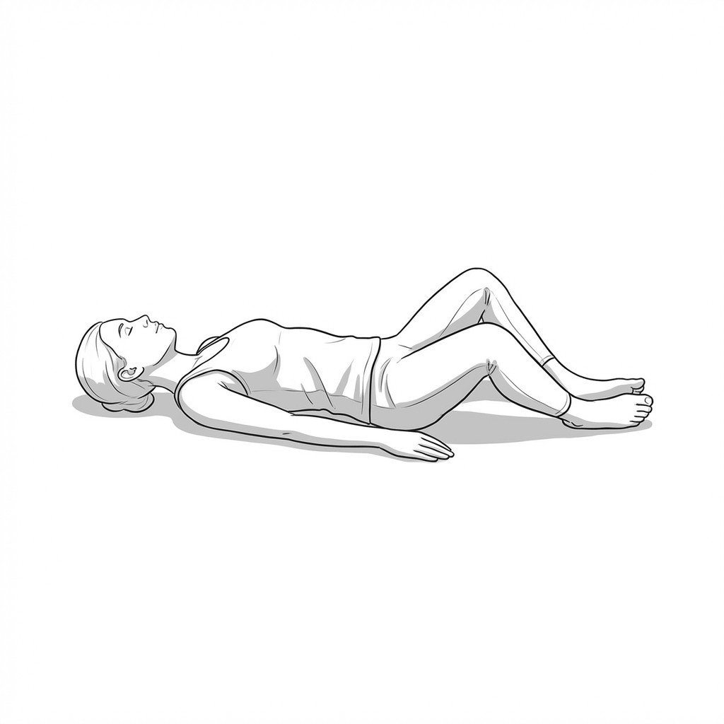
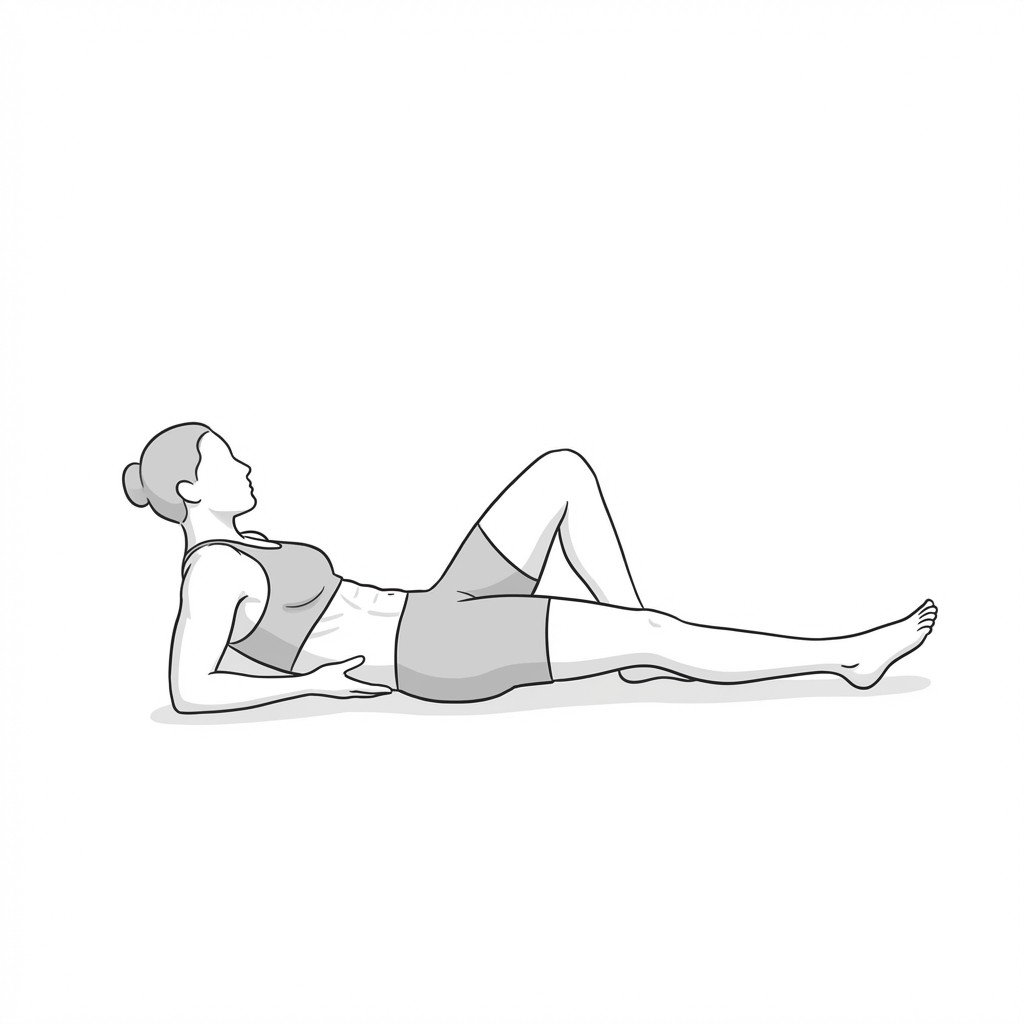
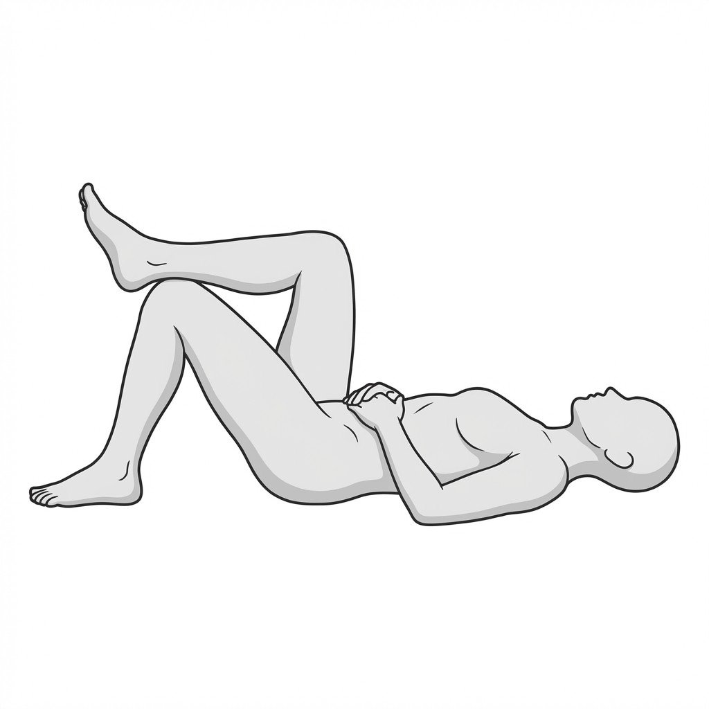
Heel slides
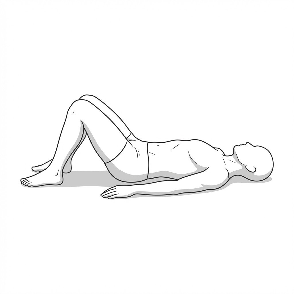
Heel slides promote early knee flexion range of motion by providing a controlled, low-friction environment for the tibiofemoral joint. When performed with proper core engagement, they challenge the deep abdominal stabilizers to control lumbopelvic alignment without generating excessive intra-abdominal pressure, making the exercise well-suited as an early diastasis recti rehabilitation exercise.
Start Exercise →Lower Leg
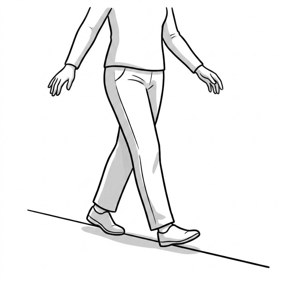
Heel-to-toe walking
Heel-to-toe walking challenges dynamic balance and proprioception by drastically reducing the base of support during the gait cycle. This forces the sensorimotor system to rapidly activate stabilizing ankle musculature, improving neuromuscular control, which may help reduce episodes of giving way in chronic ankle instability.
Start Exercise →Knee
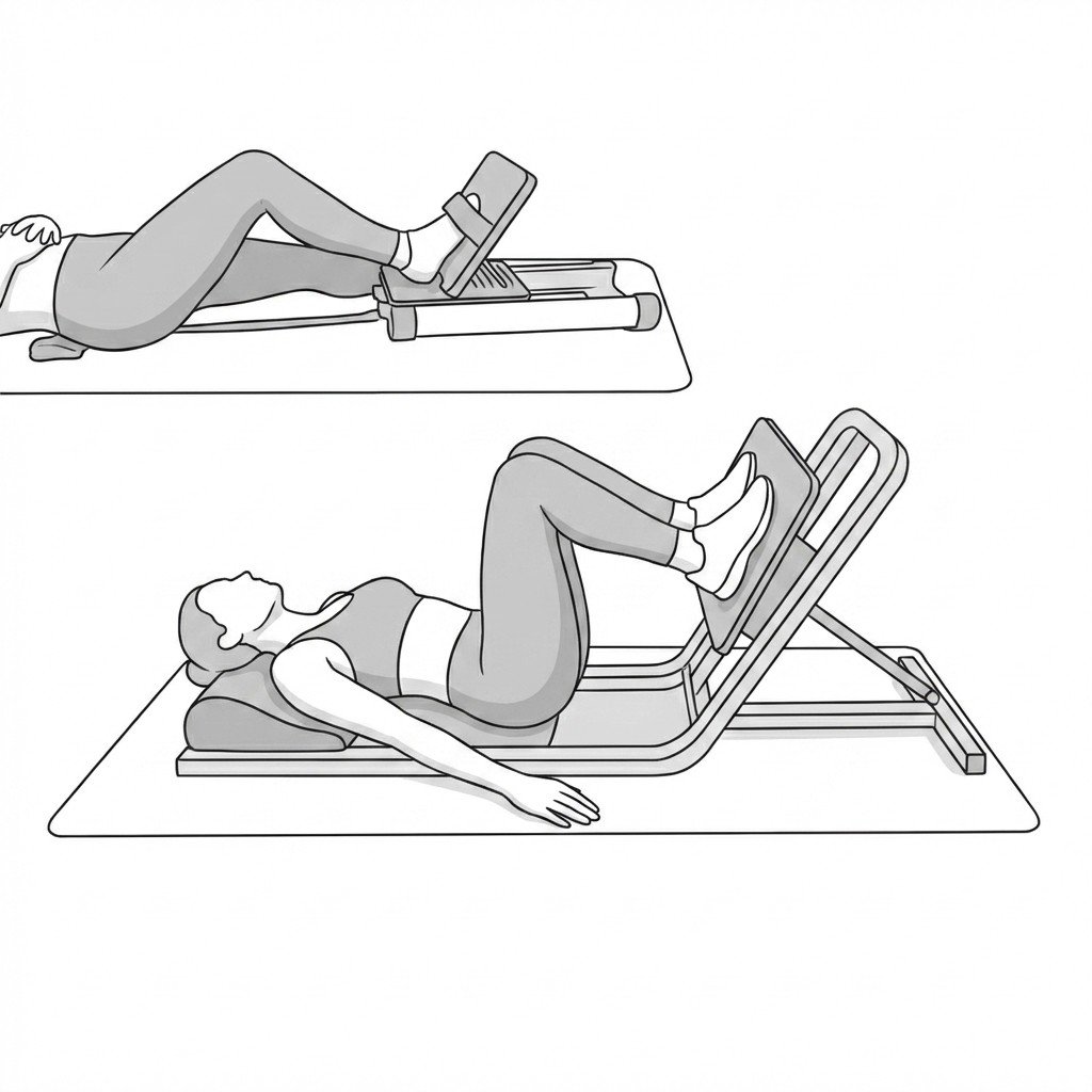
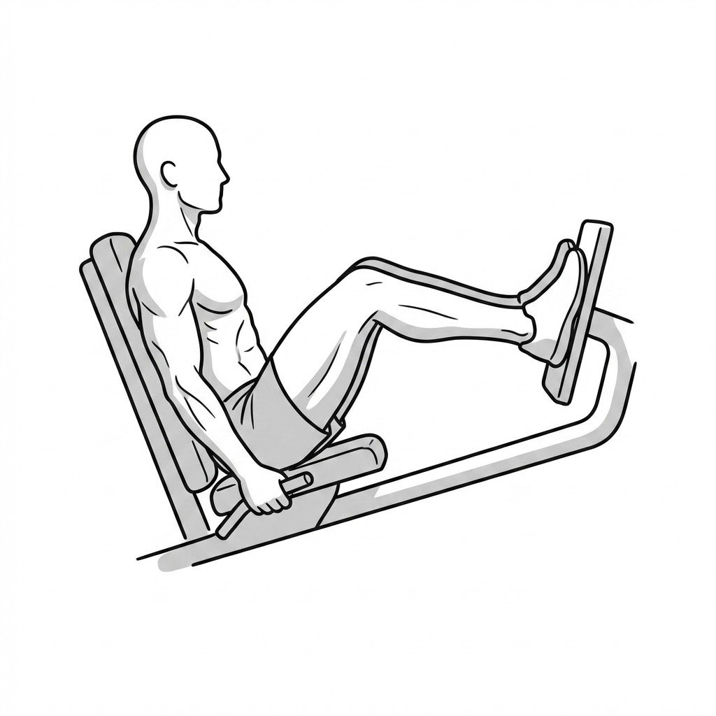
HSR leg press
The Heavy Slow Resistance (HSR) leg press induces mechanical loading on the patellar tendon, promoting collagen synthesis and structural remodeling. By utilizing slow, controlled concentric and eccentric contractions, it increases tendon time-under-tension while limiting the high strain rates that often provoke pain in reactive tendinopathies.
Start Exercise →Hip
Isometric adductor squeeze
The isometric adductor squeeze applies controlled, static tension to the inner thigh muscles without changing their length. This safely loads the adductor muscle complex to stimulate tissue remodeling and may provide short-term pain relief during early strain recovery (isometric-analgesia evidence is mixed).
Start Exercise →Cervical
Isometric cervical side-bending
Isometric cervical side-bending strengthens the lateral neck musculature without altering joint angles, providing essential stability to the cervical spine. By improving neuromuscular control and isometric endurance, it may help reduce excessive micromotion at the symptomatic segment, which is thought to help alleviate nerve root irritation associated with cervical radiculopathy.
Start Exercise →Shoulder
Isometric external rotation
Isometric external rotation recruits the posterior rotator cuff to stabilize the humeral head within the glenoid fossa without exacerbating impingement through joint motion. By producing isolated mechanical tension, it builds load capacity and may provide a short-term analgesic effect for sensitized subacromial tissues.
Start Exercise →Hip
Isometric hamstring holds
Isometric hamstring holds generate muscle tension without changing the length of the muscle, allowing for safe force generation during the early phases of tissue healing. By recruiting the muscle fibers in a static position, this exercise helps to reduce pain, maintain neuromuscular activation, and prevent muscle atrophy following a hamstring strain.
Start Exercise →Lumbar & Pelvis
Isometric hip abduction
Isometric hip abduction engages the gluteal muscles and lateral hip structures without altering joint angles, providing stabilizing compressive forces across the posterior pelvis. By recruiting these abductors, it helps produce dynamic force closure at the sacroiliac joint, which may help reduce pain and micro-instability associated with SIJ dysfunction.
Start Exercise →Lumbar & Pelvis
Isometric hip adduction
Isometric hip adduction generates tension through the adductor musculature and the anterior pelvic ring, which alters mechanical stress across the sacroiliac joint. This targeted contraction enhances force closure of the pelvis, helping to stabilize the SI joint and reduce localized pain.
Start Exercise →Hip
Isometric side-lying hip abduction
This exercise provides a low-stress, isometric load to the gluteus medius and minimus tendons without increasing compressive forces over the greater trochanter. Isometric contractions may provide short-term pain relief in irritable gluteal tendinopathy, though the analgesic evidence is mixed, while maintaining muscle capacity, making it a useful option for early-phase rehabilitation of Greater Trochanteric Pain Syndrome.
Start Exercise →Elbow & Hand
Isometric wrist extension holds
Isometric wrist extension holds involve contracting the wrist extensors against resistance without producing joint motion. This may provide a short-term analgesic effect (evidence mixed) and maintains muscular load capacity without causing the repetitive dynamic friction that exacerbates radial nerve compression.
Start Exercise →Shoulder
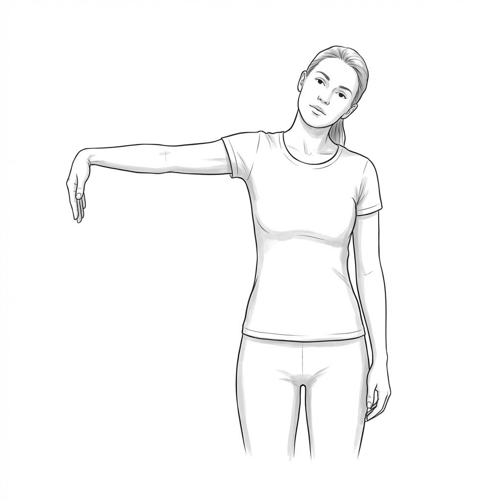
Isotonic cervical side-bending
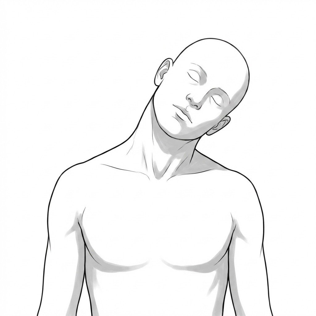
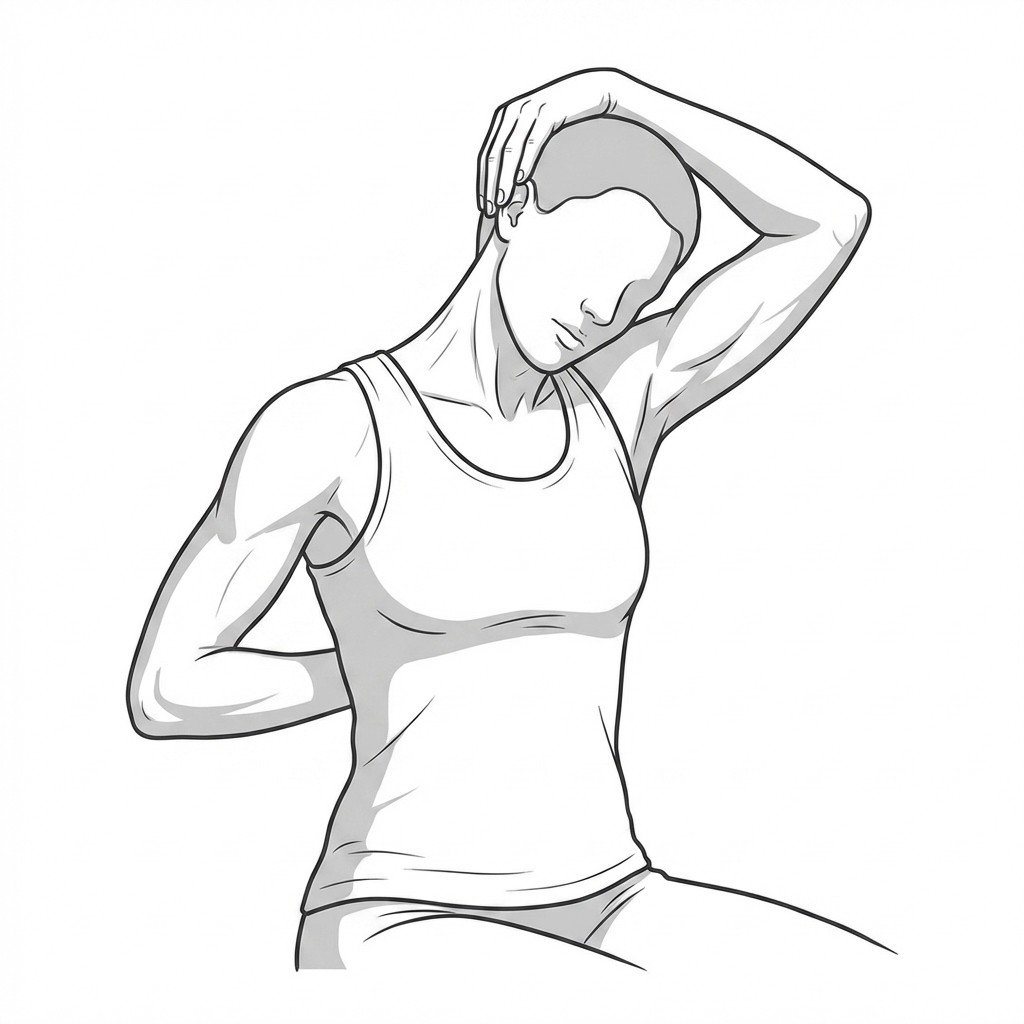
Isotonic cervical side-bending strengthens the lateral neck musculature while simultaneously providing active stretch and reciprocal inhibition to the contralateral levator scapulae. By taking the cervical spine through an active range of motion against gravity, it may help improve length-tension balance and ease protective muscle guarding. This dynamic movement helps alleviate the characteristic pain, stiffness, and restricted mobility associated with Levator Scapulae Syndrome.
Start Exercise →Shoulder
Isotonic internal rotation
Isotonic internal rotation specifically targets the subscapularis muscle to improve its force-generating capacity and endurance. By providing resistance throughout the active range of motion, it is thought to promote collagen remodeling and tendon adaptation in the anterior rotator cuff. This targeted loading is thought to address the pathological changes of tendinopathy and help improve shoulder mechanics.
Start Exercise →Lumbar & Pelvis
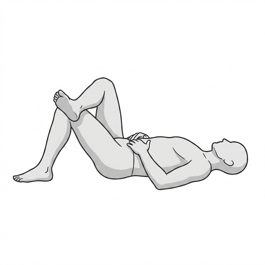
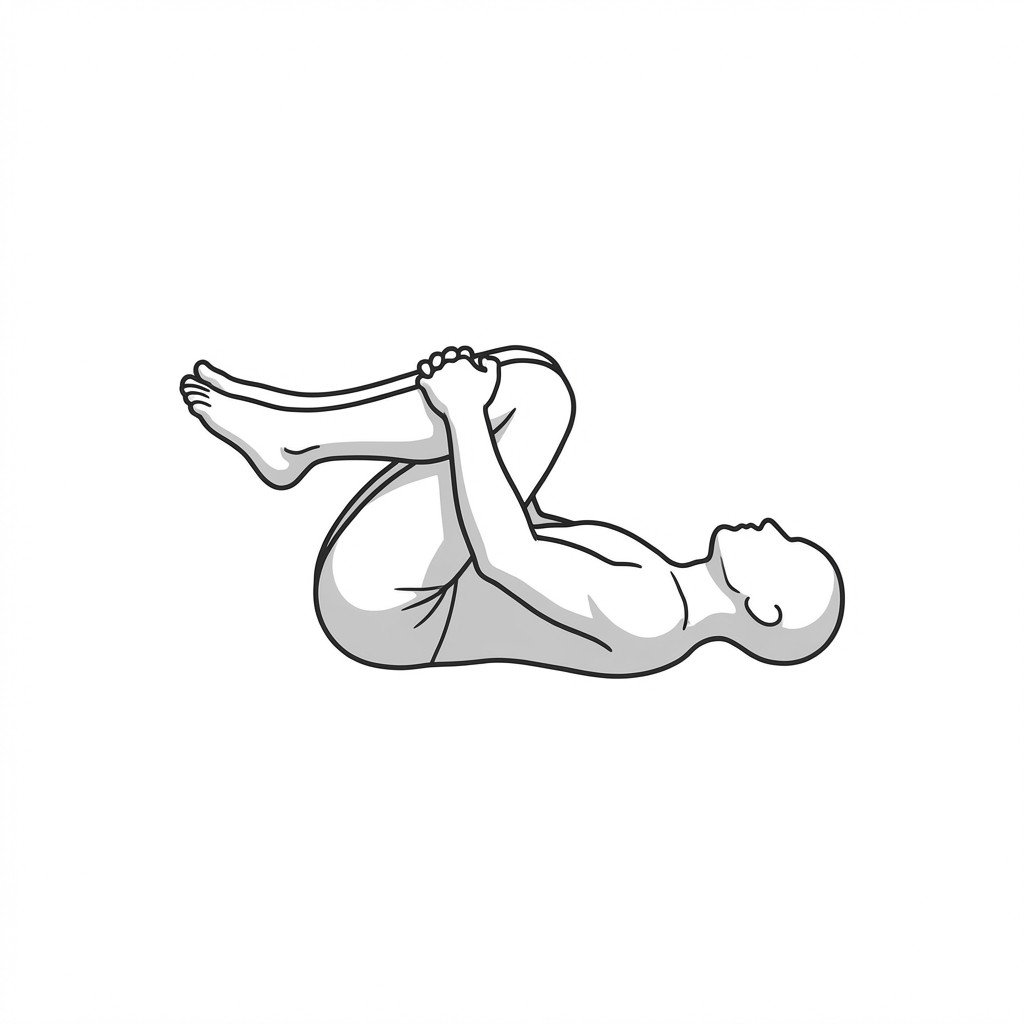
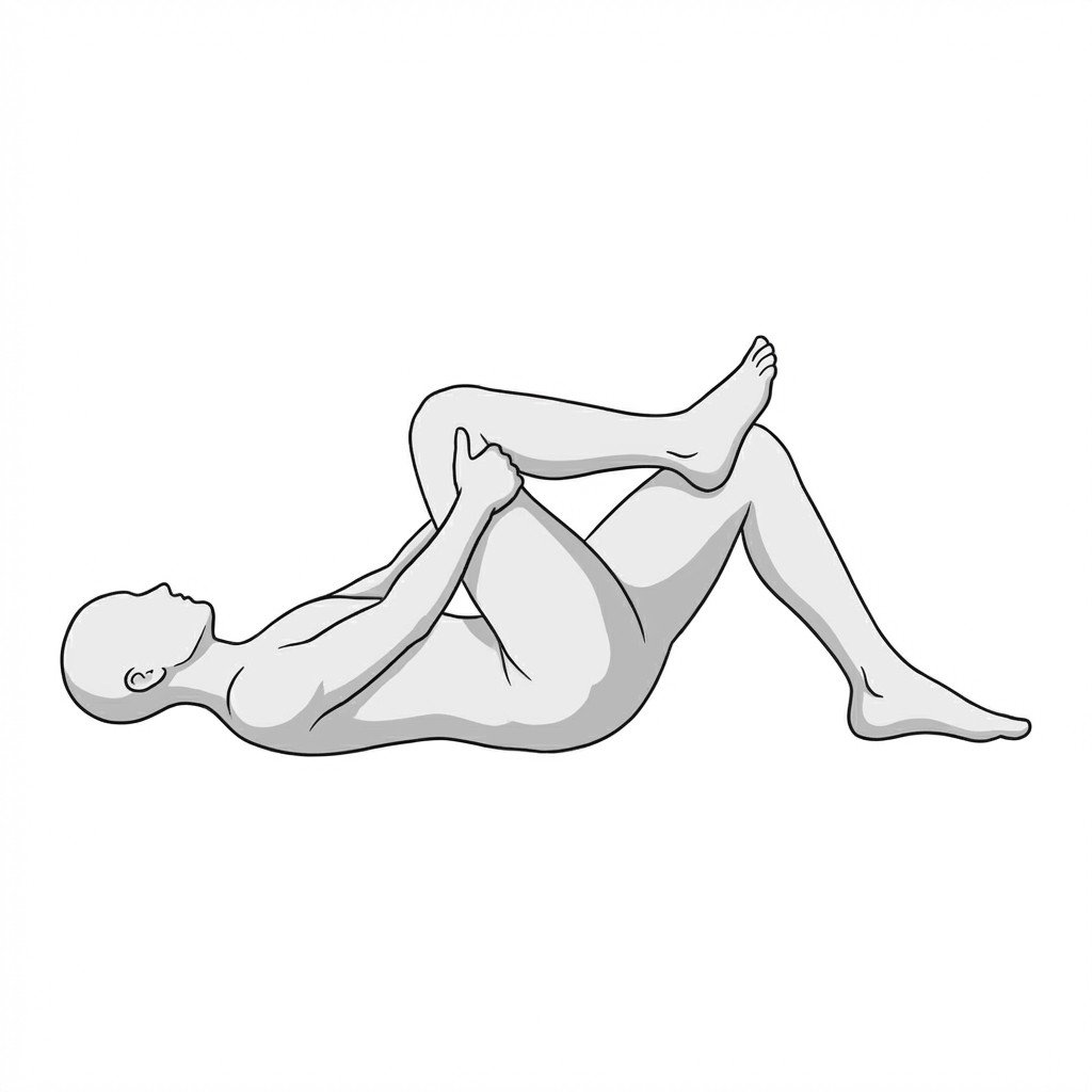
Knee-to-chest flexion series
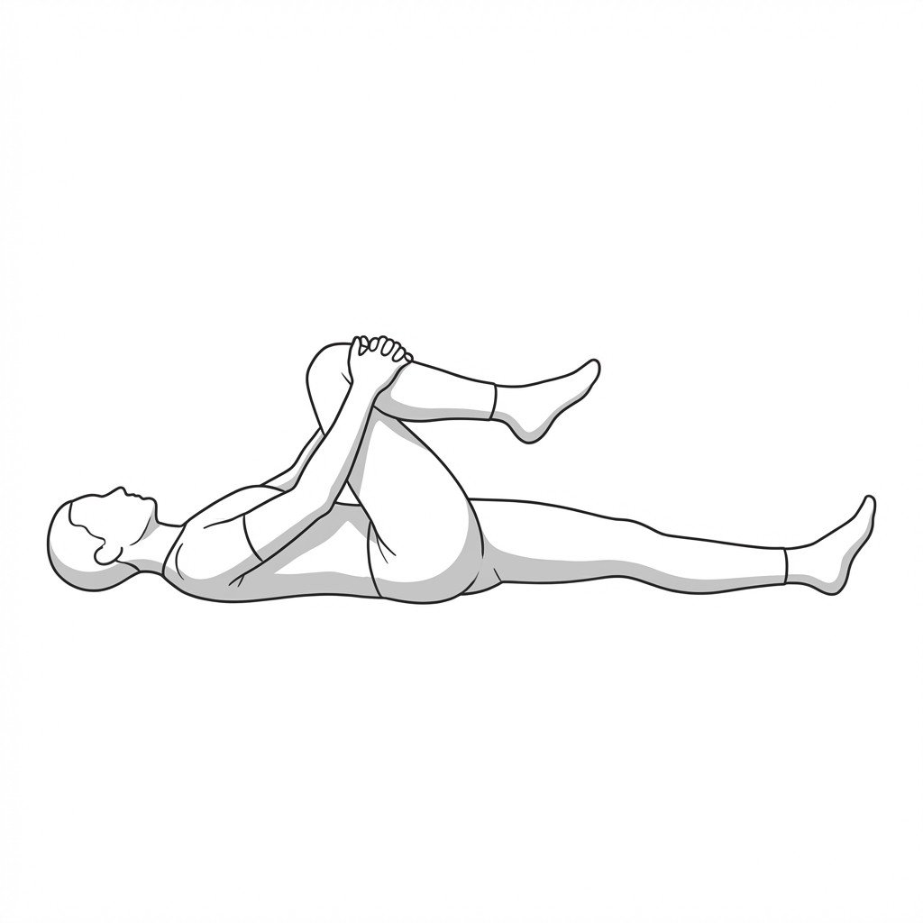
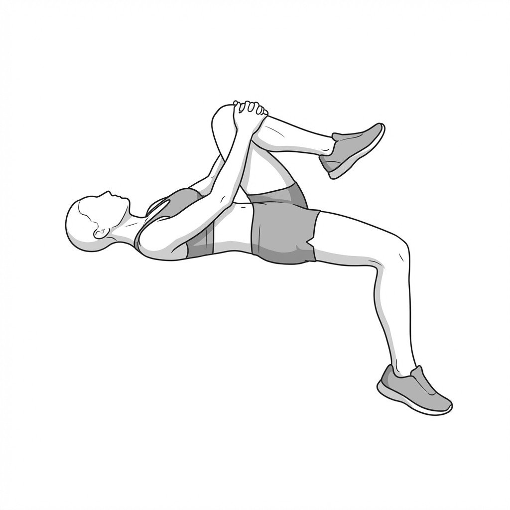
The knee-to-chest flexion series promotes lumbar spine flexion, which is thought to increase intervertebral foramen space and may reduce load on exiting spinal nerve roots. By gently stretching the lower lumbar paraspinals and gluteal musculature, it can decrease mechanical tension and may help relieve radicular symptoms in flexion-biased patients.
Start Exercise →Knee
Leg press
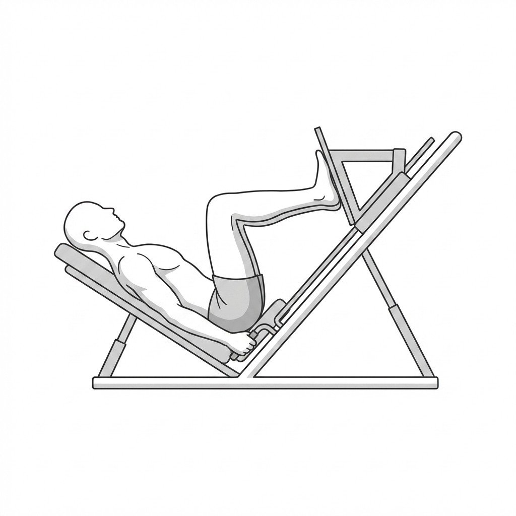
The leg press is a closed kinetic chain exercise that strengthens the quadriceps, hamstrings, and gluteal muscles while minimizing shear forces across the tibiofemoral joint. By providing controlled axial loading, it may support meniscal tissue adaptation and helps restore functional lower extremity strength required for weight-bearing activities.
Start Exercise →Cervical
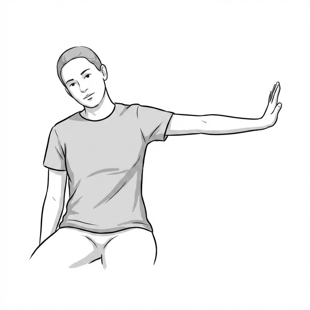
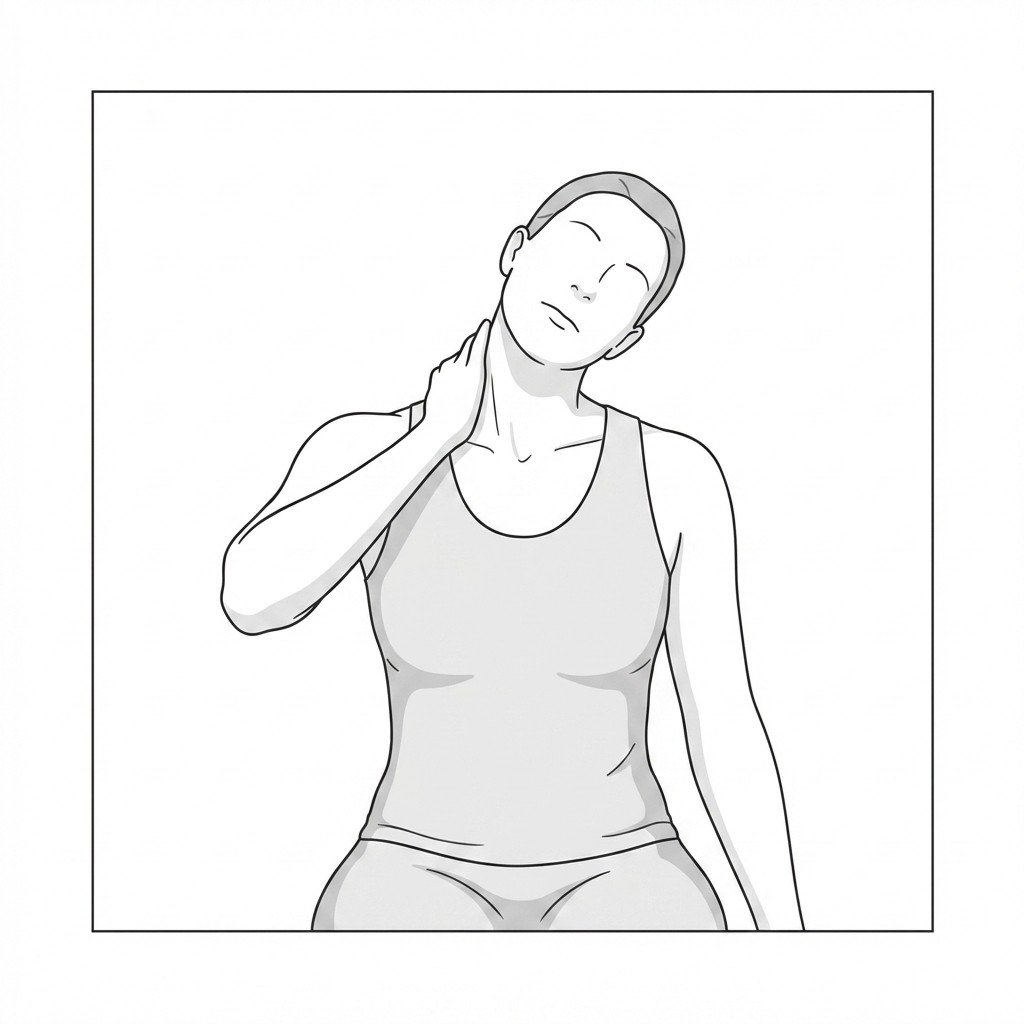
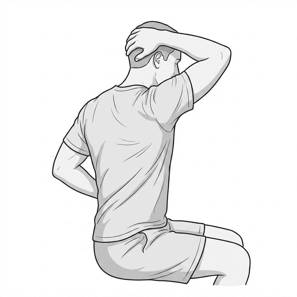
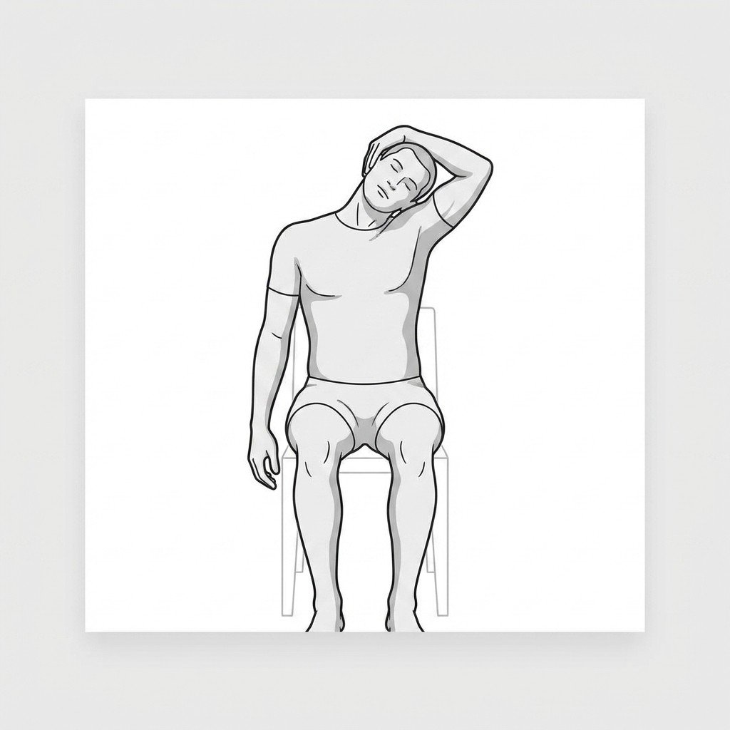
Levator scapulae stretch
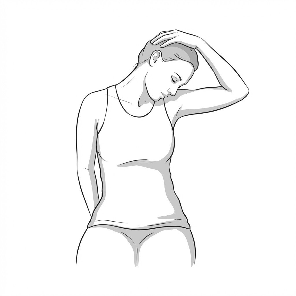
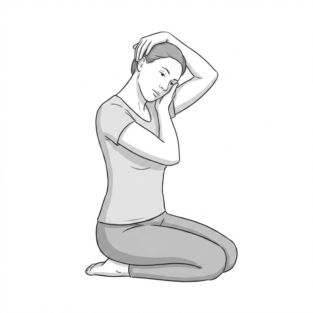
The levator scapulae stretch elongates the target muscle to reduce resting tone and alleviate tension at its attachment points on the cervical spine and superior angle of the scapula. By decreasing myofascial restriction, it may help improve cervical movement and ease the referred pain associated with cervicogenic headaches and levator scapulae syndrome.
Start Exercise →Lumbar & Pelvis
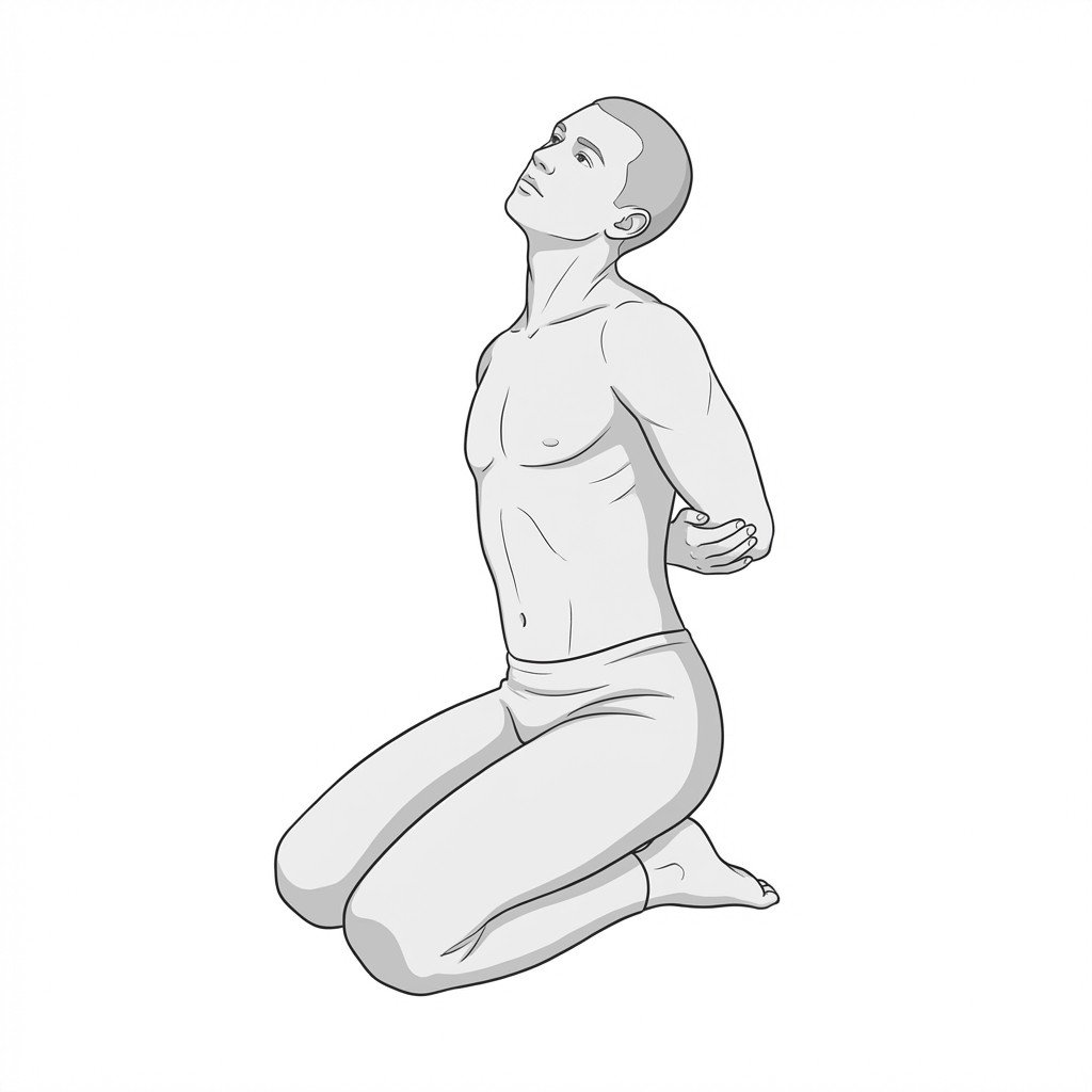
Lumbar extension progression
This exercise progression aims to promote the centralization of neural symptoms. It is thought to encourage an anterior shift of disc contents that may reduce posterior disc pressure (a proposed mechanism within the Mechanical Diagnosis and Therapy model), restore lumbar lordosis, and ease mechanical tension on the posterior annulus and surrounding pain-sensitive structures.
Start Exercise →Cervical
Mandibular resting position training
This exercise trains the proper resting posture of the jaw to unload the temporomandibular joint and reduce hypertonicity in the masticatory muscles. By maintaining a slight freeway space between the teeth, it may help reduce intra-articular pressure and parafunctional clenching.
Start Exercise →Lumbar & Pelvis
McGill curl-ups
The McGill curl-up strengthens the anterior core muscles while maintaining a neutral lumbar spine, preventing repetitive lumbar flexion. For patients with spondylolisthesis, this improves spinal stabilization and helps resist anterior shear forces on the affected vertebral segments. It effectively builds muscular endurance with minimal compressive loading on the spine.
Start Exercise →Elbow & Hand
Median nerve glides
Median nerve glides promote neurodynamic mobility by actively sliding the median nerve through its mechanical interfaces from the cervical spine down to the carpal tunnel. This intermittent excursion is thought to help reduce perineural edema, support intraneural circulation, and decrease the mechanosensitivity associated with neuropathic pain.
Start Exercise →Lower Leg
Mid-range isometric calf holds
Mid-range isometric calf holds provide sustained mechanical loading to the Achilles tendon and calf musculature without the high tensile shear forces of dynamic movement. This controlled loading induces an analgesic effect in tendinopathic tissues while stimulating collagen synthesis and improving motor unit recruitment.
Start Exercise →Knee
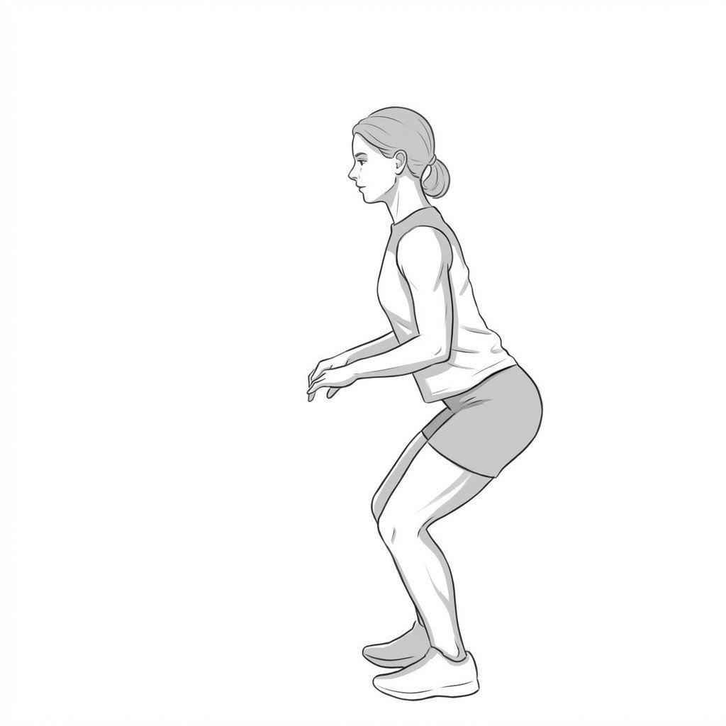
Mini-squats
Mini-squats strengthen the quadriceps and gluteal muscles through a limited, pain-free range of motion, which reduces patellofemoral compressive forces compared to deep squats. This improved muscular support helps absorb shock during gait and stabilizes the knee joint, mitigating osteoarthritis-related pain and stiffness.
Start Exercise →Thoracic & Core
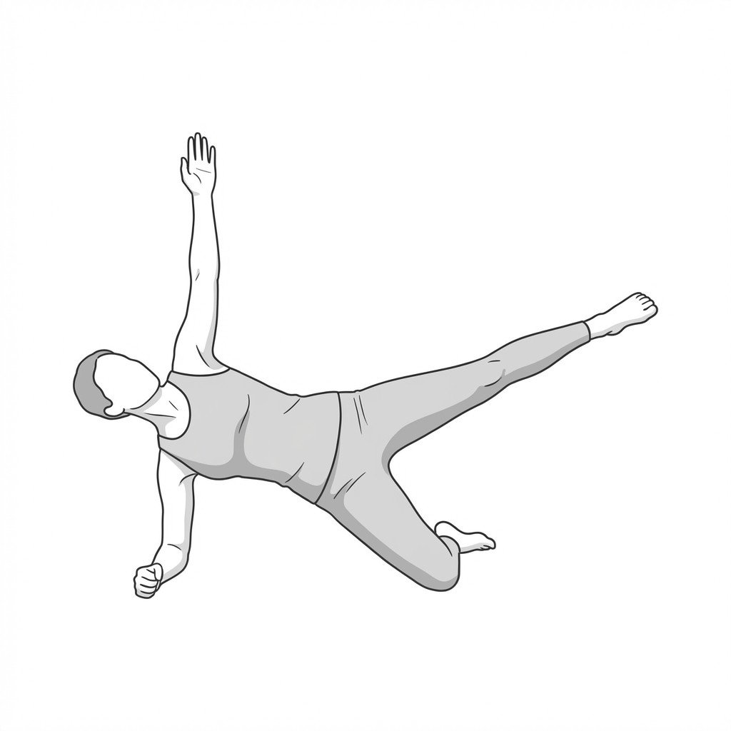
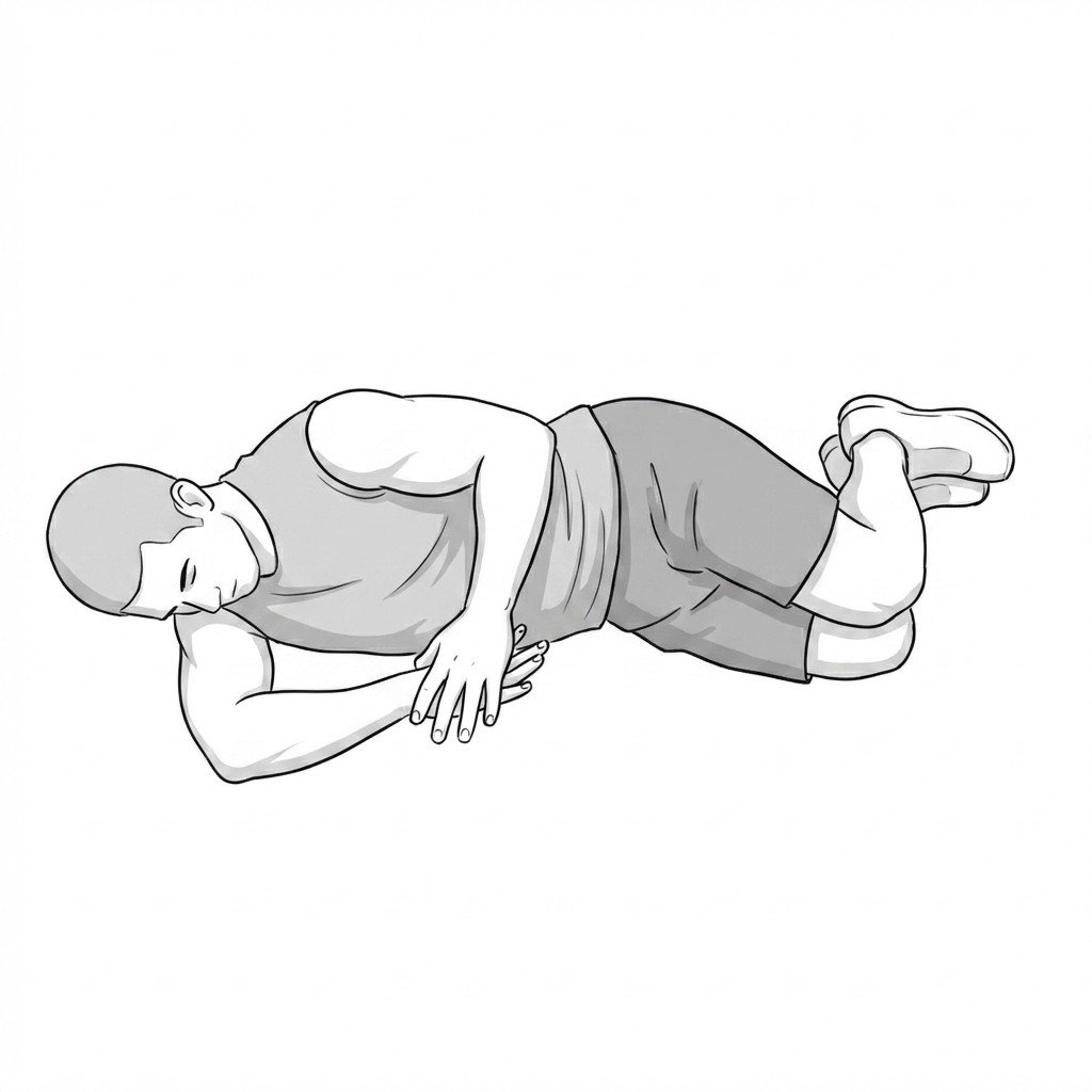
Modified side planks
The modified side plank activates the transversus abdominis and obliques while minimizing excessive intra-abdominal pressure. By placing the pivot point at the knees rather than the feet, it reduces shear forces across the linea alba. This allows for safe core stabilization to assist in narrowing the diastasis recti gap without causing midline bulging.
Start Exercise →Lumbar & Pelvis
Multifidus prone holds
Prone multifidus holds isolate and train the deep segmental stabilizers of the lumbar spine to resist anterior shear forces. By enhancing the endurance and activation of the multifidus, this exercise creates a dynamic corset that limits abnormal translation of the vertebrae. This helps reduce symptoms associated with hypermobility and mechanical instability seen in spondylolisthesis.
Start Exercise →Hip
Nordic hamstring curls
The Nordic hamstring curl is a high-load eccentric exercise that strengthens the posterior thigh muscles as they lengthen under tension. By promoting increased fascicle length and improving the muscle's ability to absorb force at longer lengths, it facilitates recovery and builds resilience against future hamstring strains.
Start Exercise →Elbow & Hand
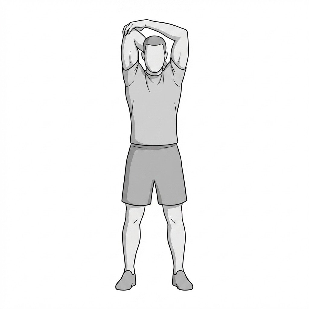
Overhead triceps stretch
This exercise elongates the triceps brachii muscle and its distal tendon by combining shoulder flexion with terminal elbow flexion. Applying a sustained, gentle tensile load is thought to support collagen organization and may reduce focal muscle hypertonicity. This improves tissue compliance and alleviates focal stress at the olecranon insertion site during functional movements.
Start Exercise →Thoracic & Core
Pallof press
The Pallof press is an anti-rotation exercise that trains the core musculature to resist dynamic rotational forces in the transverse plane. By actively engaging the trunk to prevent movement while the upper extremities move, it improves lumbopelvic stability. This helps alleviate core instability by enhancing neuromuscular control and stiffness of the spine under load.
Start Exercise →Thoracic & Core
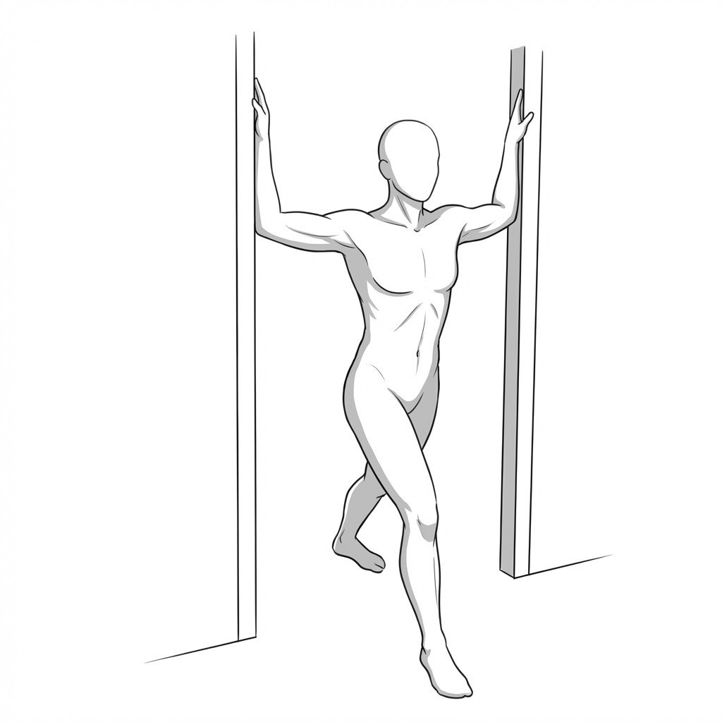
Pectoralis corner stretch
This exercise passively elongates the pectoralis major and minor muscles by utilizing the body's forward momentum against fixed anchor points. By reducing anterior chest wall tension, it helps elevate the coracoid process to decompress the neurovascular bundle in the subcoracoid space, which may help relieve symptoms associated with thoracic outlet syndrome.
Start Exercise →Shoulder
Pectoralis doorway stretch
The pectoralis doorway stretch addresses upper crossed syndrome by lengthening the tight anterior chest musculature that pulls the scapulae into protraction. By restoring normal resting length to the pectoral muscles, it reduces the chronic, eccentric strain placed on the rhomboids, alleviating interscapular pain.
Start Exercise →Cervical
Pectoralis major doorway stretch
The pectoralis major doorway stretch lengthens anterior chest wall musculature, counteracting the forward-shoulder postural faults commonly associated with axial neck pain. By reducing tension in the pectoral region, it facilitates better scapular kinematics and unloads overstressed cervical and periscapular muscles.
Start Exercise →Knee
Pelvic drops
Pelvic drops target the eccentric and concentric control of the hip abductors, particularly the gluteus medius, on the weight-bearing leg. Strengthening these muscles stabilizes the pelvis during the stance phase of gait, preventing excessive hip adduction and internal rotation that increases tension on the iliotibial (IT) band.
Start Exercise →Lumbar & Pelvis
Pelvic hikes
Pelvic hikes activate and mobilize the quadratus lumborum and gluteus medius by isolating pelvic movement in the frontal plane. This controlled concentric and eccentric loading helps break the pain-spasm cycle associated with myofascial pain syndrome while improving overall lumbopelvic stabilization.
Start Exercise →Lumbar & Pelvis
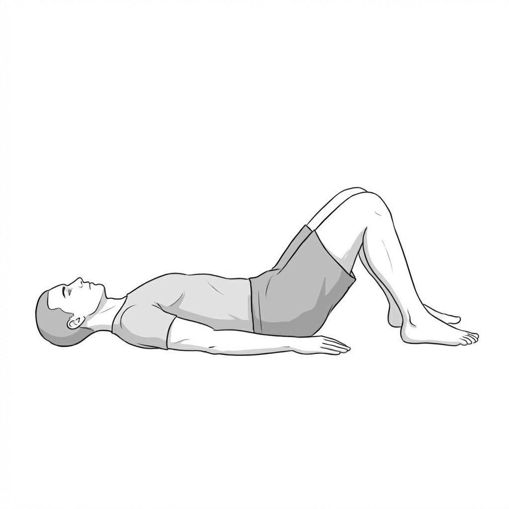
Pelvic tilts
Posterior pelvic tilts reduce lumbar lordosis by rotating the pelvis backward, which is proposed to counteract the anterior shear forces associated with spondylolisthesis. This movement may also increase intervertebral foramen space and reduce load on irritated nerve roots — a proposed mechanism — while engaging the deep core musculature to help stabilize the lower spine.
Start Exercise →Shoulder
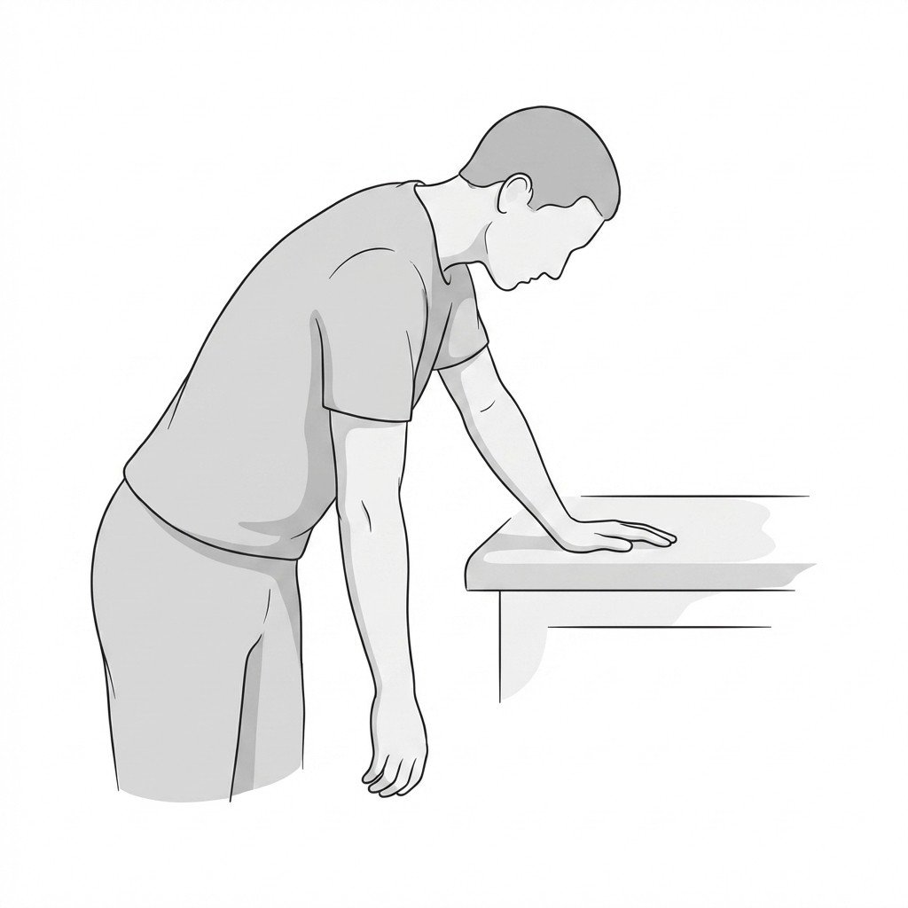
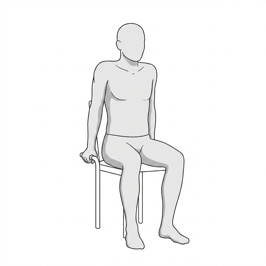
Pendulum swings
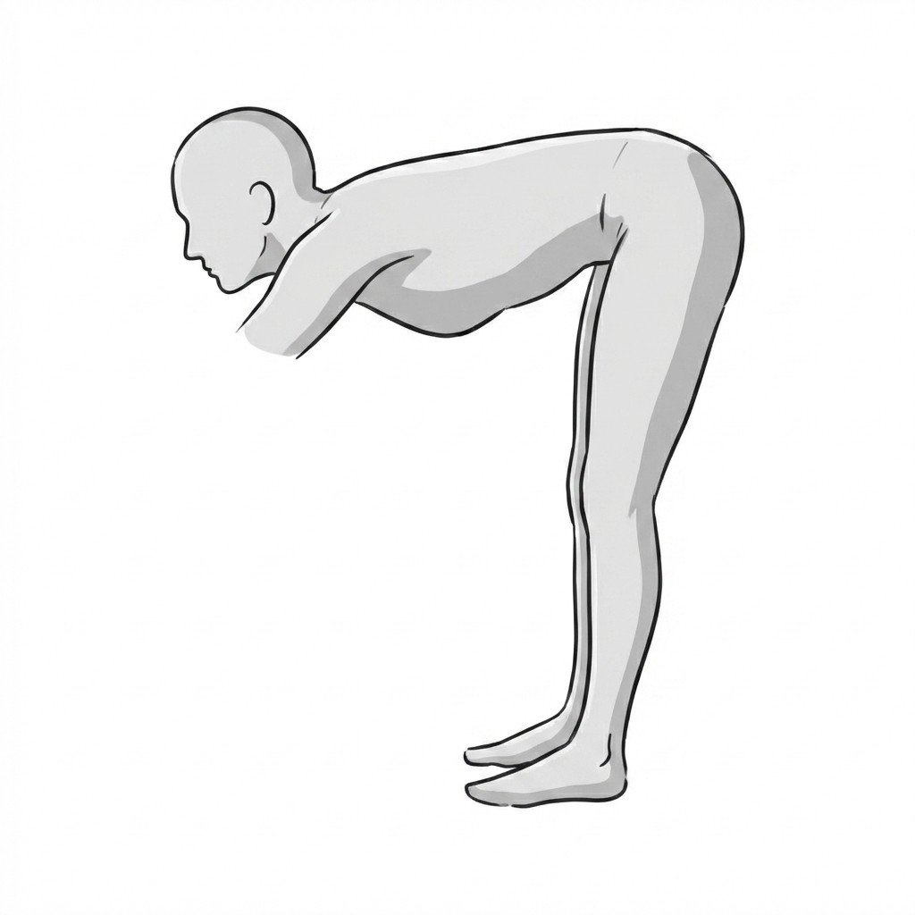
Pendulum swings utilize gravity and torso momentum to create gentle distraction and passive mobilization of the glenohumeral joint. By creating a micro-separation of the joint surfaces without active muscle contraction, this exercise may help reduce pain and maintain joint mobility during the painful stages of adhesive capsulitis.
Start Exercise →Hip
Piriformis stretch
The piriformis stretch elongates the piriformis muscle by combining hip flexion with external rotation to maximize tissue tension. This may reduce compressive forces on the underlying sciatic nerve and help ease the deep gluteal pain and related symptoms sometimes attributed to piriformis syndrome.
Start Exercise →Thoracic & Core
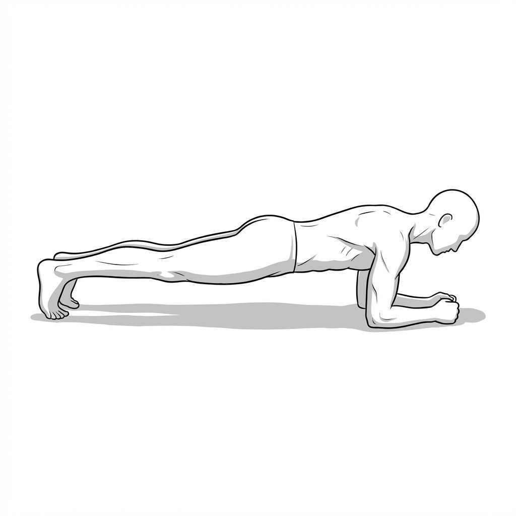
Planks
The plank is an isometric core exercise that promotes global trunk stability by demanding co-contraction of the abdominal wall and spinal stabilizers. It enhances the endurance of the lumbo-pelvic cylinder, effectively limiting excessive spinal motion and shear forces during functional tasks.
Start Exercise →Lumbar & Pelvis
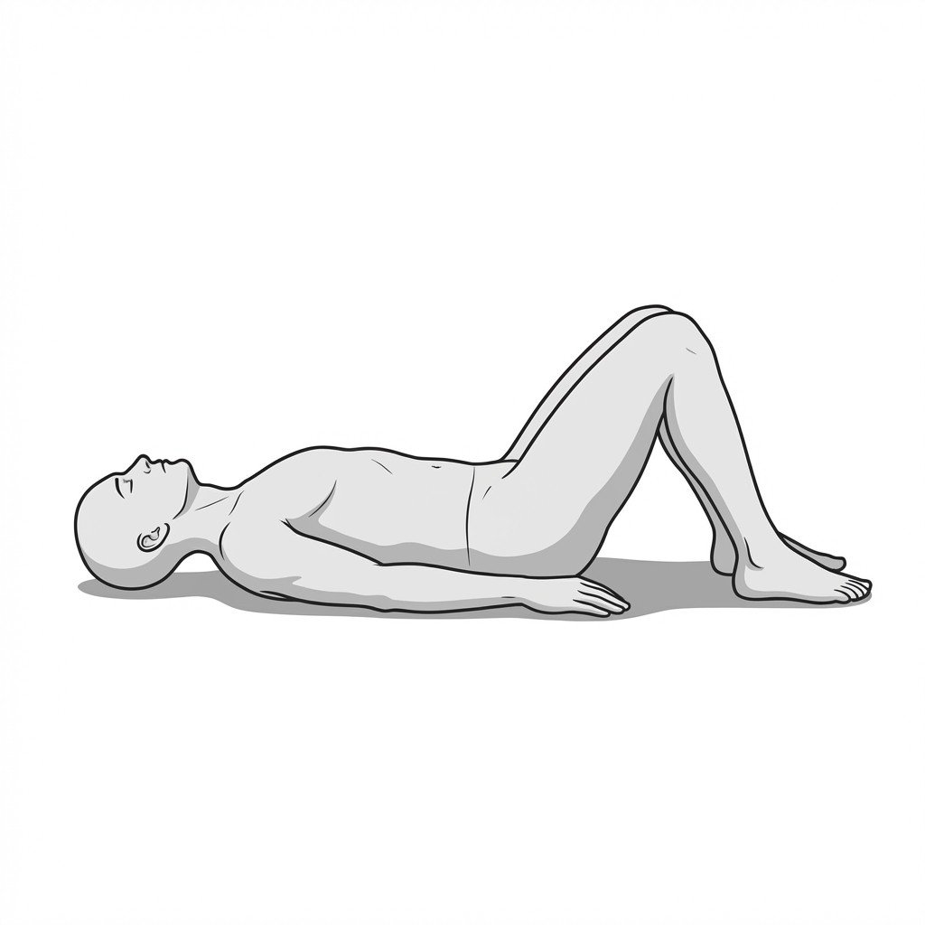
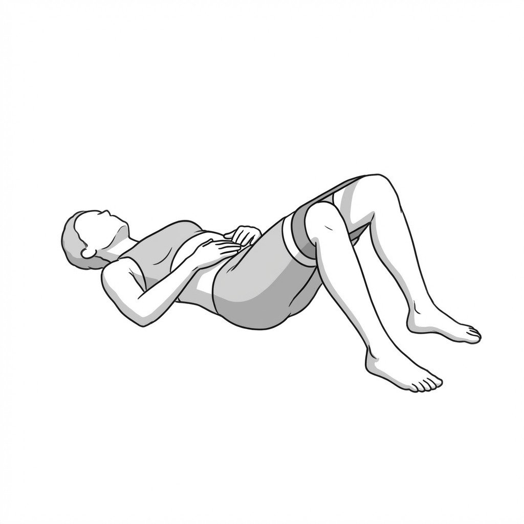
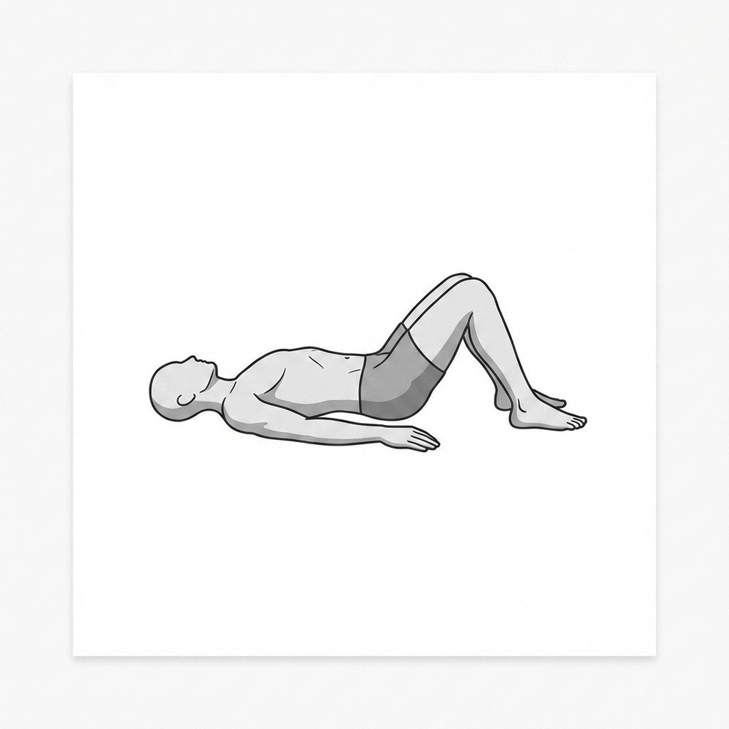
Posterior pelvic tilts
The posterior pelvic tilt engages the deep core stabilizers to rotate the pelvis backward, flattening the lumbar spine. Mechanistically, this reduces lumbar extension to decompress spinal nerve roots in radiculopathy, while simultaneously activating the transversus abdominis to gently engage the deep abdominal wall as part of diastasis recti rehabilitation.
Start Exercise →Elbow & Hand
Prayer stretch
The prayer stretch creates extension at the wrists with the elbows flexed, gently elongating the wrist and finger flexors. This stretching reduces soft tissue tension in the anterior forearm, potentially decreasing pressure within the carpal tunnel and alleviating mechanical compression on the median nerve.
Start Exercise →Elbow & Hand
Pronator strengthening
This exercise applies targeted progressive loading to the pronator teres and common flexor tendon to stimulate collagen synthesis and tissue remodeling. By increasing the load-bearing capacity of the medial epicondyle attachments, it may help reduce pain and improve functional grip strength.
Start Exercise →Elbow & Hand
Pronator teres stretch
This exercise elongates the pronator teres by placing the forearm into maximal supination with the elbow extended. By reducing tightness in this antagonist muscle, it decreases the compensatory over-activation and hypertonicity of the supinator muscle during functional tasks. Reducing supinator tension ultimately helps relieve mechanical compression on the radial nerve, alleviating symptoms of Radial Tunnel Syndrome.
Start Exercise →Thoracic & Core
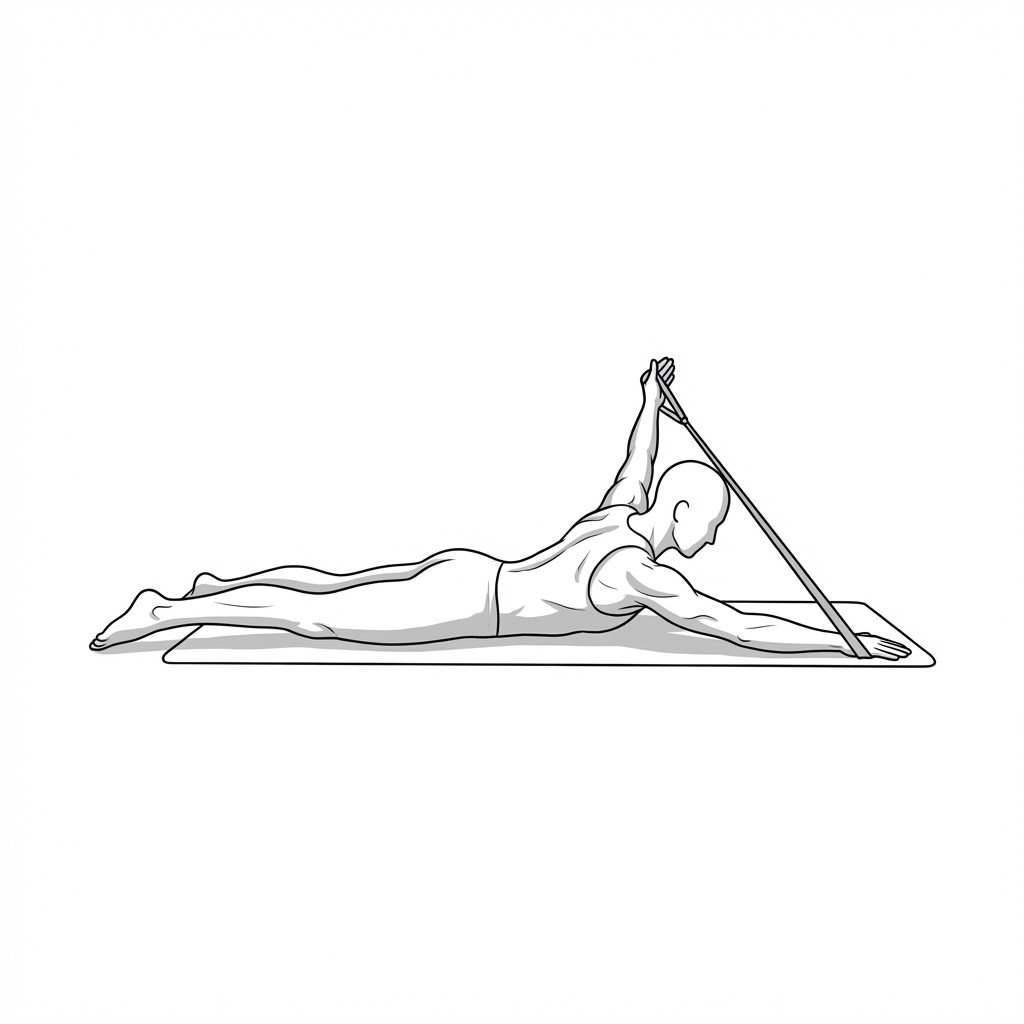
Prone cobra
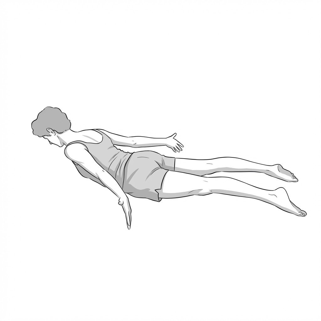
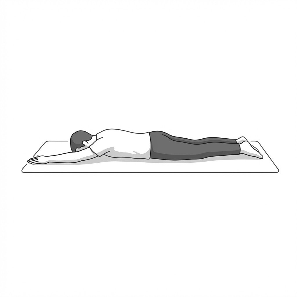
The prone cobra targets the deep spinal extensors and scapular retractors to help counteract the increased thoracic curvature associated with kyphosis. By lifting the upper trunk and externally rotating the shoulders, it is thought to support postural correction and may reduce anterior capsular tightness.
Start Exercise →Knee
Prone hamstring curls
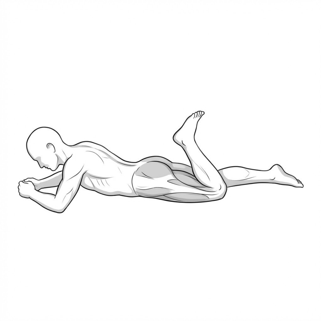
Prone hamstring curls isolate the posterior thigh musculature to improve knee flexion strength and dynamic stability of the tibiofemoral joint. In meniscal rehab, strengthening the hamstrings is thought to reduce anterior shear forces on the tibia and help unload the joint during weight-bearing activities, which may limit stress on the healing meniscus.
Start Exercise →Cervical
Prone I-raises
Prone I-raises target the lower trapezius and erector spinae to promote inferior scapular glide and thoracic extension. By strengthening these postural stabilizers, the exercise counteracts the abnormal forward head posturing and upper cervical hypertonicity commonly seen in cervical dystonia. This mechanical offloading helps reduce the compensatory workload on overactive neck muscles.
Start Exercise →Shoulder
Prone lower trapezius Y-raises
Prone lower trapezius Y-raises specifically target the lower trapezius muscle to improve scapular upward rotation and posterior tilt. By strengthening this key scapular stabilizer, the exercise helps restore optimal scapulohumeral rhythm and mitigates the compensatory overactivity commonly seen in the upper trapezius.
Start Exercise →Shoulder
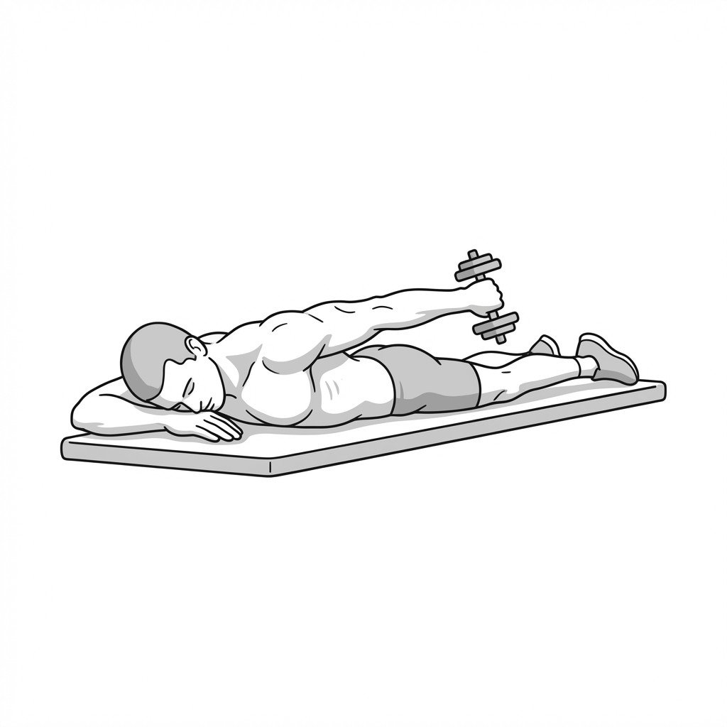
Prone shoulder extension
Prone shoulder extension actively engages the posterior shoulder musculature while placing a controlled, dynamic stretch on the anterior shoulder structures, including the long head of the biceps. By moving through this range of motion, it facilitates tendon gliding and applies mild eccentric and isometric loading to the biceps tendon, which is thought to support collagen remodeling and may improve load tolerance.
Start Exercise →Shoulder
Prone T-raises
Prone T-raises actively target the middle trapezius and rhomboids by requiring against-gravity scapular retraction. This helps alleviate interscapular pain by strengthening the posterior shoulder girdle musculature, improving postural endurance, and reducing the mechanical strain associated with protracted shoulder positions.
Start Exercise →Shoulder
Prone Y-raises
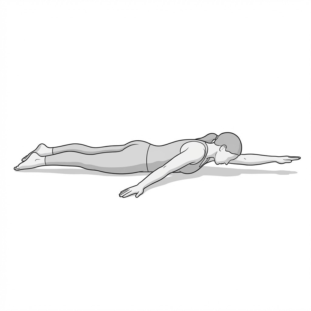
Prone Y-raises primarily target the lower trapezius to improve scapular upward rotation and posterior tilt. By strengthening the lower trapezius, this exercise may help improve the force-couple balance with the upper trapezius, reducing compensatory overactivity and associated myofascial pain.
Start Exercise →Shoulder
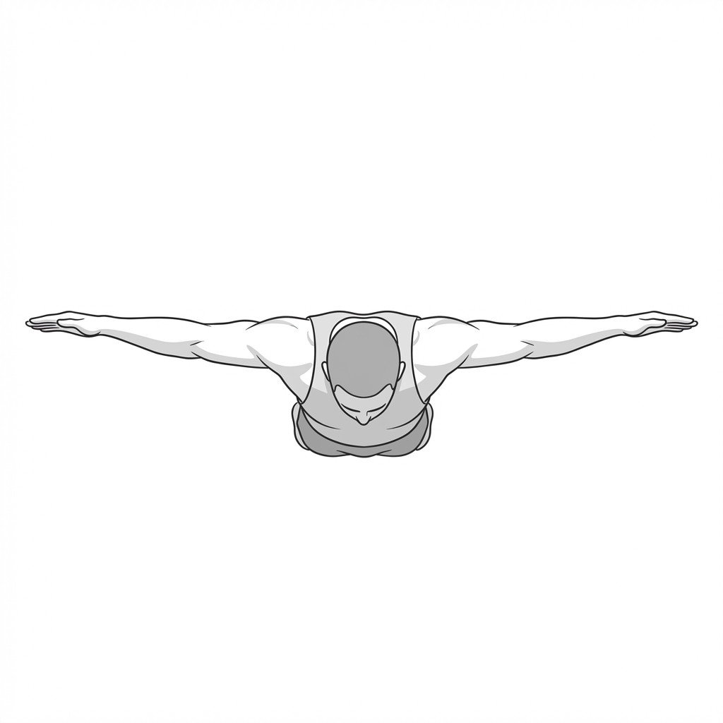
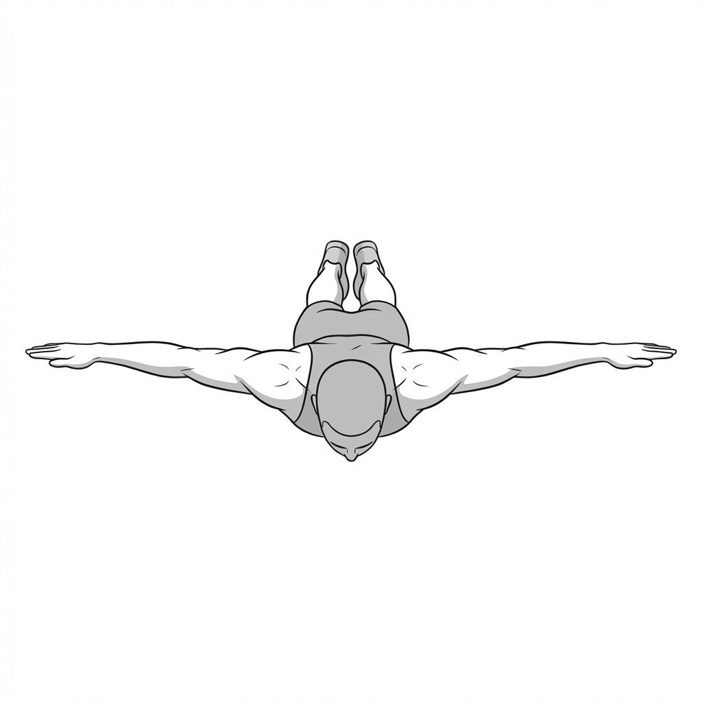
Prone Y-T-W raises
Prone Y-T-W raises comprehensively target the periscapular and rotator cuff musculature to improve scapulothoracic rhythm and dynamic stabilization. By strengthening the lower trapezius, middle trapezius, and posterior rotator cuff, this exercise may help reduce anterior scapular tipping and support subacromial space during upper extremity elevation, thereby reducing impingement symptoms.
Start Exercise →Shoulder
Pulley-assisted elevation
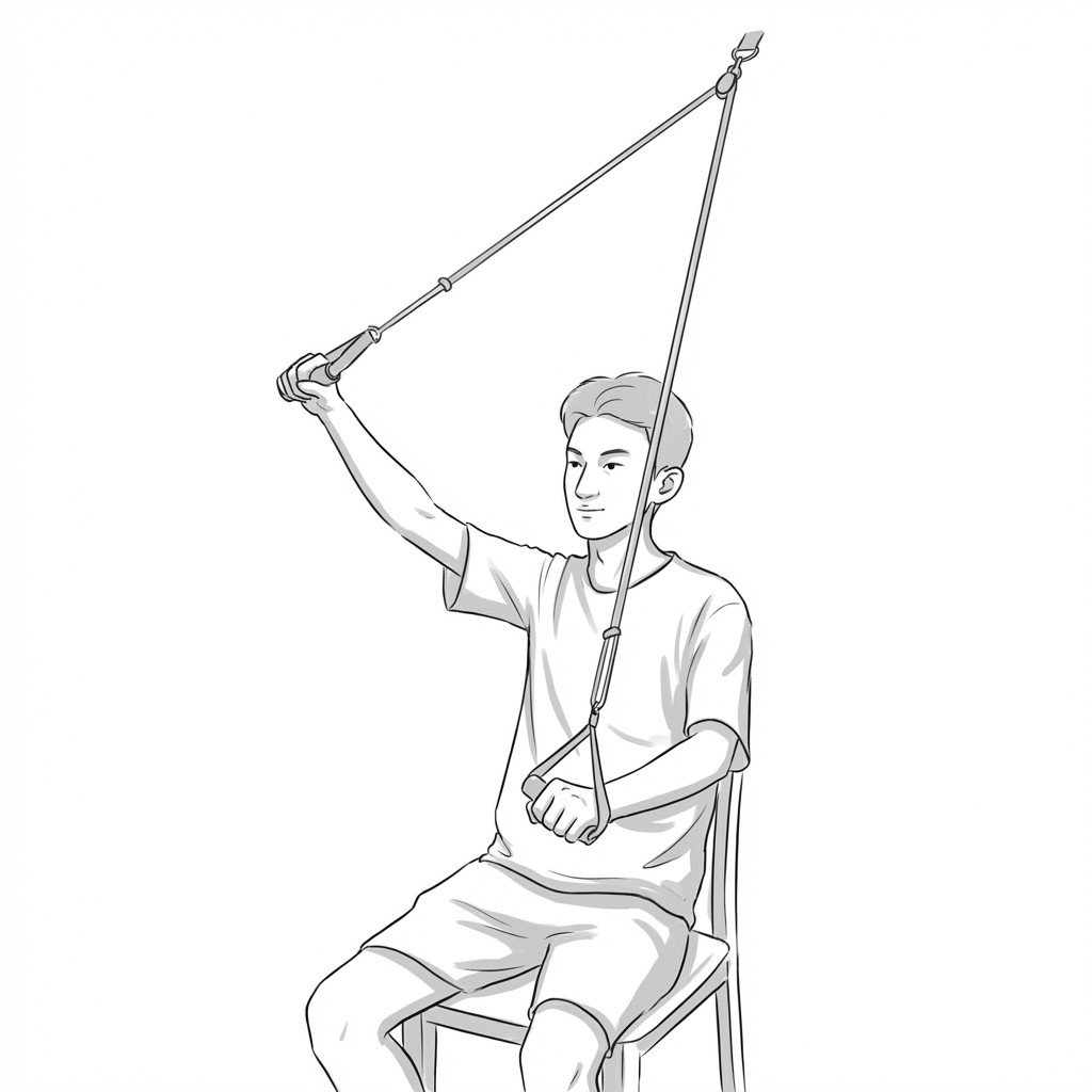
Pulley-assisted elevation is an active-assisted range of motion exercise that utilizes the unaffected arm to facilitate movement of the restricted shoulder. Mechanistically, it provides controlled stretching to the fibrotic glenohumeral joint capsule, helping to progressively restore functional overhead reach and reduce capsular contracture.
Start Exercise →Shoulder
Push-up plus
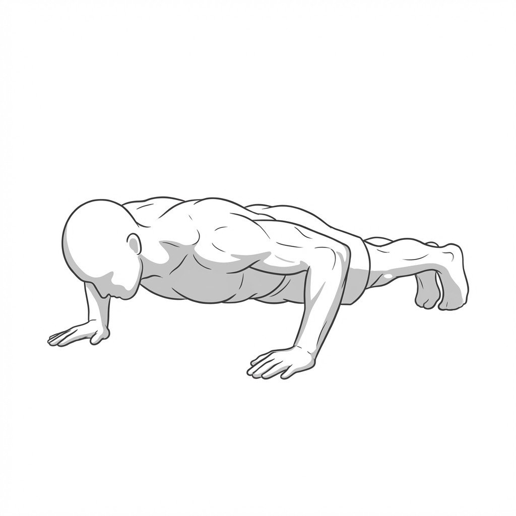
The push-up plus maximizes the activation of the serratus anterior through terminal scapular protraction at the end of a standard push-up. Strengthening this muscle improves scapulothoracic kinematics and helps correct the medial border prominence often seen in scapular dyskinesis.
Start Exercise →Elbow & Hand
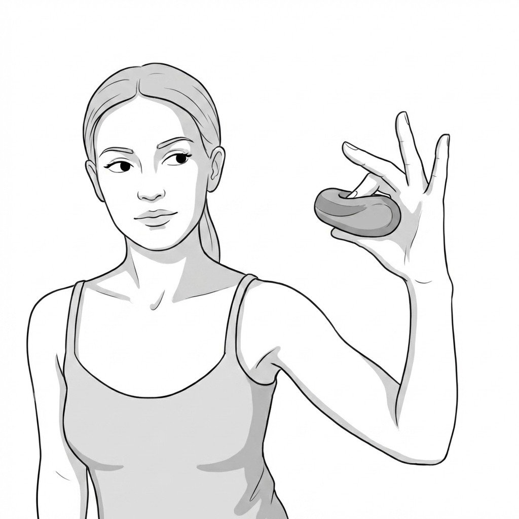
Putty grip strengthening
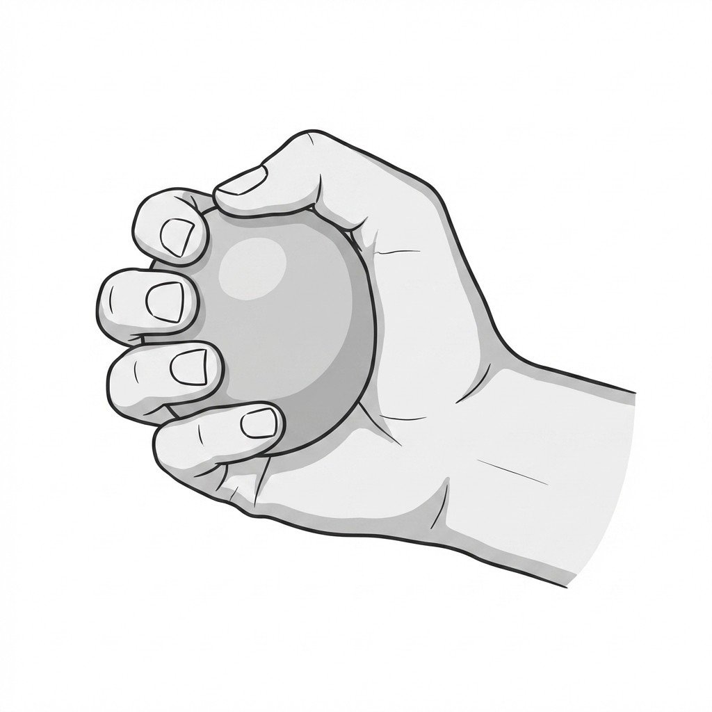
Putty grip strengthening provides resistance to the hand and forearm musculature, heavily relying on the synergistic co-contraction of the wrist extensors to stabilize the wrist during finger flexion. It helps manage lateral epicondylalgia by safely and progressively loading the common extensor tendon, improving load tolerance and supporting tissue adaptation.
Start Exercise →Lumbar & Pelvis
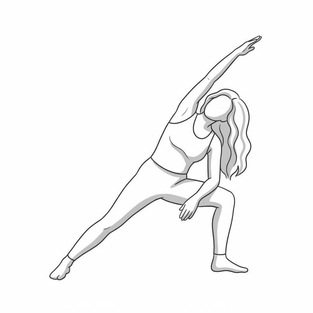
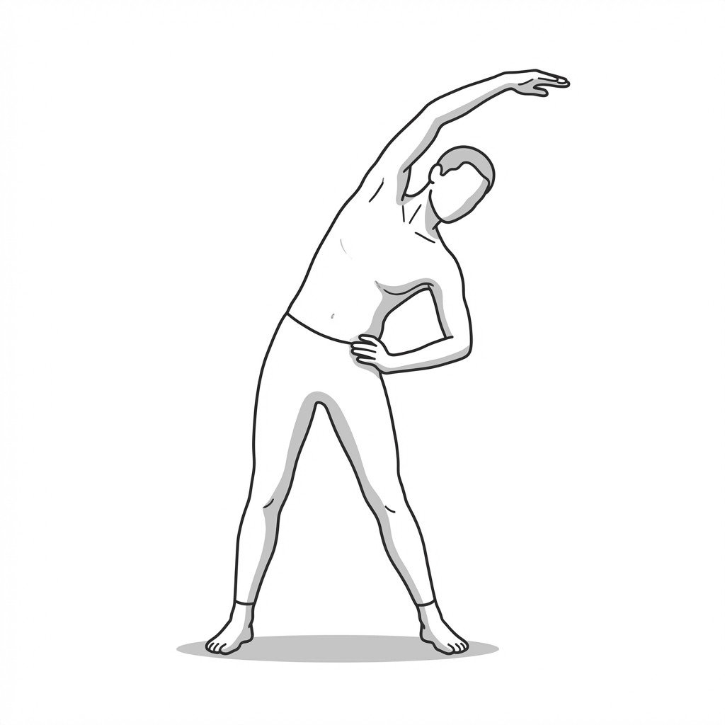
QL lateral-flexion stretch
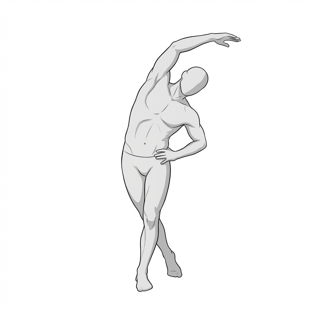
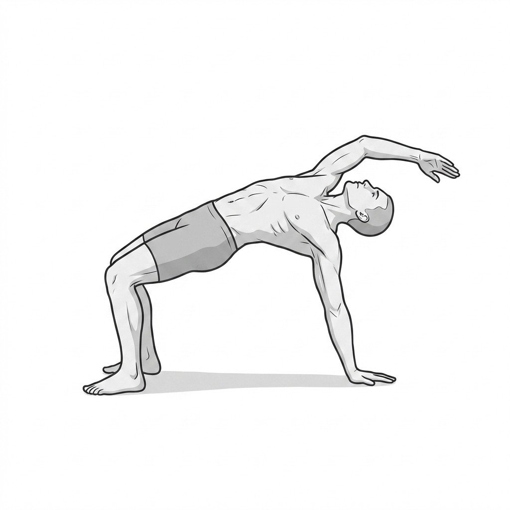
This exercise applies a sustained tensile load to the lateral core and spine stabilizers, which may help reduce muscle spindle overactivity. By mechanically lengthening the myofascial tissue, it helps alleviate ischemic pain and deactivate trigger points associated with Quadratus Lumborum Myofascial Pain Syndrome.
Start Exercise →Knee
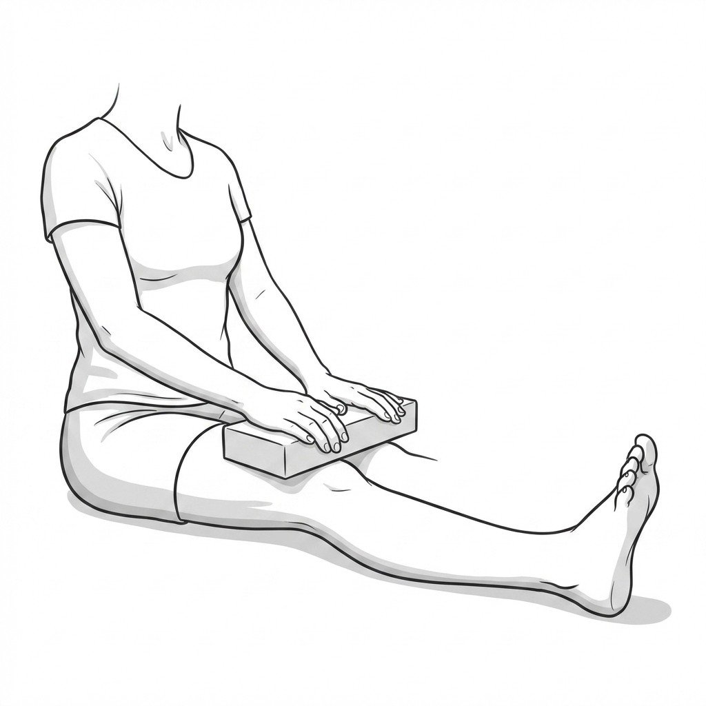
Quad sets
Quad sets are isometric contractions of the quadriceps muscles that improve neuromuscular recruitment and maintain muscle tone without loading the joint dynamically. In meniscal rehabilitation, this minimizes shear forces on the healing meniscus while preventing arthrogenic quadriceps atrophy, a common post-injury complication.
Start Exercise →Knee
Quadriceps stretch
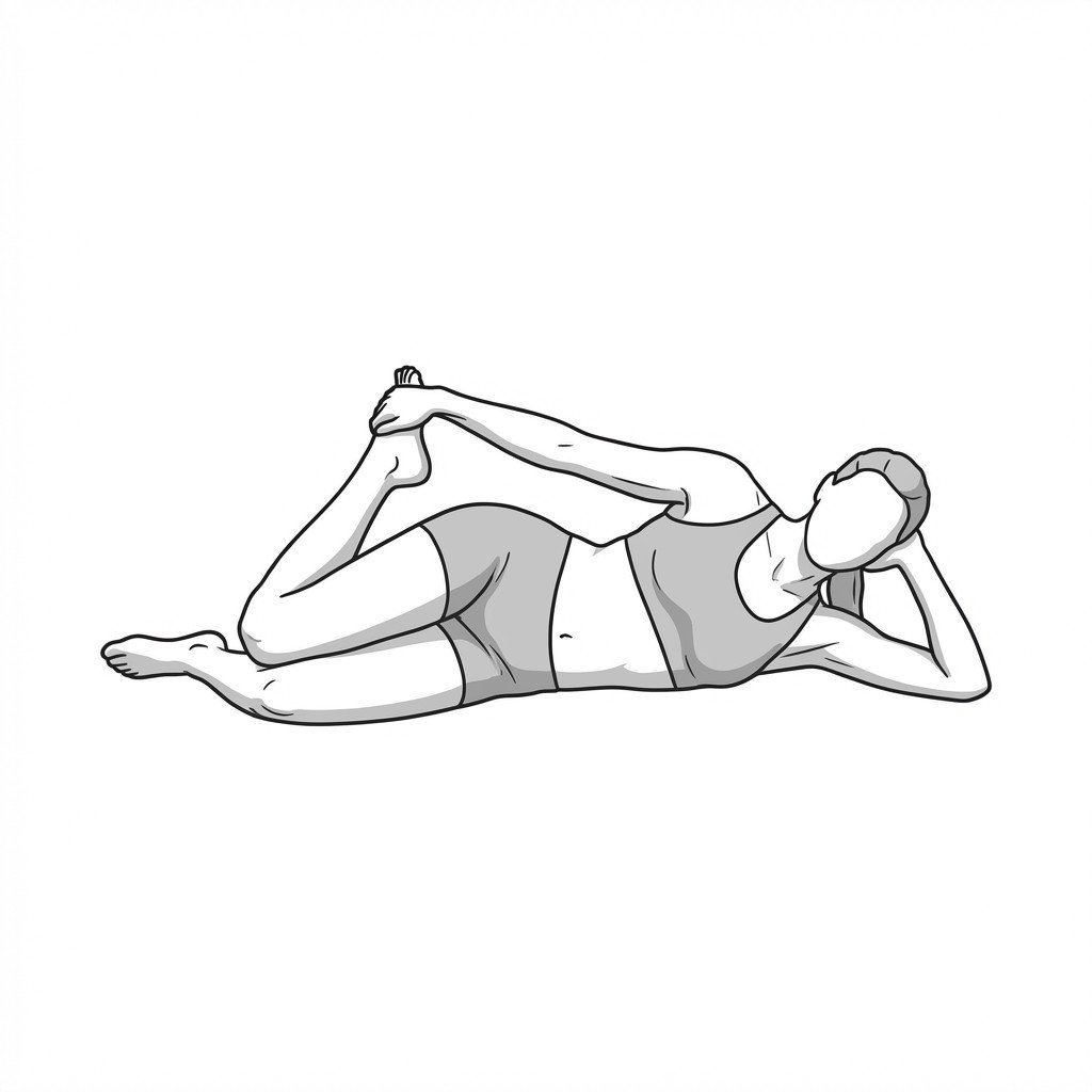
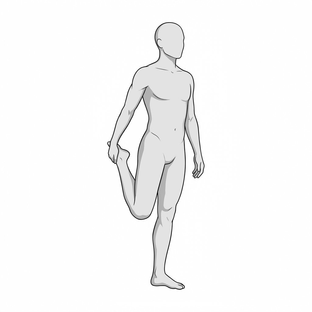
The quadriceps stretch lengthens the rectus femoris and vastus muscles, reducing passive tension on the patella. By decreasing the resting tone of these anterior thigh muscles, it mitigates compressive forces at the patellofemoral joint during movement. This improved flexibility helps normalize patellar tracking and may reduce anterior knee pain.
Start Exercise →Shoulder
Quadruped scapular push-ups plus
This exercise isolates and strengthens the serratus anterior while promoting dynamic scapular stability in a closed kinetic chain. By improving upward rotation and protraction of the scapula, it may help reduce compensatory overactivity and tension in the levator scapulae.
Start Exercise →Thoracic & Core
Quadruped thread-the-needle
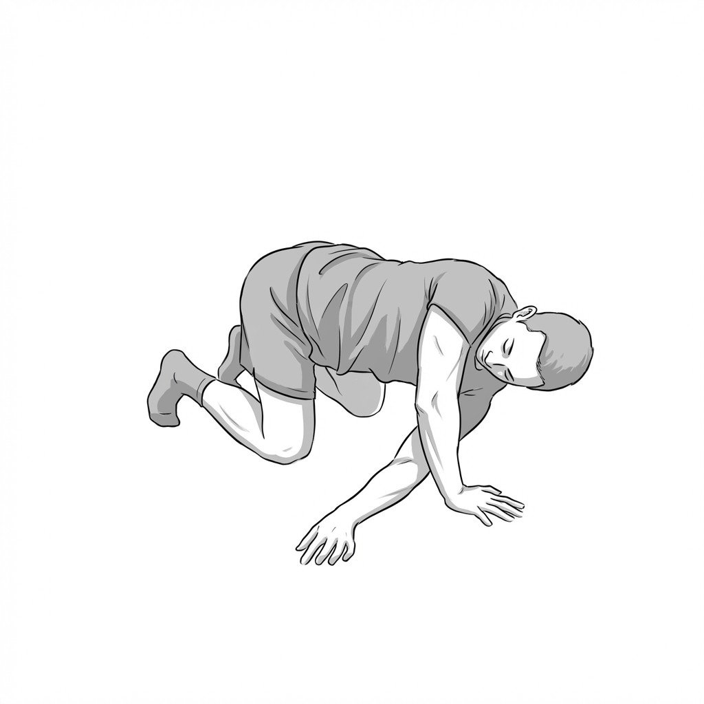
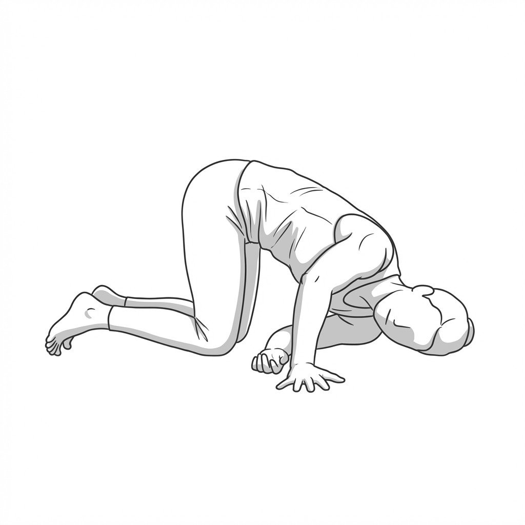
This active mobility exercise promotes thoracic rotation and extension, counteracting the prolonged flexion often seen in thoracic kyphosis. By gliding the scapula into protraction and then integrating spinal rotation, it mobilizes the costovertebral and facet joints. This helps restore physiological mobility and reduces compensatory stiffness in the cervical and lumbar spine.
Start Exercise →Elbow & Hand
Radial nerve glide
This exercise promotes the continuous excursion of the radial nerve through its mechanical interfaces in the arm, such as the radial tunnel and supinator muscle. By promoting neural mobility (thought to reduce intraneural pressure and support axoplasmic flow), it may help decrease nerve mechanosensitivity and ischemic pain associated with compressive or repetitive strain conditions.
Start Exercise →Shoulder
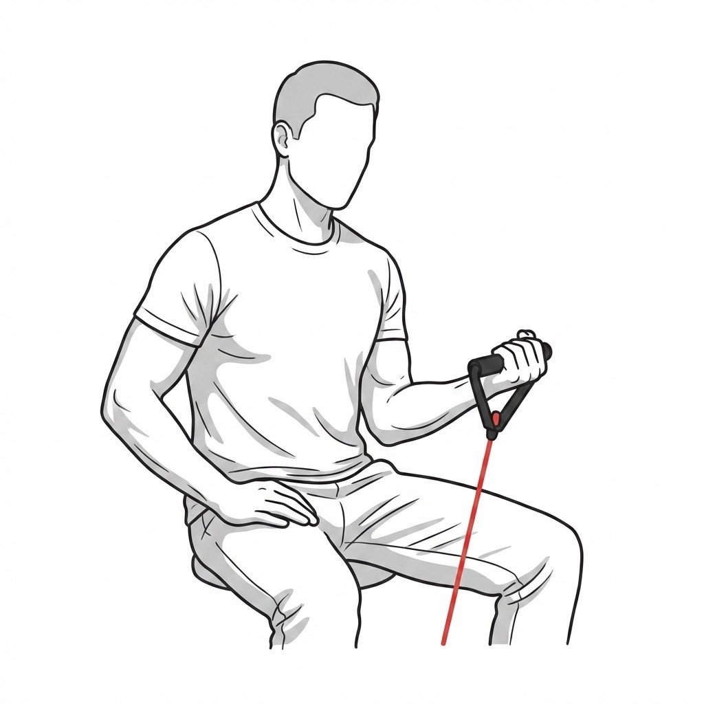
Resistance band supination
Resistance band supination targets the supinator and biceps brachii muscles, applying a controlled load to the biceps tendon insertion at the radial tuberosity. This progressive mechanical loading promotes collagen remodeling and improves tissue capacity, which is essential for recovering from tendinopathy.
Start Exercise →Shoulder
Resisted elbow flexion
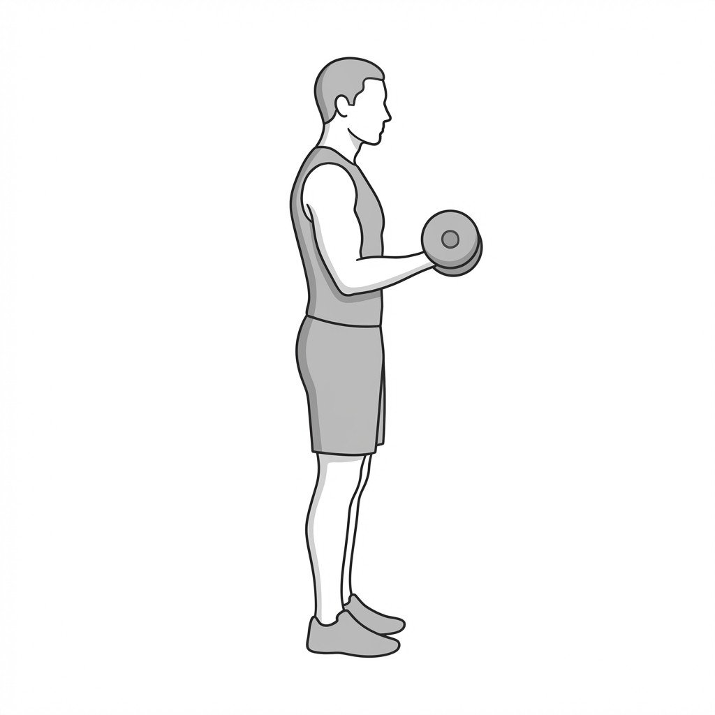
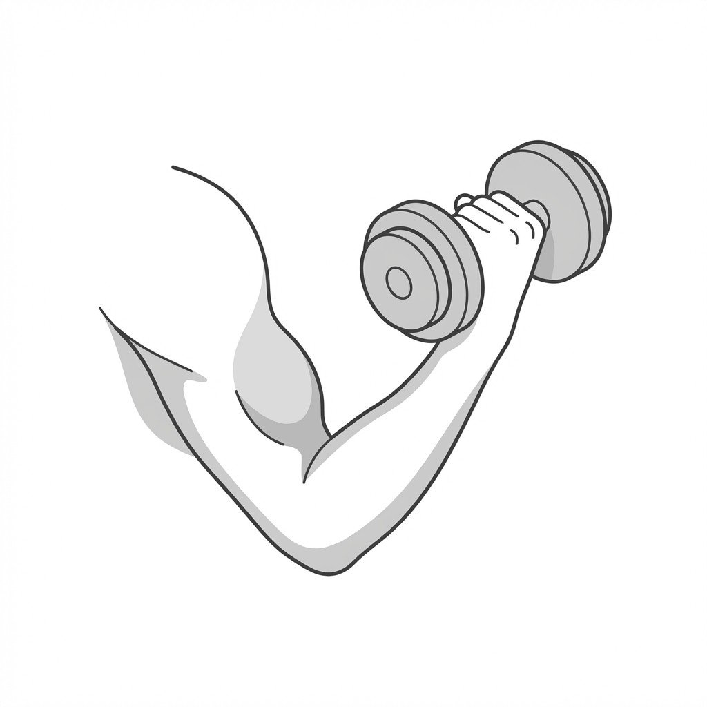
This exercise progressively loads the biceps brachii and its tendon through a controlled range of motion. Mechanical loading promotes collagen synthesis and realignment within the tendon. This gradual adaptation may help reduce pain and improve load tolerance in biceps tendinopathy.
Start Exercise →Lower Leg
Resisted eversion inversion
Resisted inversion and eversion strengthens the dynamic stabilizers of the ankle joint, primarily the peroneals and tibialis posterior. By enhancing the strength and eccentric control of these muscles, the exercise improves neuromuscular responsiveness to sudden perturbations, which may help reduce the risk of recurrent sprains in chronic ankle instability.
Start Exercise →Lower Leg
Resisted inversion band
This exercise applies concentric and eccentric loads to the tibialis posterior muscle and tendon using elastic resistance. By strengthening this musculotendinous unit, it improves dynamic support of the medial longitudinal arch and limits excessive midfoot pronation. This may help reduce load on the posterior tibial tendon and support functional recovery from tendinopathy.
Start Exercise →Lower Leg
Resisted inversion
Resisted inversion targets the tibialis posterior muscle, which plays a crucial role in controlling foot pronation and supporting the medial longitudinal arch. Strengthening this muscle helps attenuate impact forces along the medial tibial border, which may help reduce the mechanical stress thought to contribute to Medial Tibial Stress Syndrome.
Start Exercise →Cervical
Resisted isometric mouth opening
This exercise activates the jaw depressors isometrically, preventing actual movement of the temporomandibular joint (TMJ) while firing the surrounding musculature. It helps to build neuromuscular control, increase local blood flow, and relieve tension without overloading the articular disc, which may help reduce pain and improve control in TMJ dysfunction.
Start Exercise →Elbow & Hand
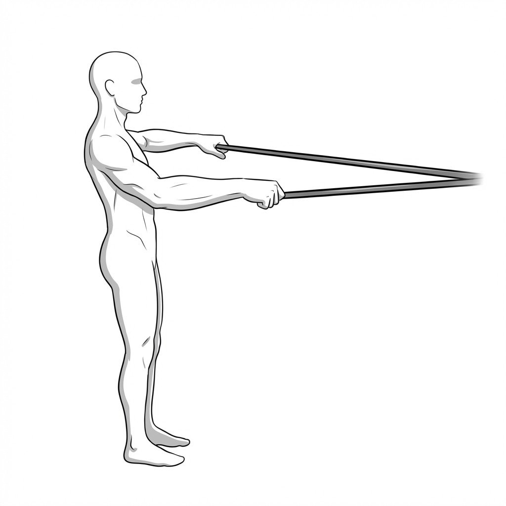
Reverse Tyler Twist
The Reverse Tyler Twist is an eccentric strengthening exercise specifically designed to load the wrist flexors and pronators using a flexible rubber resistance bar. By lengthening the contracted muscle-tendon unit under tension, it is thought to promote collagen remodeling and may help reduce pain at the medial epicondyle.
Start Exercise →Hip
Romanian deadlifts light
Light Romanian deadlifts provide controlled, eccentric loading to the posterior chain, specifically targeting the hamstrings. By lengthening the muscle under a sub-maximal load, this exercise is thought to support favorable alignment of regenerating muscle tissue and to increase the tolerance of the musculotendinous junction to tensile forces, which is an important goal in hamstring strain rehabilitation.
Start Exercise →Elbow & Hand
Rubber band finger extensions
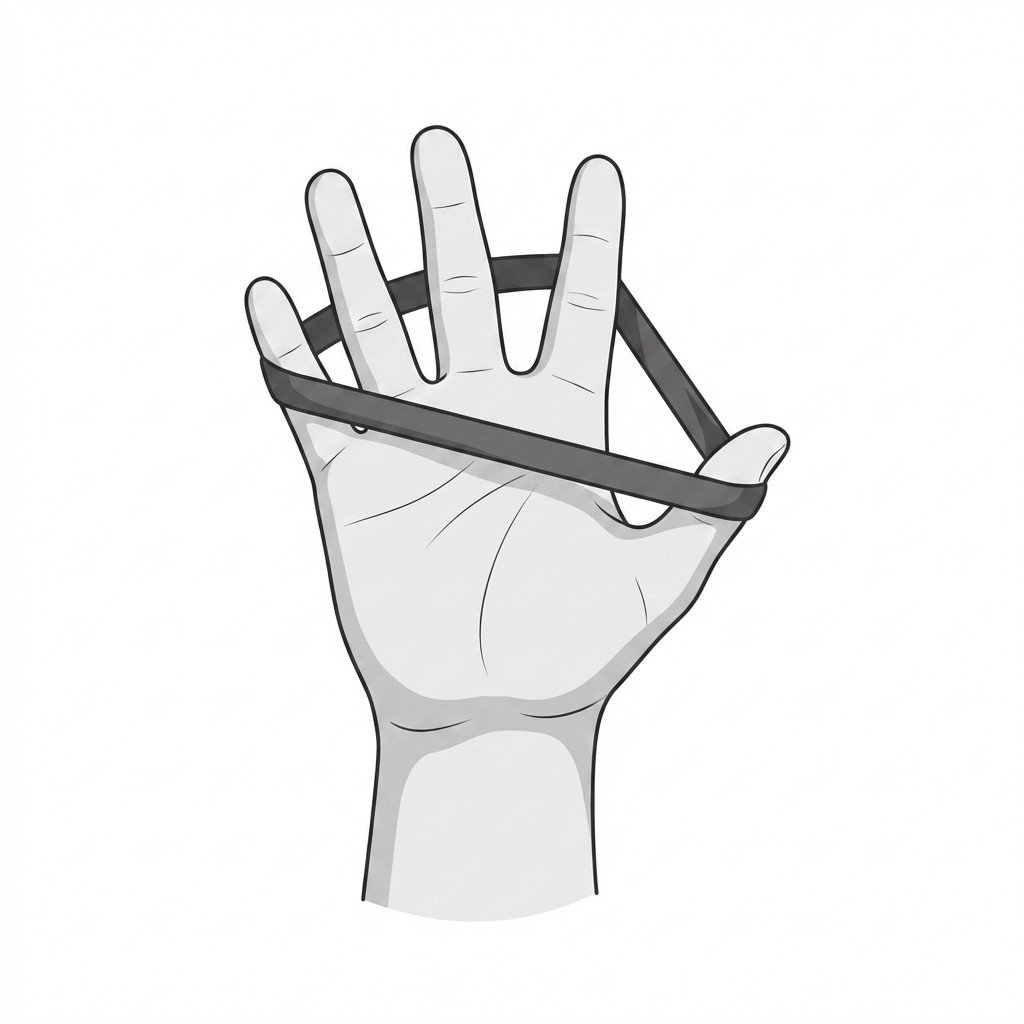
Rubber band finger extensions strengthen the finger and wrist extensor muscles to improve global forearm muscle balance. In medial epicondylalgia, strengthening the antagonistic extensor group helps stabilize the wrist during grip activities, which may help reduce load on the overloaded common flexor tendon.
Start Exercise →Thoracic & Core
Scalene stretch
The scalene stretch elongates the anterior, middle, and posterior scalene muscles, which frequently hypertrophy or spasm and compress the neurovascular bundle in the interscalene triangle. By restoring normal resting length, this stretch may help reduce compression on the brachial plexus and subclavian vessels, which can ease neurological and vascular symptoms associated with Thoracic Outlet Syndrome.
Start Exercise →Shoulder
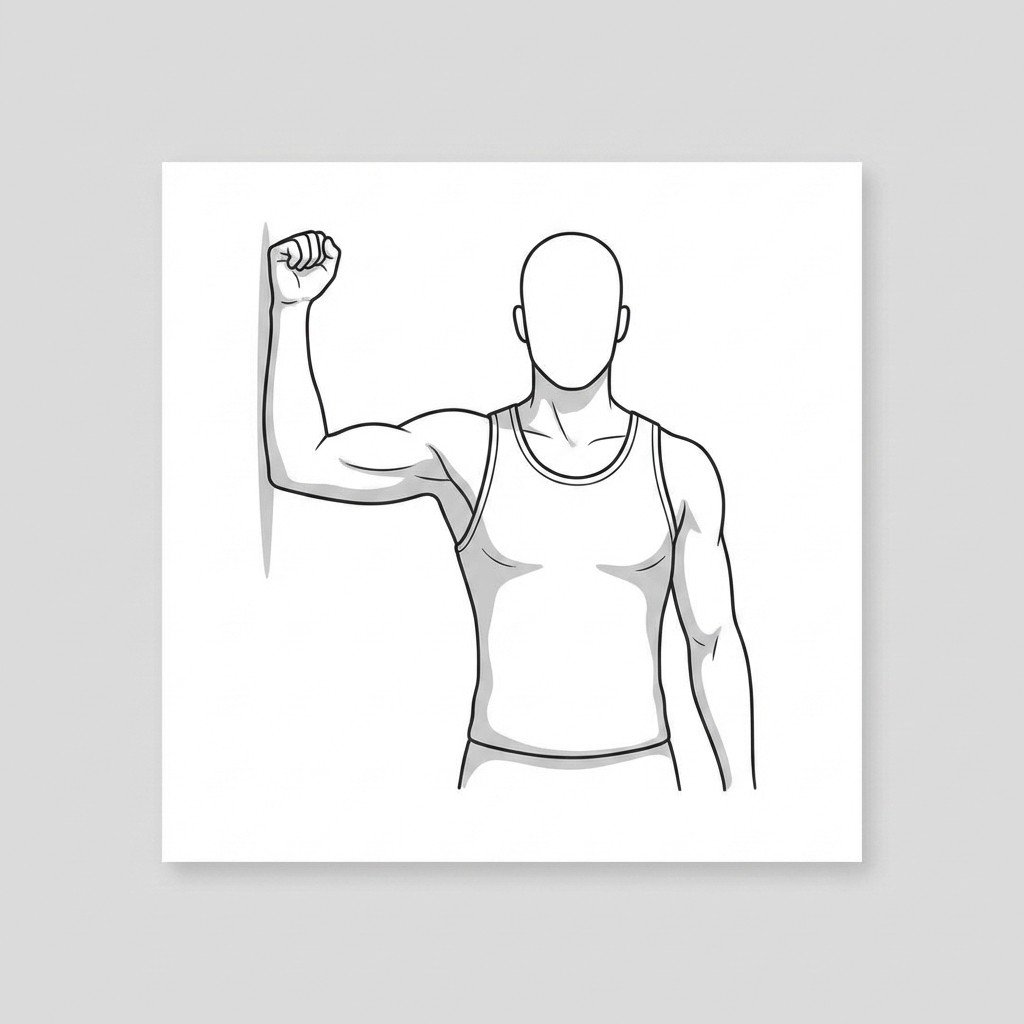
Scapular clock
The scapular clock exercise focuses on improving neuromuscular control and proprioception of the scapulothoracic joint. By actively moving the scapula through various planes of motion against a wall, it enhances the synergistic activation of the periscapular muscles. This helps address scapular dyskinesis, which may improve subacromial space and ease impingement-related pain.
Start Exercise →Cervical
Scapular retraction with depression
This exercise facilitates the activation of the lower trapezius and rhomboids while inhibiting the often-overactive upper trapezius. By restoring normal scapular kinematics and expanding the costoclavicular space, it reduces neurovascular tension and compression in the brachial plexus.
Start Exercise →Cervical
Scapular retraction
Scapular retraction activates the periscapular musculature to counteract the forward head and rounded shoulder posture commonly associated with axial neck pain. By improving scapulothoracic endurance and restoring neutral resting posture, this exercise reduces excessive mechanical load on the cervical spine and limits compensatory overactivity of the upper cervical extensors.
Start Exercise →Hip
Sciatic nerve glide
This exercise safely mobilizes the sciatic nerve through its anatomical sheath to reduce mechanical tension and improve neural excursion. By alternately applying and releasing tension at the neck and knee, it prevents nerve tethering and reduces ectopic discharge, alleviating radicular symptoms common in Piriformis Syndrome.
Start Exercise →Cervical
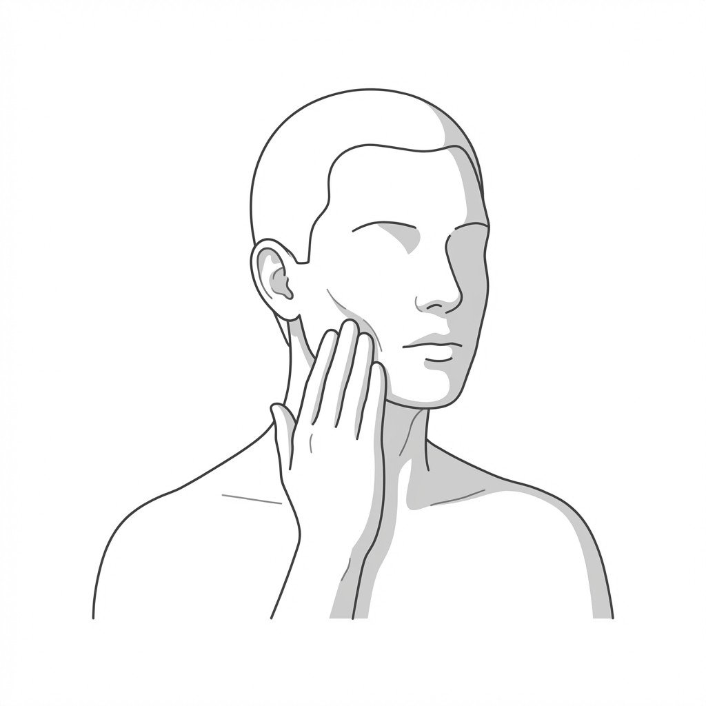
SCM gentle stretch
The SCM gentle stretch lengthens the sternocleidomastoid muscle, reducing anterior cervical tension and fascial pull on the cranium. By relieving trigger points and restoring normal resting length to the anterior neck, it decreases referred pain patterns often implicated in cervicogenic headaches.
Start Exercise →Lower Leg
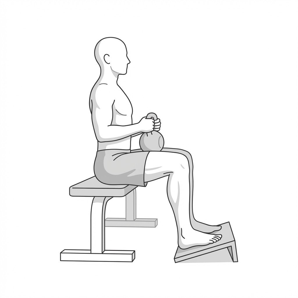
Seated heavy soleus raises
Seated calf raises preferentially target the soleus muscle by placing the gastrocnemius in a shortened, mechanically disadvantaged position via knee flexion. This isolated heavy loading stimulates collagen synthesis and improves the load-bearing capacity of the Achilles tendon to directly manage tendinopathy.
Start Exercise →Hip
Seated hip rotation
Seated hip rotation actively engages the deep hip rotators and gluteal muscles to improve joint lubrication and capsular mobility. By moving the femur rotationally within the acetabulum, it reduces joint stiffness and preserves functional range of motion critical for managing hip osteoarthritis.
Start Exercise →Knee
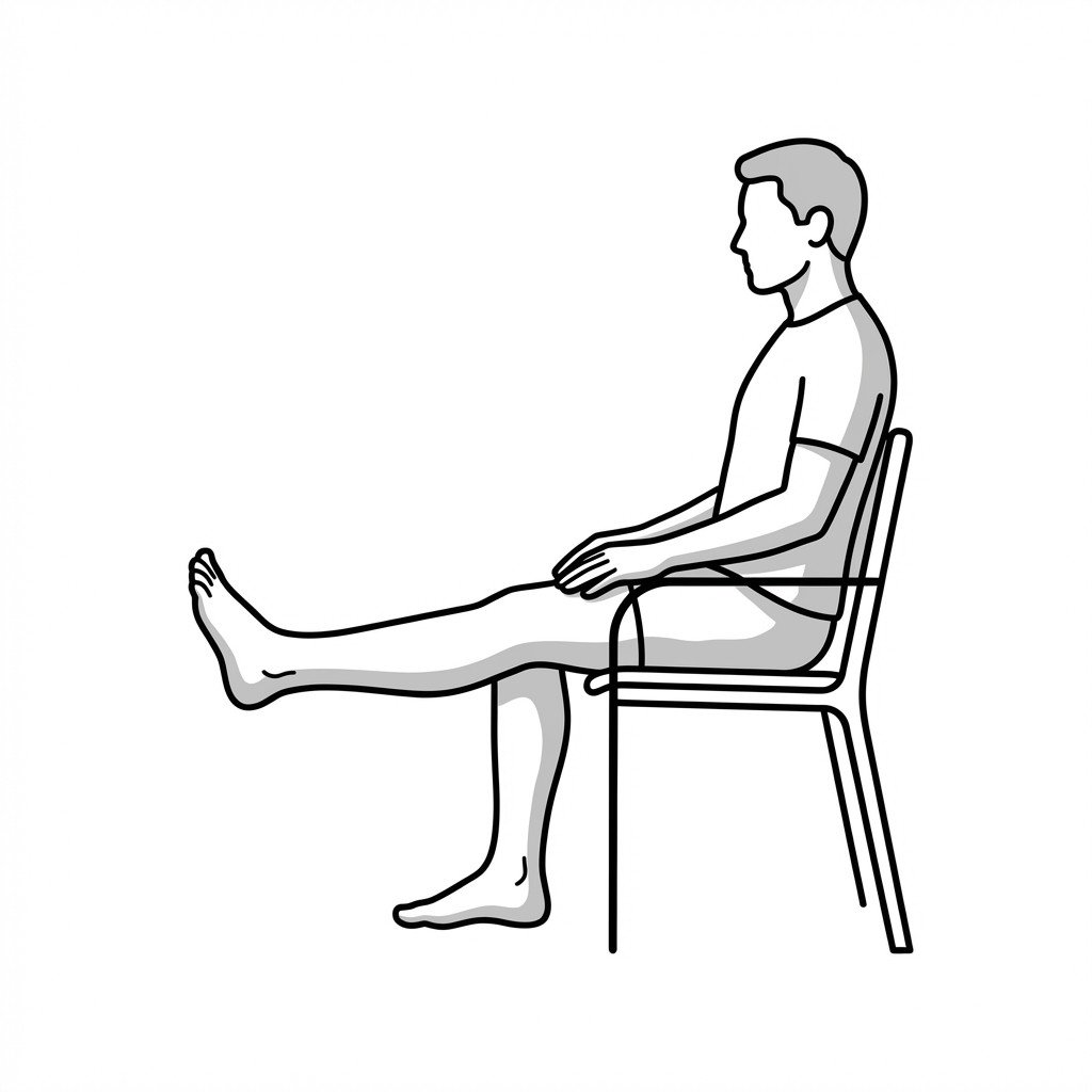
Seated knee extensions
Seated knee extensions isolate and strengthen the quadriceps muscles by moving the knee against gravity through an open-kinetic chain arc. In knee osteoarthritis, enhancing quadriceps strength improves joint stability, increases shock absorption, and reduces abnormal intra-articular loading, thereby mitigating pain.
Start Exercise →Thoracic & Core
Seated rib mobilization
Seated rib mobilization applies targeted overpressure to the posterolateral rib angles combined with active trunk rotation and deep breathing to restore costovertebral joint arthrokinematics. By improving posterior rib mobility, this technique reduces the compensatory mechanical stress placed on the inflamed anterior costochondral junctions.
Start Exercise →Shoulder
Seated scapular depression
Seated scapular depression actively engages the lower trapezius and latissimus dorsi to pull the shoulder blades downward. This mechanism provides reciprocal inhibition to the overactive levator scapulae, reducing its resting tone and alleviating the focal neck pain associated with the syndrome.
Start Exercise →Thoracic & Core
Seated thoracic rotations
Seated thoracic rotations mobilize the facet joints of the mid-back and activate the intrinsic spinal rotators. By actively rotating the spine while the pelvis is stabilized, this exercise helps counteract the rigid flexed posture of thoracic kyphosis, restoring transverse plane mobility and promoting upright spinal alignment.
Start Exercise →Cervical
Sensory trick motor training
Sensory trick motor training leverages a unique neurological phenomenon in cervical dystonia where a specific tactile stimulus temporarily reduces involuntary muscle spasms. By voluntarily holding the corrected posture while gradually fading the tactile cue, it helps retrain sensorimotor integration in the basal ganglia to improve midline head control.
Start Exercise →Shoulder
Serratus anterior punches
Serratus anterior punches isolate and strengthen the serratus anterior muscle through targeted scapular protraction. By improving serratus anterior function, this exercise restores proper upward rotation and posterior tilt of the scapula, thereby increasing the subacromial space and reducing tendon compression in Subacromial Pain Syndrome.
Start Exercise →Lower Leg
Short foot exercise
The short foot exercise targets the intrinsic muscles of the foot to elevate and support the medial longitudinal arch. By enhancing arch stability and neuromuscular control, it reduces excessive tension on the medial tibial border and posterior tibial tendon.
Start Exercise →Shoulder
Side-lying wiper
The side-lying wiper exercise selectively targets the posterior rotator cuff by utilizing gravity to resist shoulder external rotation in an adducted position. This isolated strengthening enhances dynamic stability of the glenohumeral joint, which improves load tolerance in the tendon and reduces tendinopathic pain.
Start Exercise →Lumbar & Pelvis
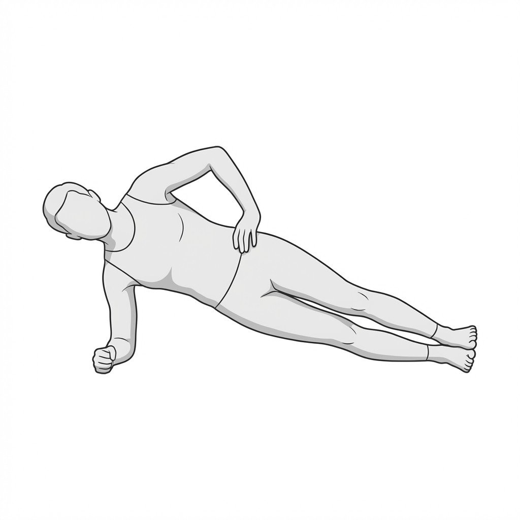
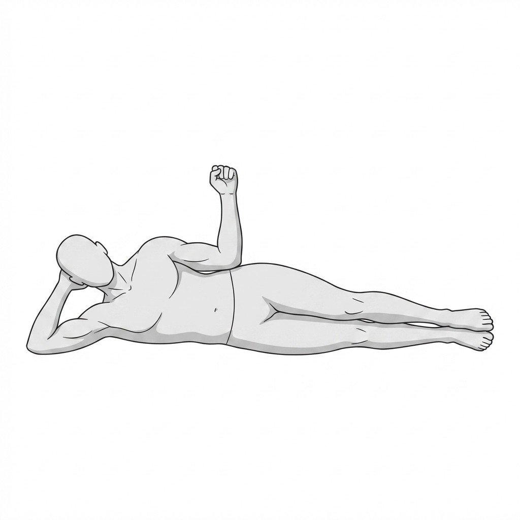
Side planks
The side plank is an isometric stabilization exercise that engages the lateral core musculature to resist lateral spinal flexion. For Quadratus Lumborum (QL) Myofascial Pain Syndrome, it builds endurance and load tolerance in the QL, reducing compensatory overactivity and spasm. This improved neuromuscular control promotes local tissue desensitization and functional lumbopelvic stability.
Start Exercise →Lower Leg
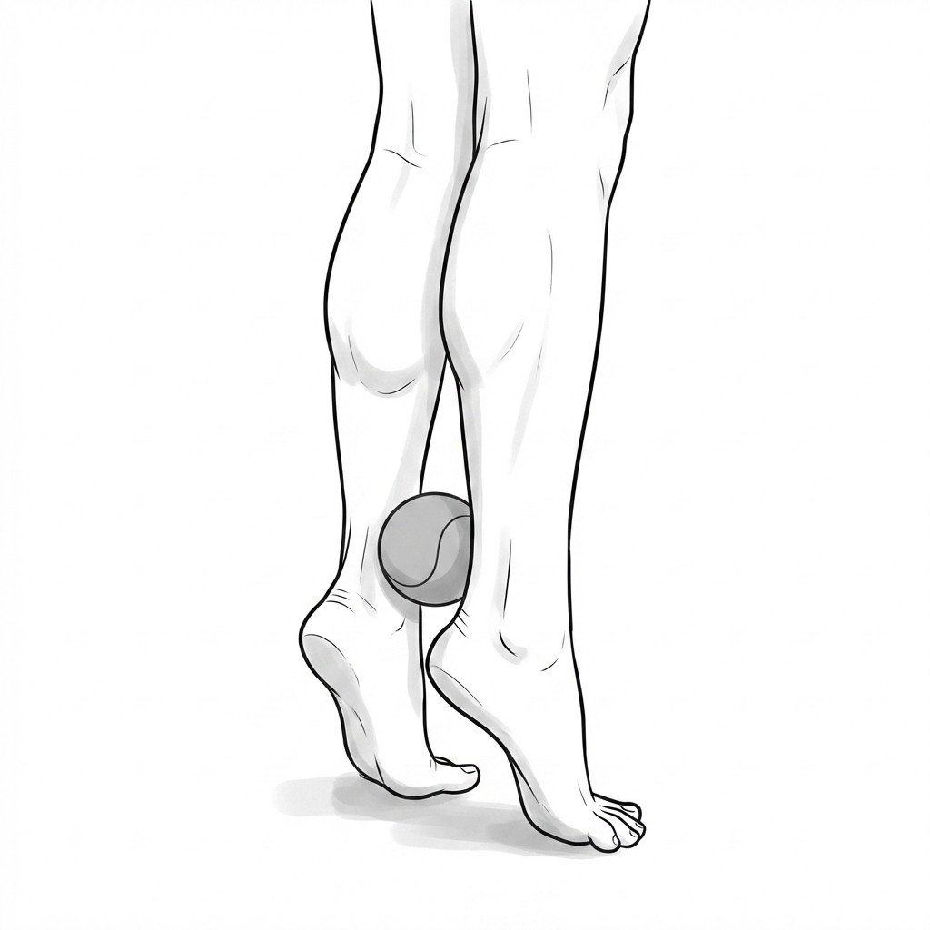
Single-leg heel raise with ball
Placing a ball between the heels or ankles during a heel raise forces the foot into slight inversion, which preferentially loads the tibialis posterior muscle. This mechanistic activation counteracts the pathological valgus collapse seen in Posterior Tibial Tendon Dysfunction (PTTD) by strengthening the primary dynamic stabilizer of the medial longitudinal arch.
Start Exercise →Knee
Single-leg stance
The single-leg stance improves proprioception and neuromuscular control by challenging the body's static balance on a narrow base of support. Mechanistically, it facilitates co-contraction of the lower extremity stabilizers, which enhances joint stability and afferent feedback necessary for rehabilitating ankle and knee pathologies.
Start Exercise →Shoulder
Sleeper stretch
The sleeper stretch targets the posterior capsule and posterior rotator cuff muscles of the shoulder, aiming to increase glenohumeral internal rotation. By restoring internal rotation and posterior tissue extensibility, it helps center the humeral head in the glenoid fossa during arm elevation, thereby reducing subacromial impingement mechanics.
Start Exercise →Hip
Slider eccentric curls
Slider eccentric curls apply tension to the hamstrings as they lengthen under load. This targeted eccentric stress is thought to promote the addition of sarcomeres in series, a proposed adaptation associated with increased fascicle length and an improved ability to absorb high forces, which may help reduce the risk of strain recurrence.
Start Exercise →Hip
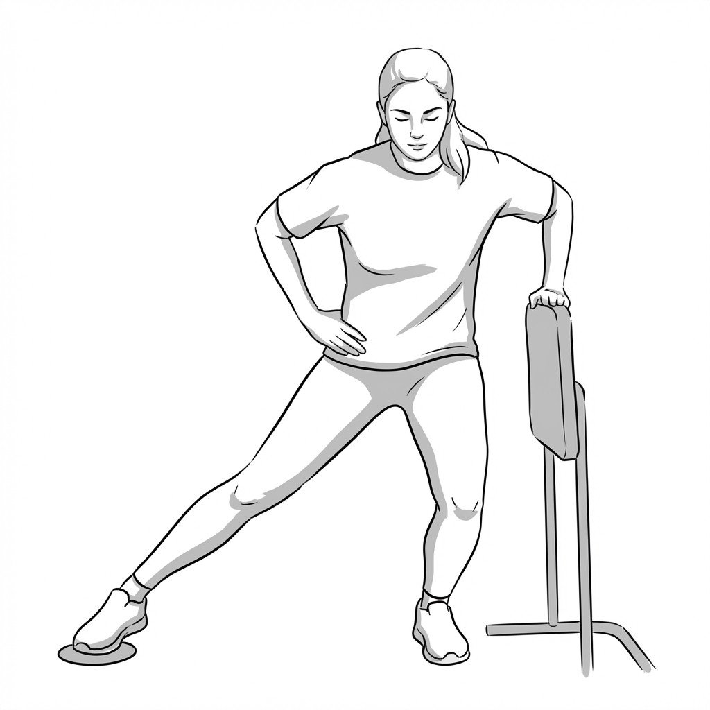
Sliding lateral lunges
Sliding lateral lunges provide eccentric loading to the hip adductors while demanding simultaneous dynamic stabilization from the gluteals and quadriceps. This eccentric lengthening phase helps remodel regenerating muscle fibers and tendon tissue in a recovering adductor strain, progressively increasing tissue load tolerance.
Start Exercise →Cervical
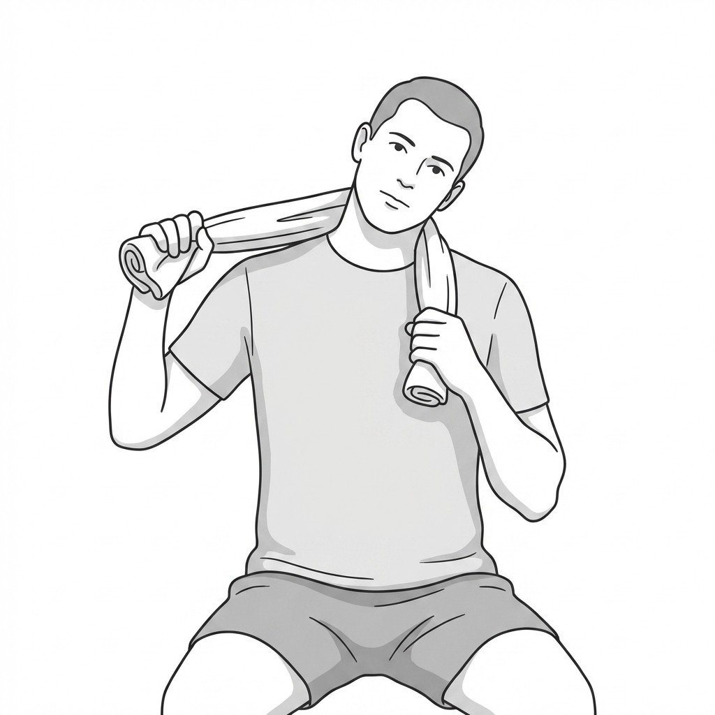
SNAGs with towel
Sustained Natural Apophyseal Glides (SNAGs) utilize a towel to provide a pain-free accessory glide to the upper cervical facet joints during active physiological movement. This facilitates normal arthrokinematics, reduces capsular restriction, and decreases afferent nociceptive input from the upper cervical spine, which may help address musculoskeletal contributors to cervicogenic headache.
Start Exercise →Lower Leg
Soleus stretch
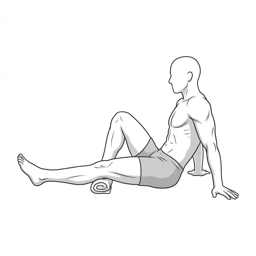
This exercise isolates the deeper calf muscle by flexing the knee, effectively placing the two-joint gastrocnemius muscle on slack. Improving the tissue extensibility of the soleus increases ankle dorsiflexion range of motion, which reduces abnormal tensile stresses on the Achilles tendon and medial tibial border during the stance phase of gait.
Start Exercise →Hip
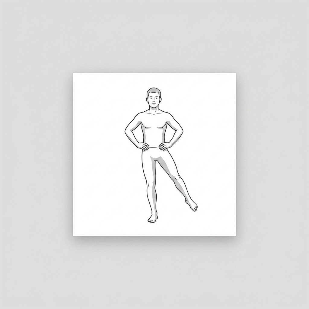
Standing hip abduction
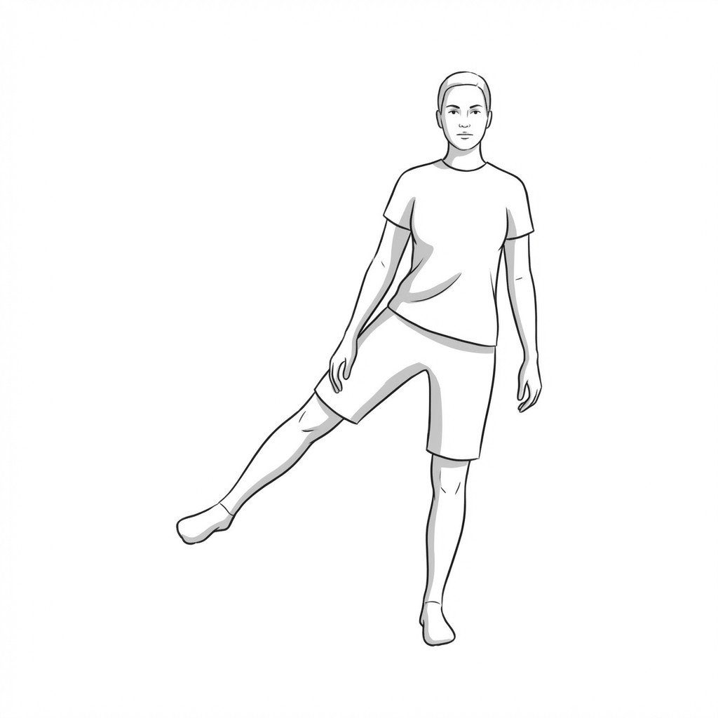
Standing hip abduction isolates and loads the gluteus medius and minimus tendons, progressively improving their load-bearing capacity. By performing this upright, it avoids excessive compressive forces across the greater trochanter while simultaneously challenging the stabilizing muscles of the stance leg.
Start Exercise →Hip
Standing hip extension
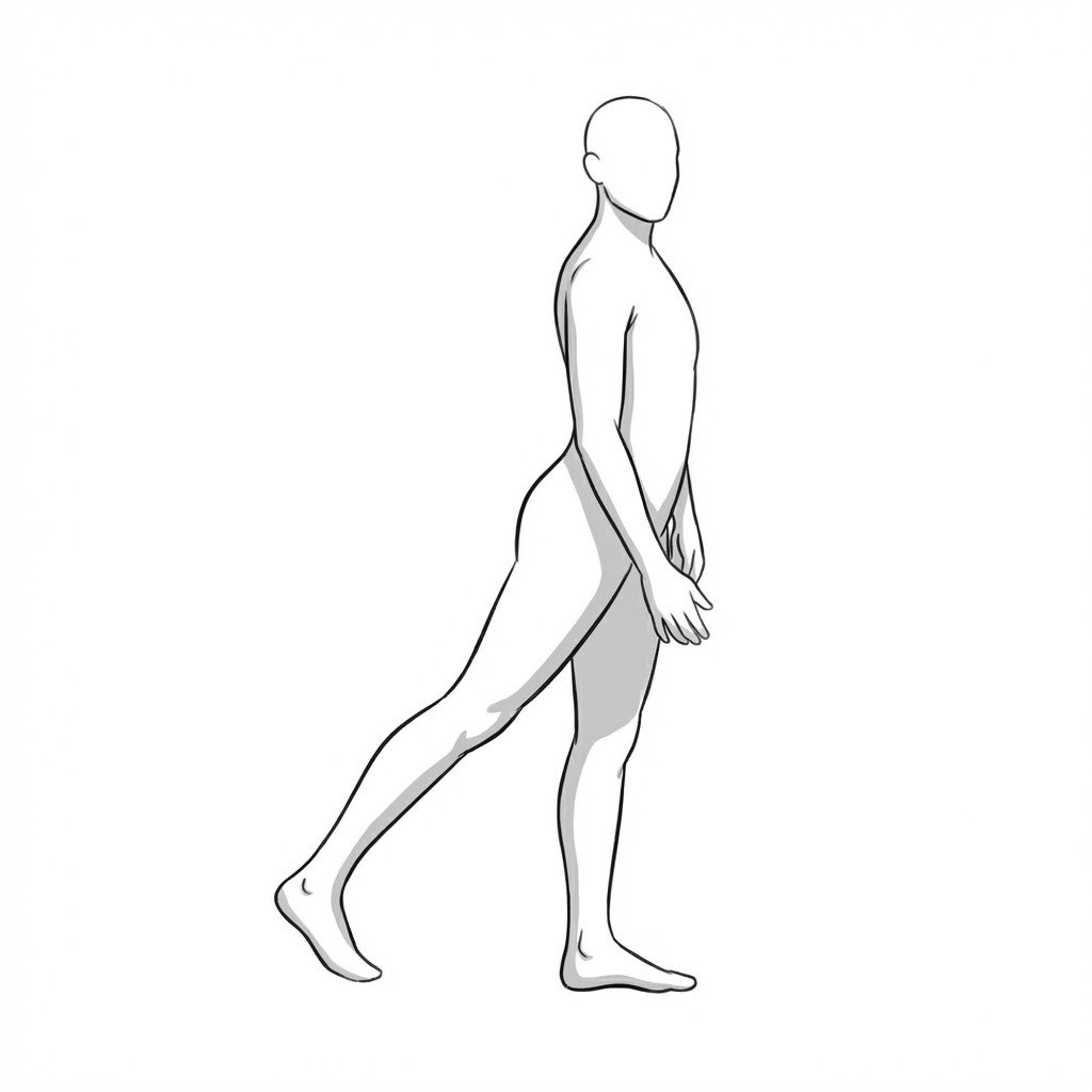
Standing hip extension actively engages the posterior chain to improve hip joint stability and counter the flexed posture commonly adopted by patients with hip osteoarthritis. By strengthening the gluteal muscles, this exercise helps reduce excessive joint loading during ambulation and mitigates capsular stiffness.
Start Exercise →Knee
Standing quad stretch
This exercise lengthens the rectus femoris and vastus muscles, reducing resting tension on the patellar tendon. By improving quadriceps flexibility, it may help reduce mechanical strain across the extensor mechanism during load-bearing, which aids in mitigating symptom exacerbation in patellar tendinopathy.
Start Exercise →Lower Leg
Star excursion training
Star excursion training challenges the sensorimotor system by requiring single-leg dynamic balance while voluntarily shifting the center of mass. It enhances neuromuscular control, proprioception, and functional stability of the ankle joint. This helps mitigate the sensorimotor deficits commonly associated with chronic ankle instability and may help reduce recurrent giving-way episodes.
Start Exercise →Hip
Stationary cycling
Stationary cycling provides low-impact cardiovascular conditioning and promotes synovial fluid circulation in the hip and knee joints. By avoiding high-impact loading, it supports joint health through gentle repetitive motion and strengthening of the surrounding musculature, which may improve joint stability and help reduce osteoarthritic pain.
Start Exercise →Knee
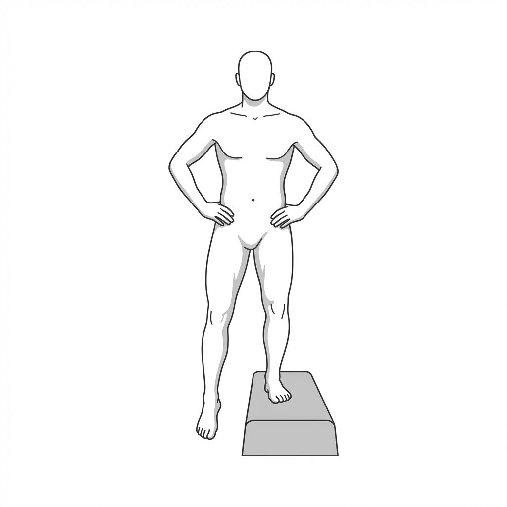
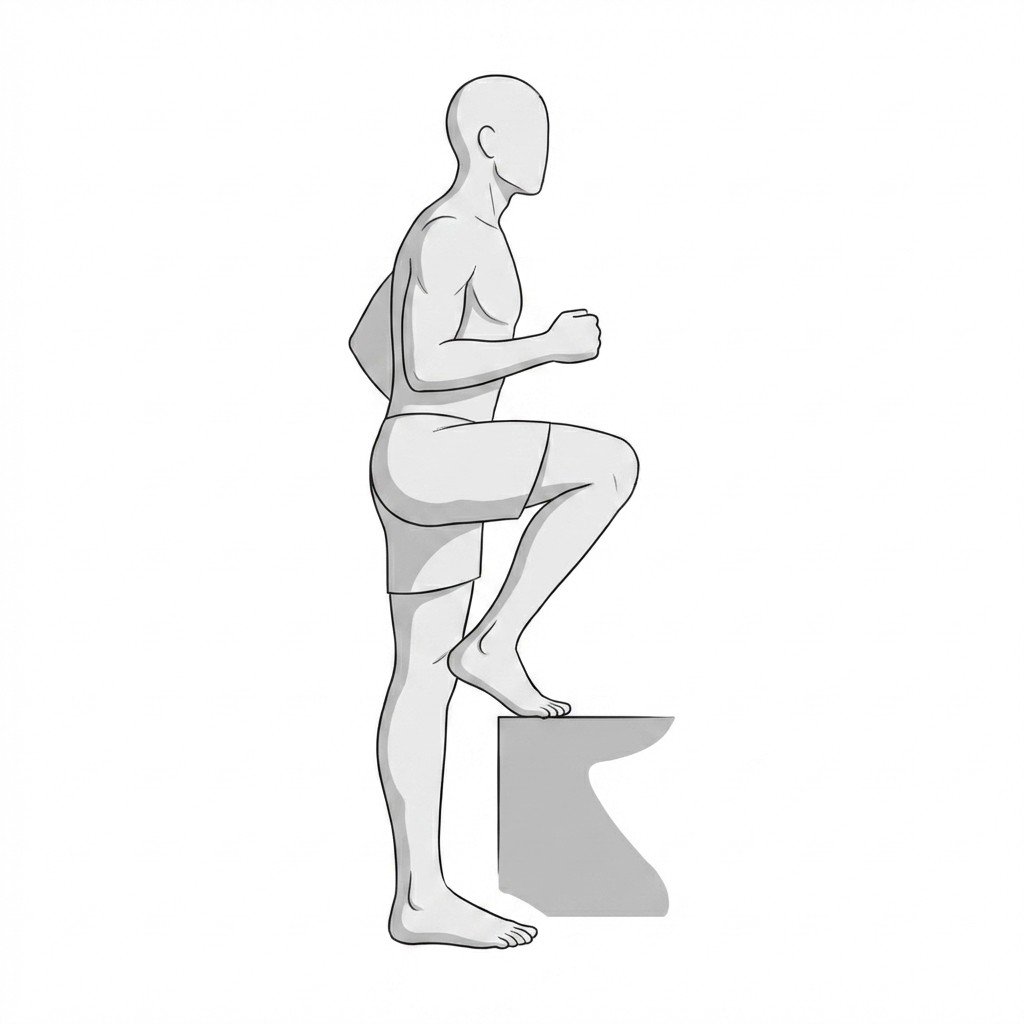
Step-downs
Step-downs focus on the eccentric control of the quadriceps while demanding dynamic stabilization from the hip abductors. By improving neuromuscular control of the lower extremity, this exercise helps control medial knee collapse, which may reduce abnormal contact stress on the patellofemoral joint.
Start Exercise →Knee
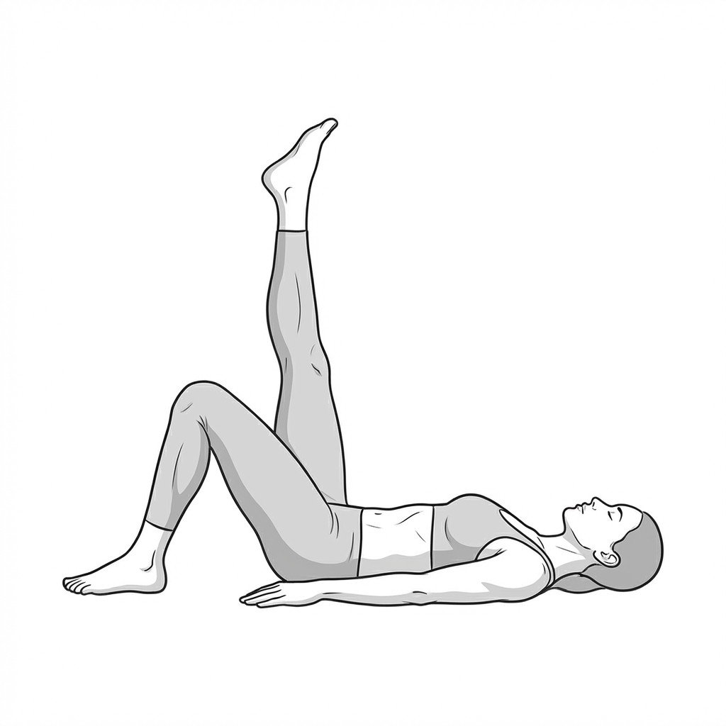
Straight leg raises
Straight leg raises activate the quadriceps complex without placing compressive load on the patellofemoral joint. By performing this exercise with a fully extended knee, it strengthens the thigh and hip flexor musculature while minimizing patellar tracking stress, making it highly tolerable for early-stage rehab.
Start Exercise →Cervical
Suboccipital gentle stretch
This exercise provides targeted elongation of the suboccipital musculature at the base of the skull, which often becomes hypertonic and shortened in cervical dystonia. By applying a gentle, sustained stretch, it is thought to reduce local muscle spindle sensitivity and to improve upper cervical mobility. This may help reduce tension-related pain and assist in modulating abnormal cervical posturing.
Start Exercise →Elbow & Hand
Supinator strengthening
Supinator strengthening targets the muscles responsible for turning the forearm palm-up, which share fascial connections and load requirements with the lateral elbow tendons. By improving the load capacity of the supinator, this exercise may help reduce excessive stress on the common extensor origin during gripping and twisting, supporting tissue adaptation in lateral epicondylalgia.
Start Exercise →Lumbar & Pelvis
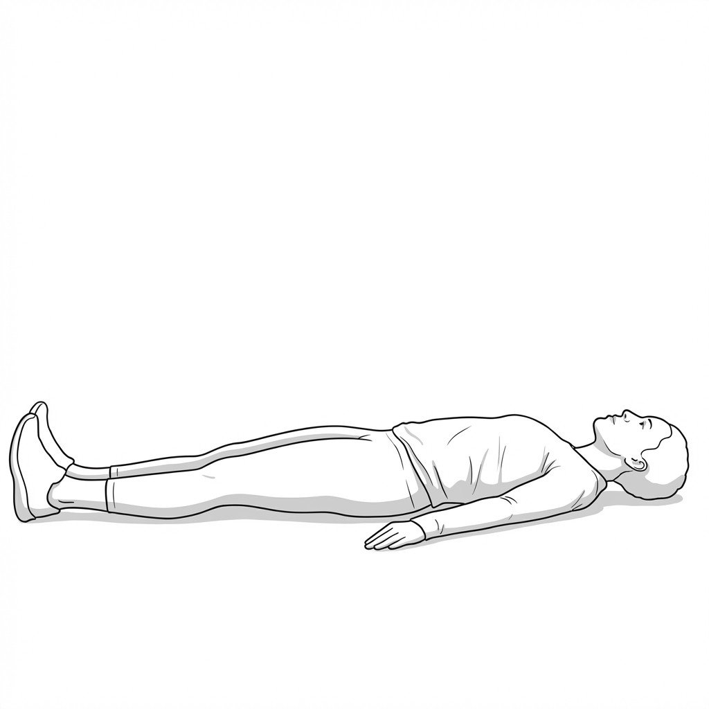
Supine core bracing
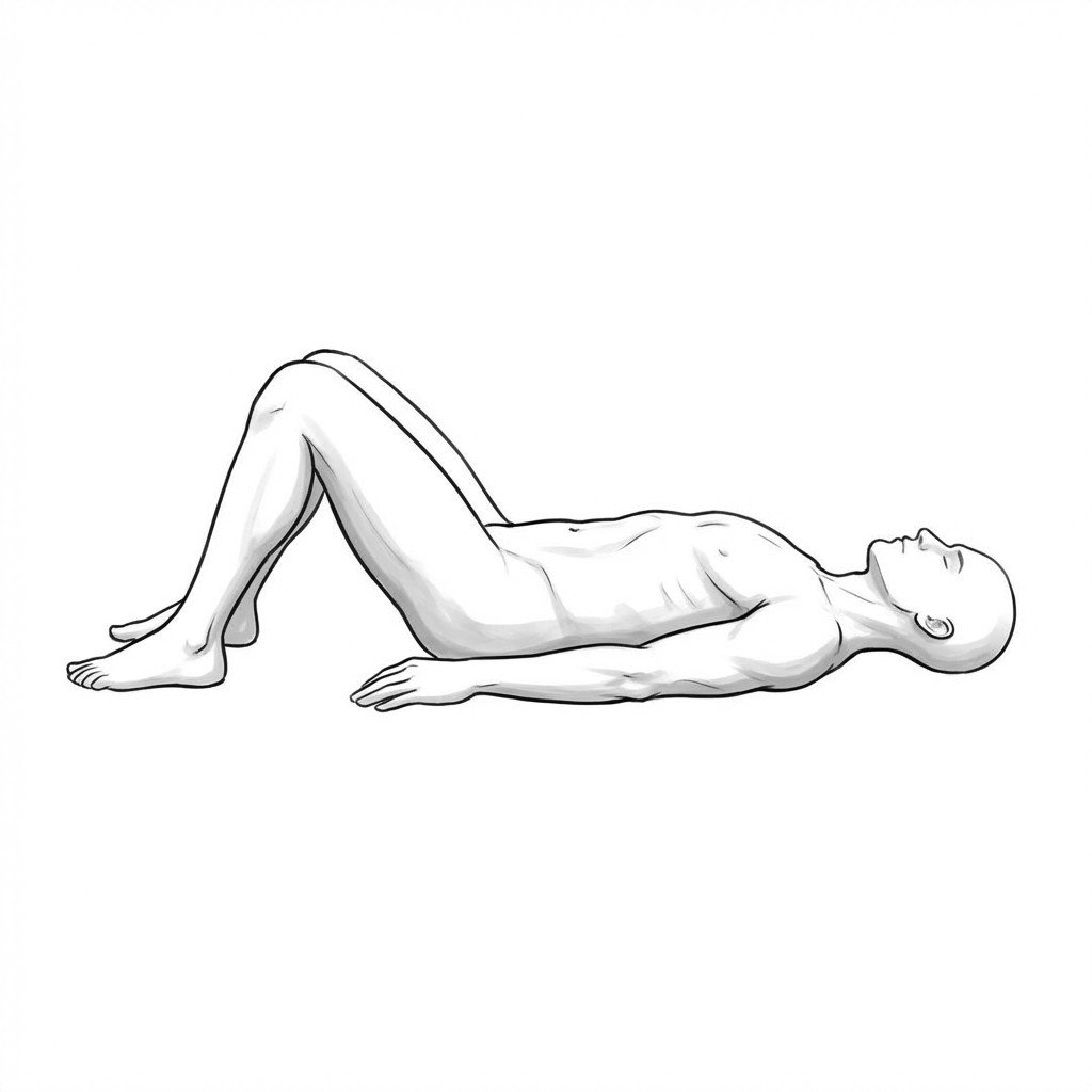
Supine core bracing activates the deep abdominal muscles to create an internal corset around the spine, increasing intra-abdominal pressure. This limits excessive anterior shear forces and stabilizes the lumbar spine in a neutral position. For spondylolisthesis, this dynamic stabilization may help limit excessive segmental motion and reduce associated nerve-root or facet irritation.
Start Exercise →Lumbar & Pelvis
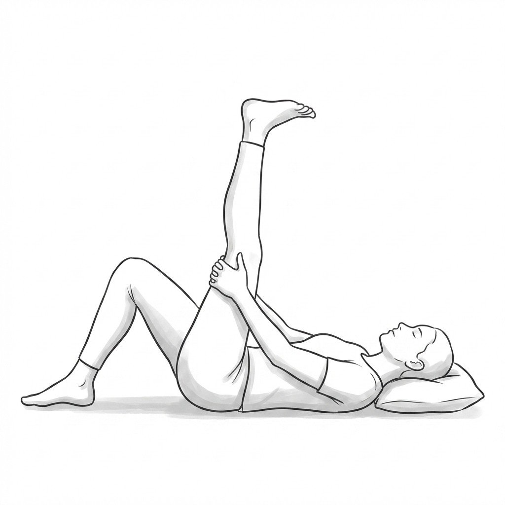
Supine hamstring stretches
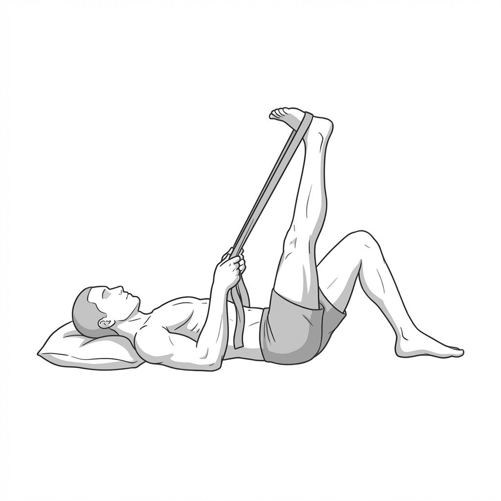
Supine hamstring stretches help lengthen the posterior thigh muscles, which frequently become short and tight as a compensatory pelvic mechanism in spondylolisthesis. By improving hamstring flexibility, this exercise helps restore normal pelvic biomechanics and reduces secondary mechanical stress on the lower back. Performing it in a supine position protects the lumbar spine by keeping it supported and in a neutral alignment.
Start Exercise →Thoracic & Core
Supine marches
Supine marches focus on engaging the deep core musculature, particularly the transversus abdominis, to stabilize the pelvis and lumbar spine while moving the lower extremities. By maintaining appropriate intra-abdominal pressure without outward bulging, this exercise may help support the abdominal wall and improve neuromuscular control in individuals with diastasis recti.
Start Exercise →Elbow & Hand
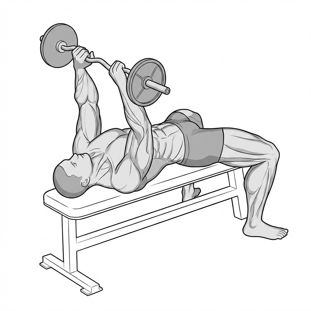
Supine skull crushers
This exercise provides controlled eccentric and concentric loading to the triceps brachii while the shoulder is in a flexed position. Mechanistically, this targeted loading is thought to stimulate collagen synthesis and tenocyte activity, helping to increase the load-bearing capacity of the triceps tendon in tendinopathy.
Start Exercise →Cervical
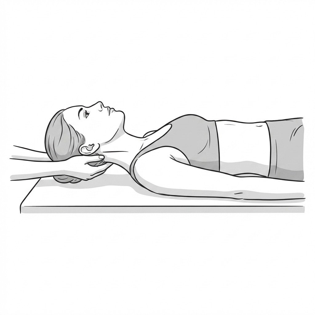
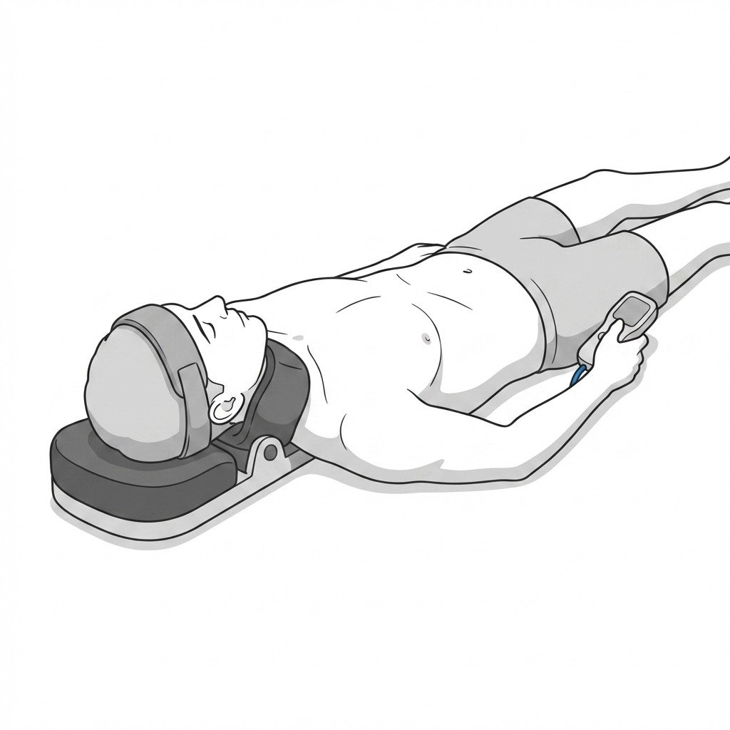
Supine supported cervical traction
Supine supported cervical traction applies a gentle longitudinal stretch to the cervical spine, which may transiently increase the intervertebral foraminal space. This gentle traction is thought to reduce load on irritated cervical nerve roots and, for some people, can help centralize symptoms and ease radiating arm pain associated with cervical radiculopathy.
Start Exercise →Lumbar & Pelvis
Supine TrA activation
Supine transversus abdominis (TrA) activation isolates the deep core musculature to create a musculofascial corset around the lower abdomen. By generating tension through the thoracolumbar fascia, it enhances force closure across the sacroiliac joint. This increased lumbopelvic stability may help reduce shear forces and pain associated with SIJ dysfunction.
Start Exercise →Hip
Supine wall straddle
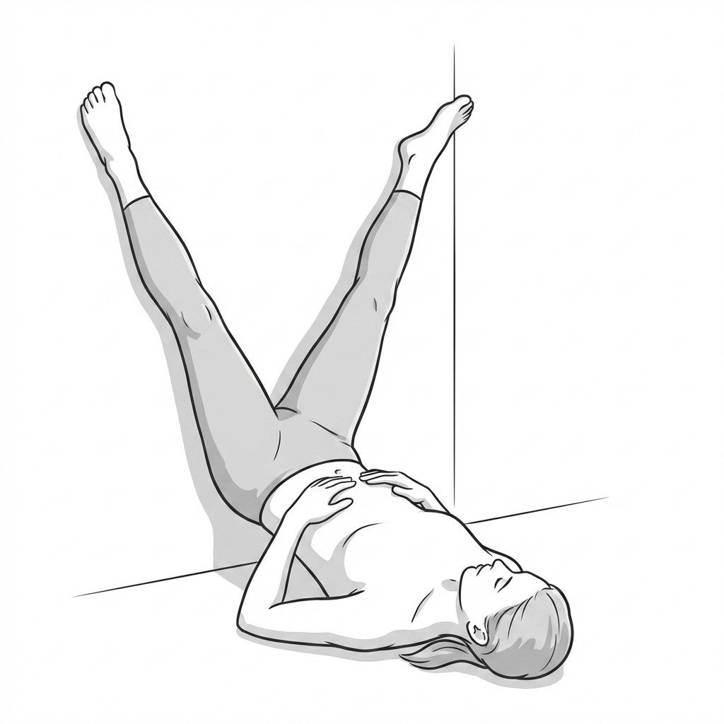
The supine wall straddle utilizes gravity to provide a gentle, prolonged passive stretch to the medial thigh musculature. By fully supporting the legs against a wall, the patient can safely control the range of motion without active eccentric loading, promoting tissue relaxation and elasticity in healing adductor strains.
Start Exercise →Lower Leg
Tandem stance
The tandem stance narrows the base of support to challenge postural control and directly loads the ankle stabilizers. For Posterior Tibial Tendon Dysfunction, this exercise builds isometric strength and proprioceptive capacity in the tibialis posterior to better control pronation and support the medial longitudinal arch.
Start Exercise →Cervical
Targeted antagonistic isometrics
This exercise utilizes the neurological principle of reciprocal inhibition by voluntarily activating the muscles that oppose the dystonic spasm. Isometric contraction of these antagonist muscles reflexively decreases the neural drive to the overactive, spasming tissues. This helps temporarily reduce abnormal neck posturing, alleviate pain, and restore voluntary neuromuscular control.
Start Exercise →Elbow & Hand
Tendon glides
Tendon glides promote the differential gliding of the flexor digitorum superficialis and profundus tendons through the carpal tunnel. This active mobilization is thought to reduce tenosynovial adhesions and localized edema and may help relieve mechanical pressure on the median nerve.
Start Exercise →Knee
Terminal knee extensions
Terminal knee extensions isolate the final degrees of knee extension, preferentially engaging the quadriceps and heavily recruiting the vastus medialis. By strengthening these structures and improving motor control in the terminal range, this exercise may help improve patellar tracking and reduce patellofemoral compressive stress.
Start Exercise →Knee
TFL stretch
This exercise applies a prolonged tensile load to the tensor fasciae latae and the proximal iliotibial band complex to help reduce myofascial resting tone. Decreasing tension transmitted through the IT band may help reduce the compressive forces at the lateral femoral condyle, a proposed mechanism in IT Band Syndrome.
Start Exercise →Hip
Thomas stretch
The Thomas stretch elongates the anterior hip flexors, specifically the iliopsoas and rectus femoris, which frequently become adaptively shortened in hip osteoarthritis. By restoring anterior hip flexibility, this stretch may help reduce capsular tightness and improve functional hip extension during the gait cycle.
Start Exercise →Cervical
Thoracic extension mobility
Thoracic extension mobility addresses upper back stiffness, which often drives compensatory lower cervical flexion and upper cervical hyperextension. Improving thoracic mobility may help reduce mechanical load on the upper cervical joints, which may in turn ease the referred pain patterns associated with cervicogenic headaches.
Start Exercise →Thoracic & Core
Thoracic rotation with strap
This exercise utilizes a strap to provide a localized mobilization-with-movement to the thoracic spine and posterior rib joints. By improving posterior costovertebral and costotransverse joint mobility, it may help reduce compensatory hypermobility and mechanical strain at the anterior costochondral junctions inflamed in costochondritis.
Start Exercise →Shoulder
Thread-the-needle
Thread-the-needle is a quadruped mobility exercise that promotes thoracic spine rotation while dynamically stretching the posterior shoulder and periscapular musculature. By uncoupling the movement of the ribcage from the scapula, it may help mobilize stiff spinal segments and reduce tension in tight interscapular muscles. This may help ease rhomboid pain often associated with prolonged static postures or scapular dyskinesis.
Start Exercise →Elbow & Hand
Thumb band strengthening
This exercise provides targeted progressive load to the abductor pollicis longus and extensor pollicis brevis tendons. By gradually exposing these structures to tension, it is thought to support collagen remodeling and improve tissue tolerance in the tendinopathy of De Quervain's Tenosynovitis.
Start Exercise →Lower Leg
Tibialis anterior raises
Tibialis anterior raises strengthen the primary dorsiflexor of the ankle to improve shock absorption during gait and running. By increasing the load capacity of the anterior compartment, this exercise may help reduce eccentric load on the tibia and surrounding fascia, supporting recovery from Medial Tibial Stress Syndrome.
Start Exercise →Lower Leg
Toe yoga
Toe yoga involves alternating the lifting of the great toe and the lesser toes to isolate and strengthen the intrinsic and extrinsic foot muscles. By enhancing neuromuscular control of the 'foot core', this exercise helps support the medial longitudinal arch and may reduce mechanical tension on the plantar fascia. This improved load-bearing capacity may help reduce symptoms of plantar fasciitis.
Start Exercise →Lower Leg
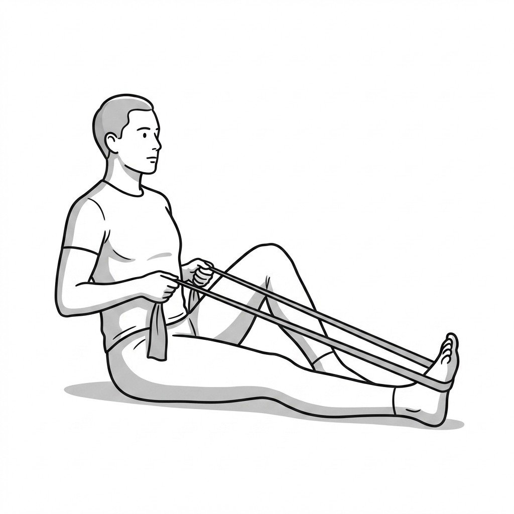
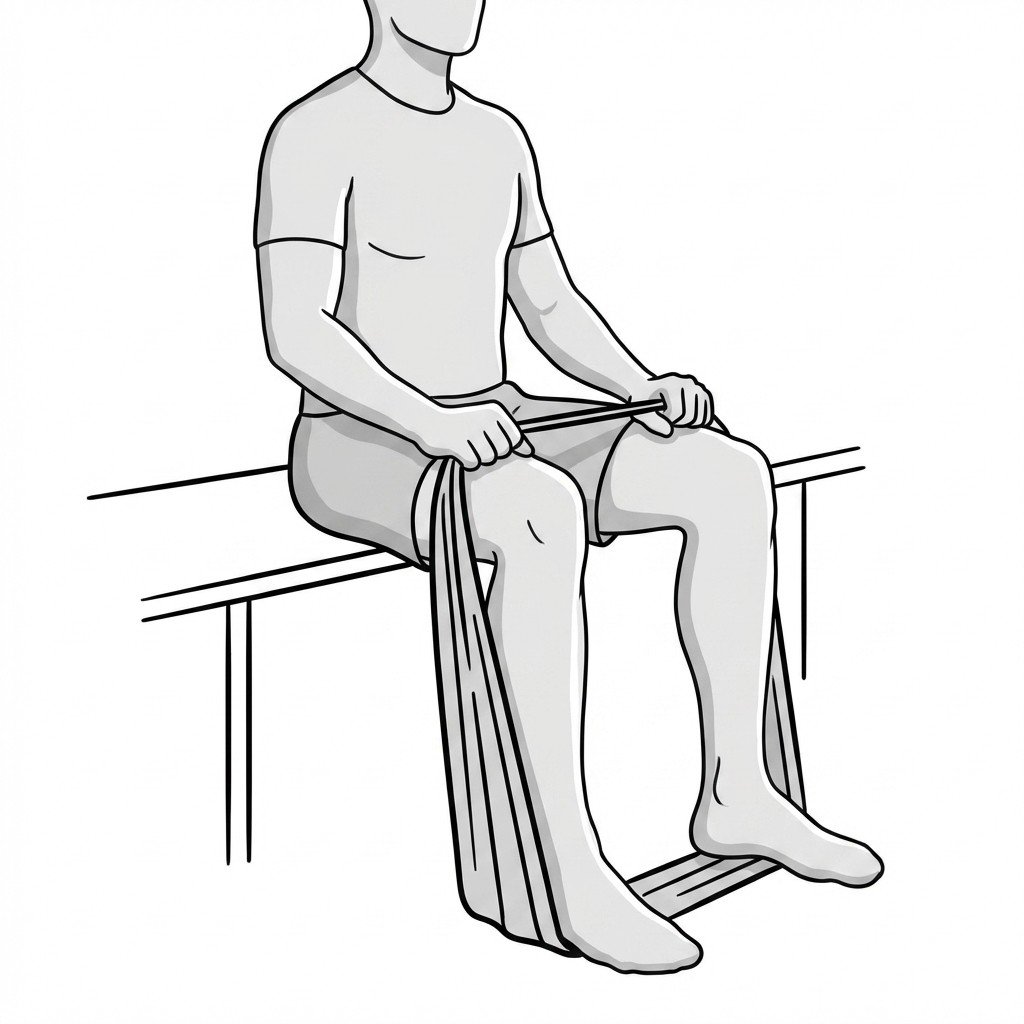
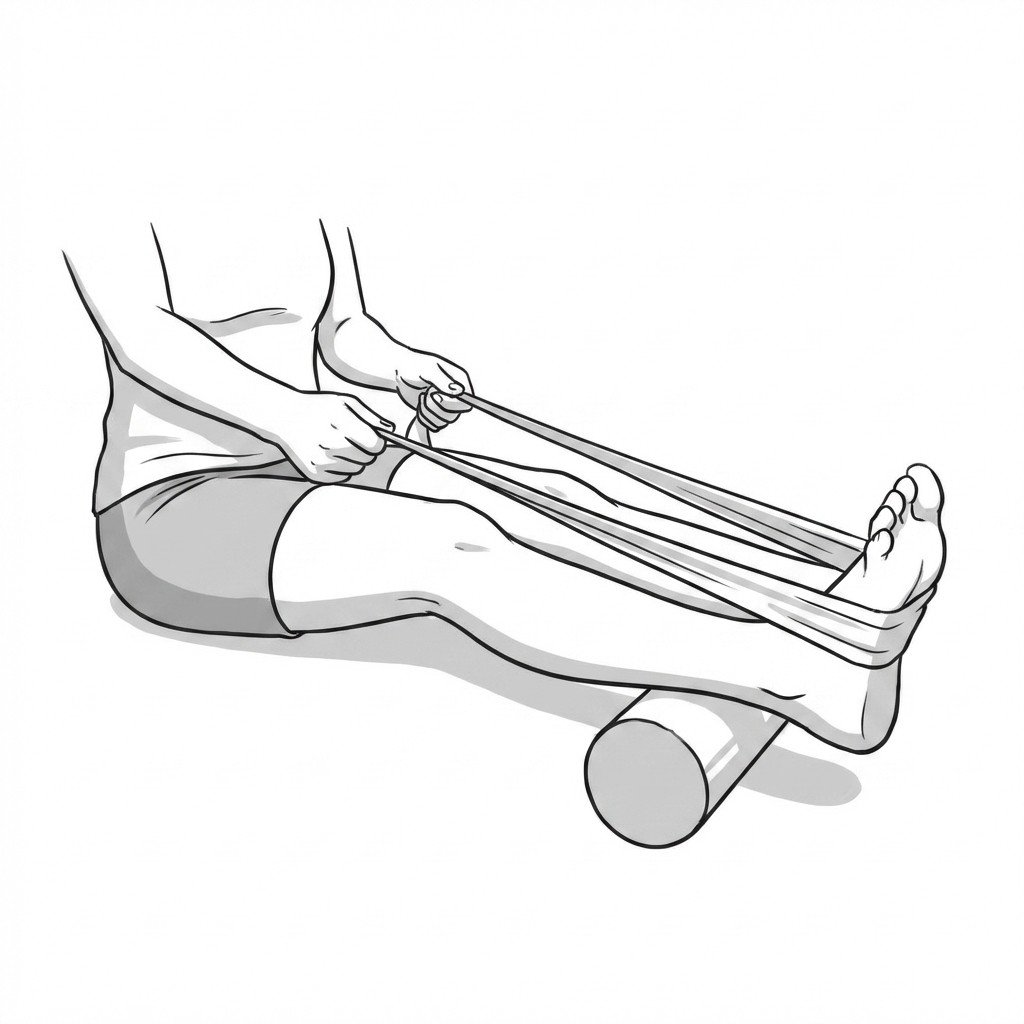
Towel pull plantar fascia stretch
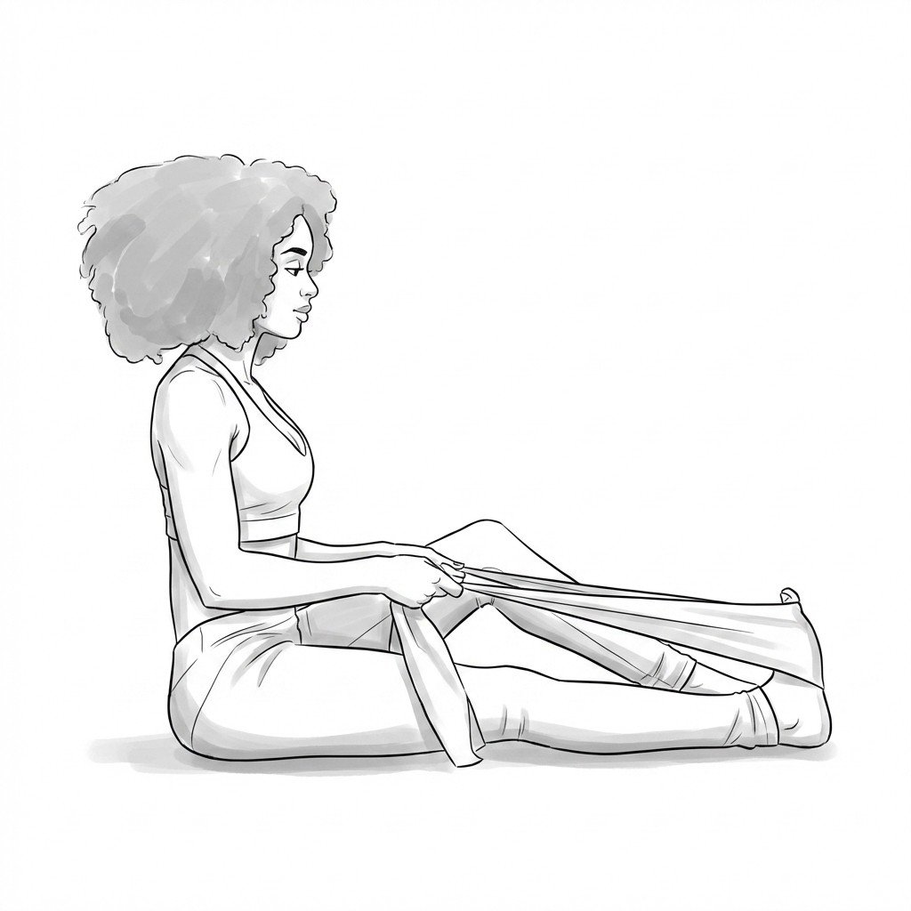
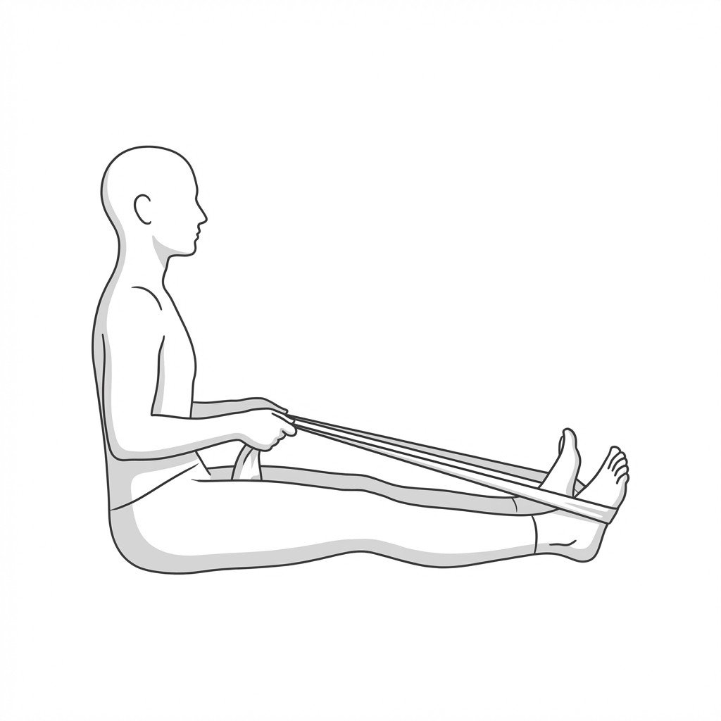
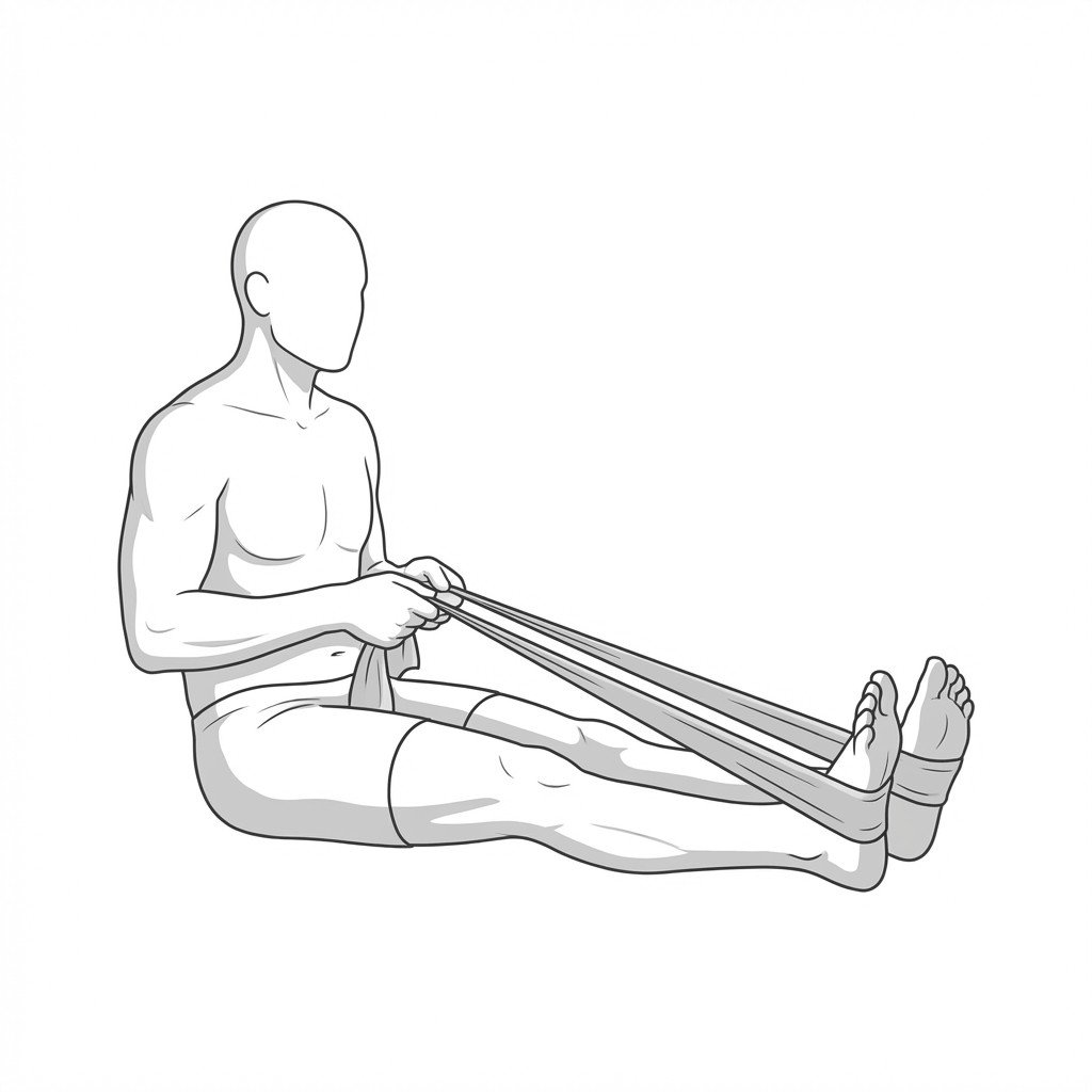
The towel pull plantar fascia stretch applies a tensile load across the longitudinal arch of the foot and the Achilles tendon complex. By passively dorsiflexing the ankle and extending the toes, it elongates the plantar aponeurosis and calf musculature, which may help reduce tissue stiffness and ease tension at the calcaneal insertion.
Start Exercise →Lower Leg
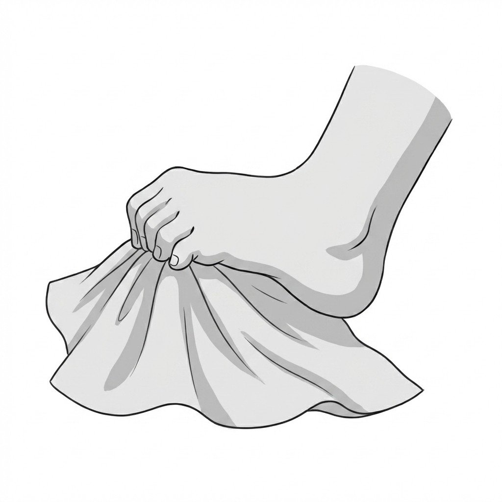
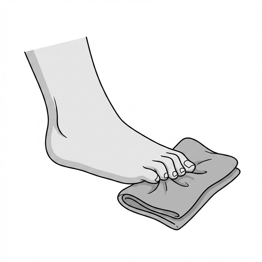
Towel scrunches
Towel scrunches target the intrinsic muscles of the foot to strengthen the medial longitudinal arch. By enhancing dynamic muscular support of the foot, this exercise may help reduce tension and load on the plantar fascia during weight-bearing activities.
Start Exercise →Thoracic & Core
Transversus abdominis draw-in
The transversus abdominis draw-in maneuver selectively activates the deepest abdominal muscle to increase intra-abdominal pressure and tension the thoracolumbar fascia. This is proposed to create a natural corset effect that may improve dynamic spinal stability and help reduce lumbar shear forces in people with poor lumbopelvic control.
Start Exercise →Elbow & Hand
Triceps isometric holds
Triceps isometric holds create static tension in the triceps muscle without changing the muscle length or moving the elbow joint. This minimizes sheer forces on the triceps tendon and ulnar nerve, providing an analgesic effect while safely loading the musculotendinous unit. It can be useful in early rehabilitation for building tissue capacity, typically without aggravating the cubital tunnel.
Start Exercise →Elbow & Hand
Tyler Twist
The Tyler Twist is an eccentric strengthening exercise specifically designed to load the common extensor tendon using a flexible rubber resistance bar. By emphasizing the controlled eccentric (lengthening) phase of wrist extension, it is thought to stimulate collagen synthesis and tendon remodeling, which may help reduce pain and improve function in lateral epicondylalgia.
Start Exercise →Elbow & Hand
Ulnar nerve glide
This exercise dynamically mobilizes the ulnar nerve through its anatomical sheaths, including the cubital tunnel and Guyon's canal. By gliding the nerve rather than statically stretching it, the exercise may help reduce intraneural edema, minimize scar tissue tethering, and improve neural mobility.
Start Exercise →Cervical
Upper trapezius stretch
The upper trapezius stretch specifically elongates hypertonic muscle fibers by increasing the distance between the cervical spine and the acromion. Relieving tension in this region may help reduce nociceptive input from myofascial trigger points, which is a common driver of axial neck pain.
Start Exercise →Shoulder
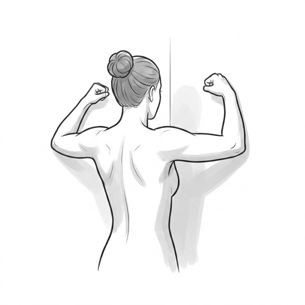
Wall angels
Wall angels mechanically enforce thoracic extension and scapular retraction while simultaneously gliding the arms overhead against a rigid surface. This activates the scapular retractors and depressors while actively mobilizing the anterior chest wall, helping to reverse postural deficits associated with thoracic kyphosis and interscapular strain.
Start Exercise →Shoulder
Wall biceps stretch
The wall biceps stretch elongates the long and short heads of the biceps brachii, reducing resting muscle tension and improving tissue extensibility. By alleviating contractile tightness pulling on the proximal or distal tendon, it helps decrease focal pain and mechanical stress associated with biceps tendinopathy.
Start Exercise →Knee
Wall sit isometric
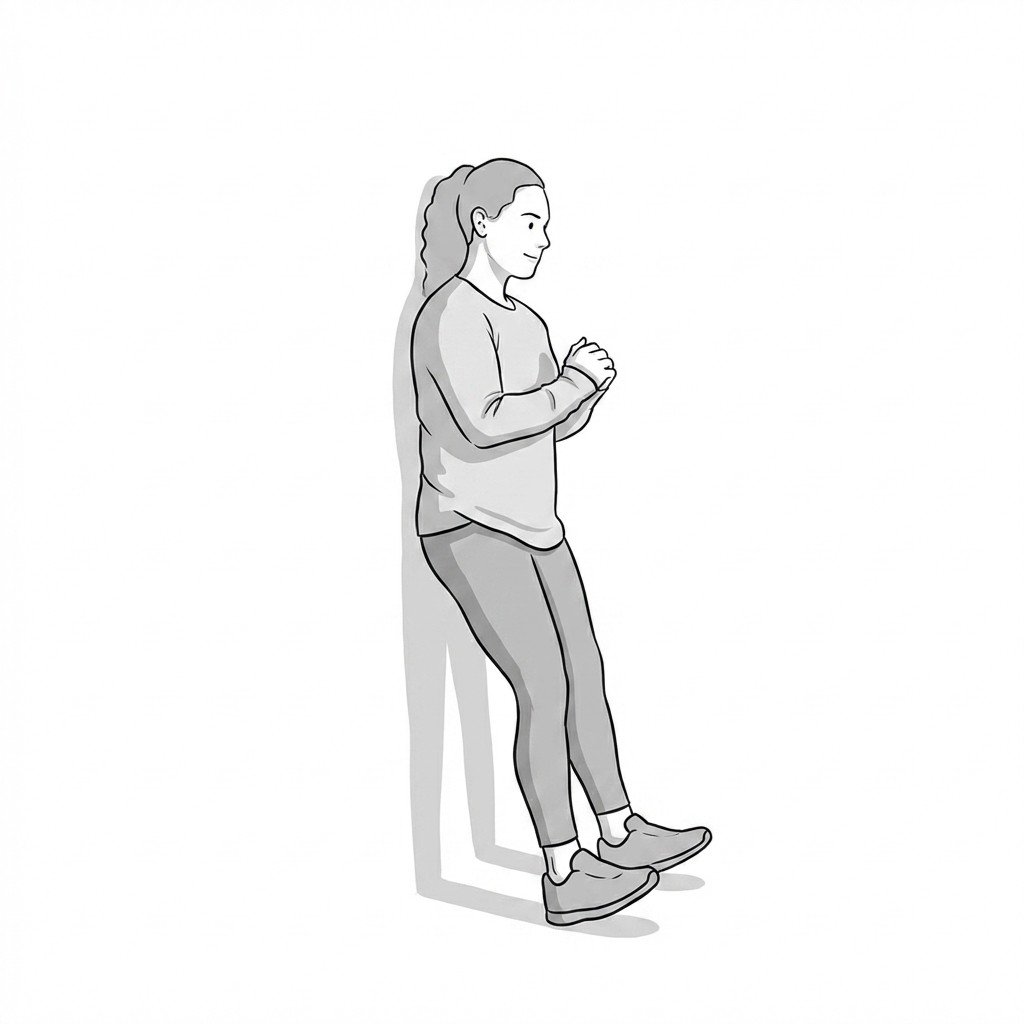
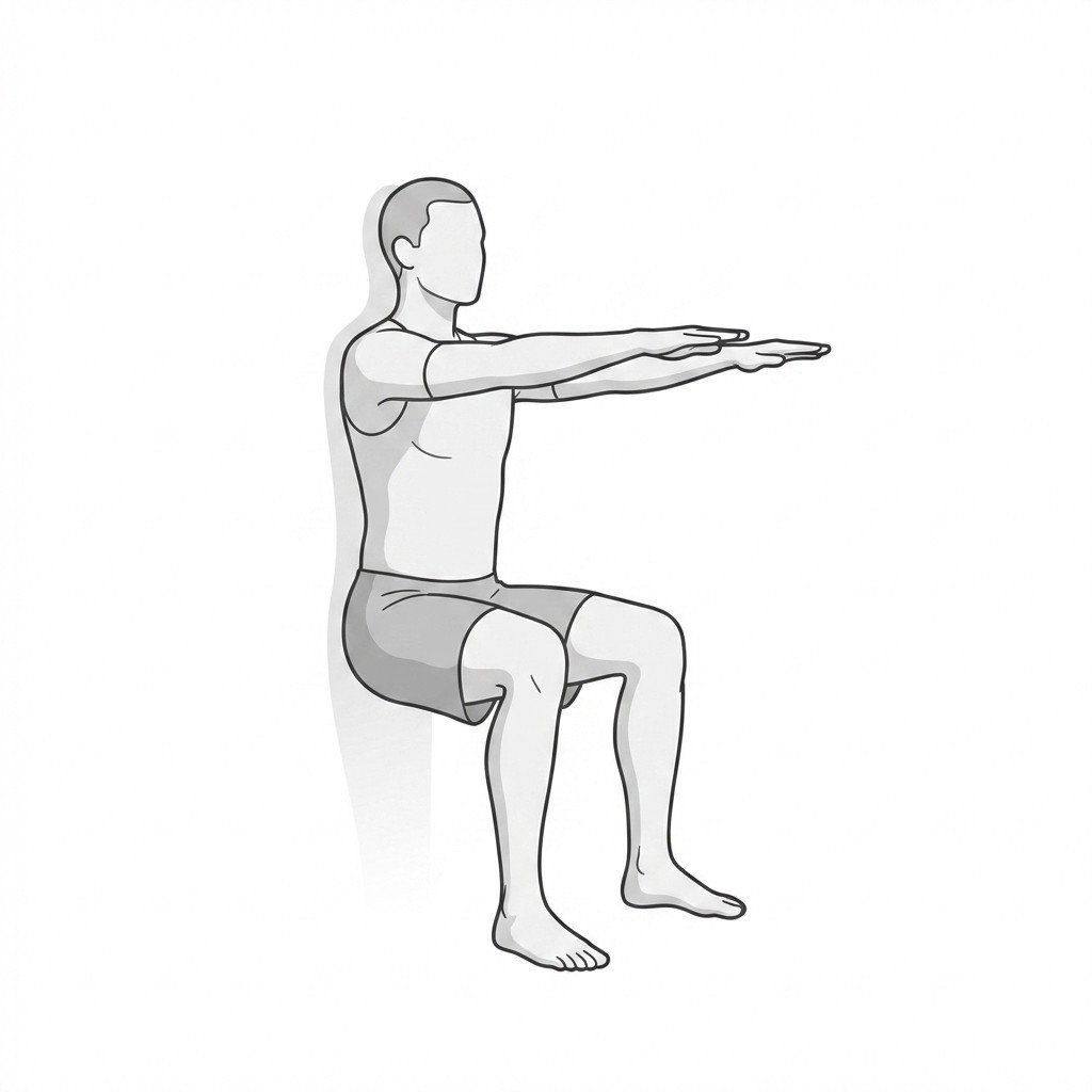
The wall sit provides a heavy isometric load to the quadriceps and patellar tendon without the compressive shear forces of isotonic movement. This sustained contraction helps induce central pain modulation and normalizes motor unit recruitment. It can be a useful tool to help manage tendon-related pain while maintaining local load capacity.
Start Exercise →Knee
Wall sits
Wall sits provide a closed-kinetic chain isometric contraction of the quadriceps, targeting the vastus muscles while minimizing shear forces on the patellofemoral joint. This isometric loading may provide a short-term local analgesic effect and builds the baseline muscular endurance necessary for dynamic knee stability.
Start Exercise →Lower Leg
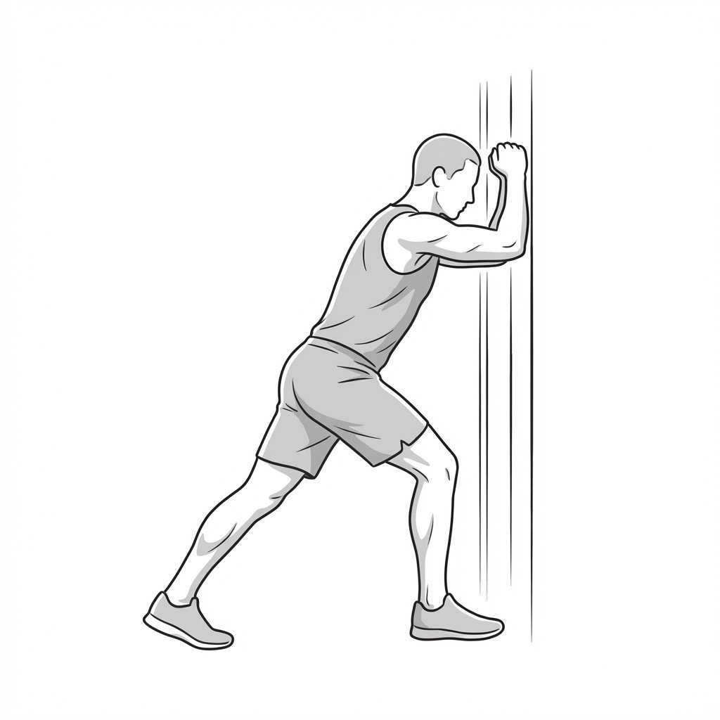
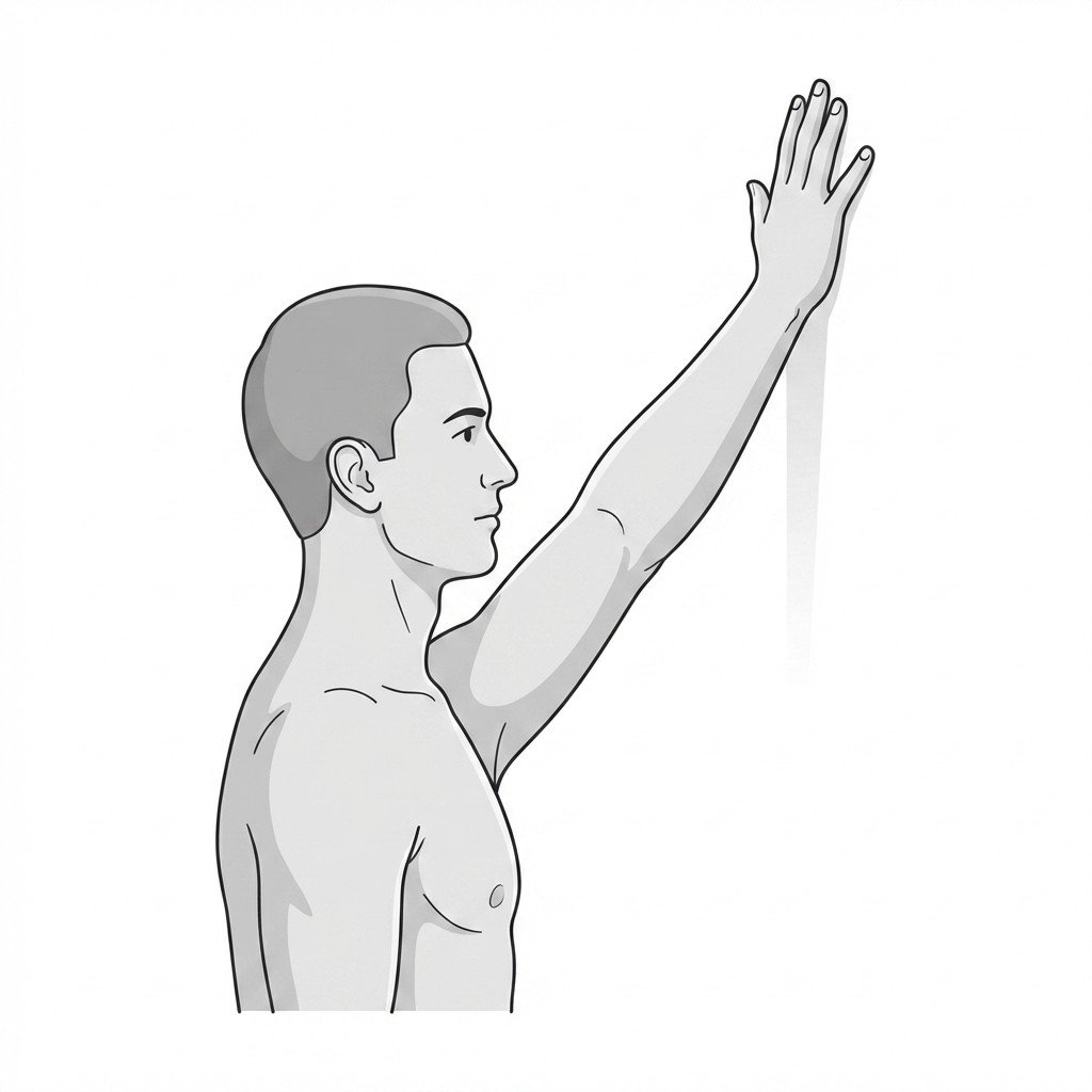
Wall slant calf stretch
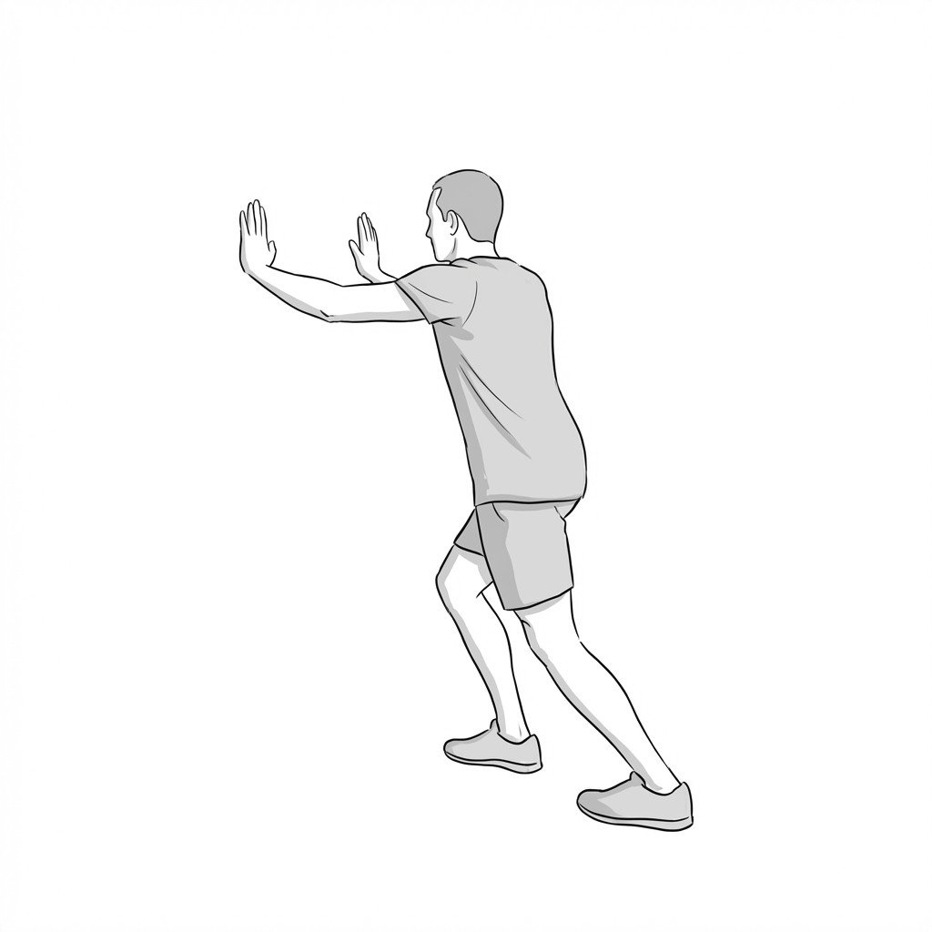
The wall slant calf stretch provides a prolonged, passive stretch to the gastrocnemius and soleus muscles, increasing tissue extensibility in the posterior lower leg. By improving ankle dorsiflexion, it may help reduce mechanical tension on the plantar fascia during gait. This helps alleviate heel pain associated with plantar fasciitis.
Start Exercise →Shoulder
Wall slides with liftoff
Wall slides with liftoff facilitate scapular upward rotation and posterior tilt, helping to correct altered movement patterns characteristic of scapular dyskinesis. The sliding motion heavily engages the serratus anterior, while the terminal liftoff emphasizes lower trapezius activation.
Start Exercise →Shoulder
Wall walk
The wall walk acts as an active-assisted range of motion (AAROM) exercise that progressively stretches the tightened glenohumeral joint capsule. By using the fingers to incrementally elevate the arm, it may help improve shoulder flexion and reduce capsular tightness in patients with adhesive capsulitis.
Start Exercise →Elbow & Hand
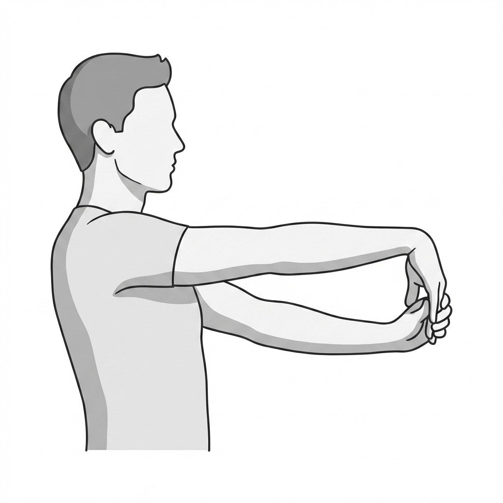
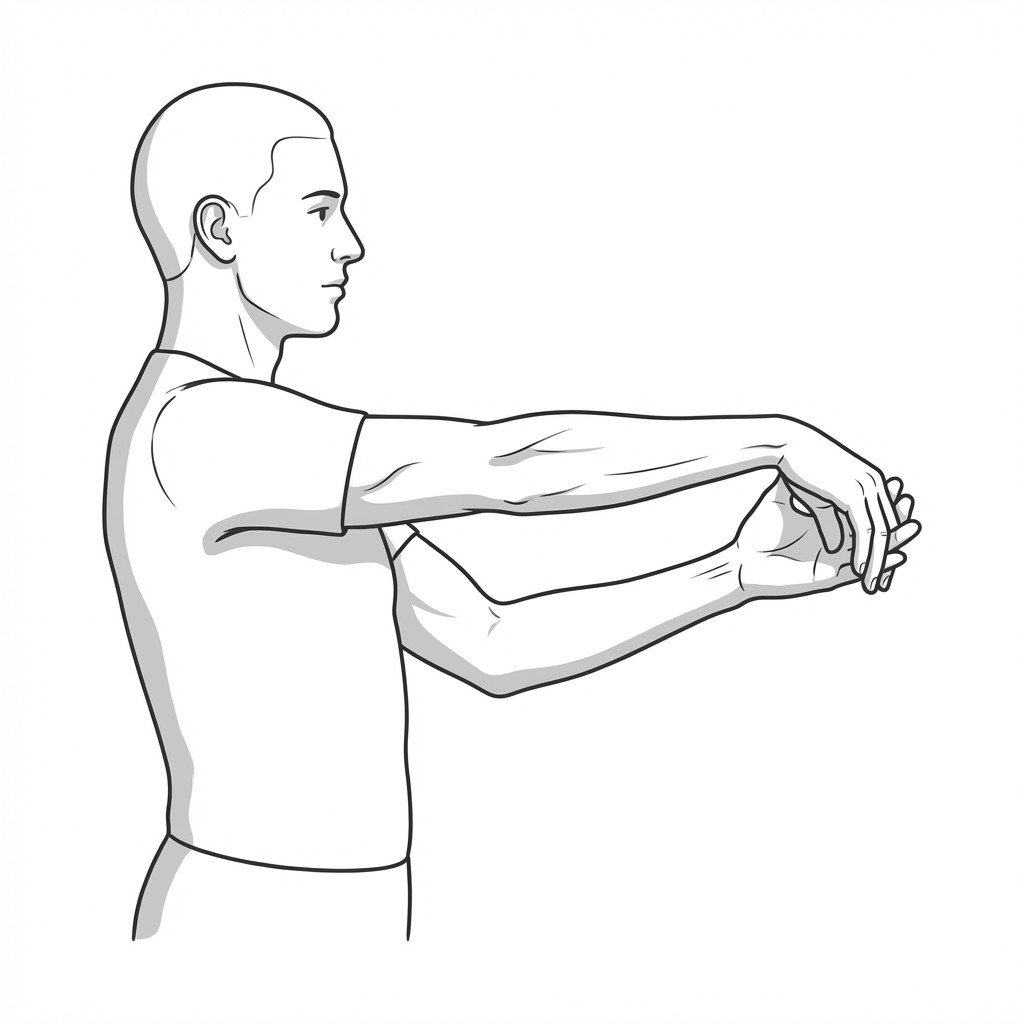
Wrist extensor stretch
This exercise lengthens the common extensor tendon and wrist extensor musculature, reducing resting tissue tension at the lateral epicondyle. By reducing sensitivity and tension in the extensor carpi radialis brevis and increasing tissue compliance, it may help alleviate the localized pain and stiffness associated with lateral epicondylalgia.
Start Exercise →Elbow & Hand
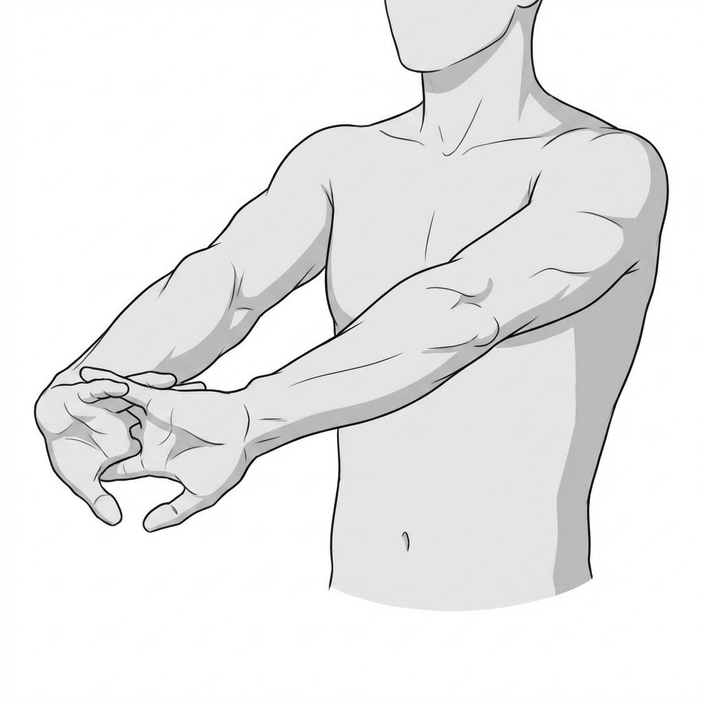
Wrist flexion stretch
This exercise elongates the extensor muscles and fascia on the dorsal aspect of the forearm by moving the wrist into a sustained flexed position. Restoring flexibility to the wrist extensors helps balance overall forearm muscle tension, reducing strain on the flexor tendons that pass through the carpal tunnel.
Start Exercise →Elbow & Hand
Wrist flexor stretch
The wrist flexor stretch elongates the muscle-tendon unit of the forearm flexors and pronators originating at the medial epicondyle. This mechanical tension is thought to support collagen organization during early tendon remodeling, which may reduce tissue stiffness and help alleviate pain associated with medial epicondylalgia.
Start Exercise →