Skip to main content
Atlas · Neck

Semispinalis Cervicis

Semispinalis cervicis is a deep posterior neck muscle linking the upper thoracic spine to cervical vertebrae. It contributes to neck extension and is relevant to cervical strength and endurance assessment, while ordinary neck pain rarely identifies one deep muscle as its sole source.

Semispinalis Cervicis
Body region
Neck
Reference type
Anatomy & examination
Trigger-point map not yet documented
Common symptoms
2
patterns cataloged
Contributors
2
Factors to assess

Muscle overview

Symptoms and first steps

Practical first steps

  • Try comfortable support and varied positions during prolonged tasks.
  • Keep comfortable head movement rather than holding the neck rigid.
  • Avoid forceful attempts to press through superficial muscles to reach a deep point.

When to seek assessment

  • New or progressive arm weakness, hand clumsiness or balance difficulty needs medical assessment.
  • Sudden unusual neck pain or headache with visual, speech, balance or other neurological symptoms requires urgent assessment.

Semispinalis cervicis attaches to cervical vertebrae; semispinalis capitis reaches the skull. The distinction matters anatomically, but a head-lifting task does not isolate either muscle.

Examination, palpation and treatment evidence

Illustrated overview

Semispinalis Cervicis · Anatomy

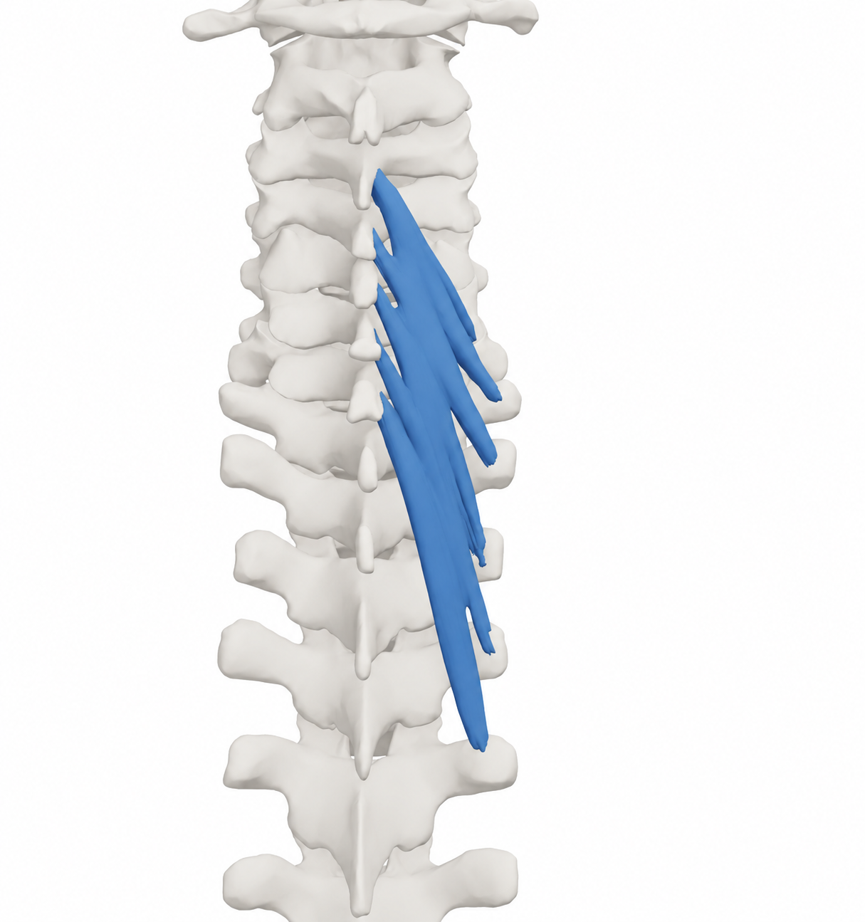
Right semispinalis cervicis with the source skeletal context. Source model rendered and material restyled; blue identifies the model structure, including its tendon-like continuations.

BodyParts3D, © The Database Center for Life Science. Adapted by MyofascialPain: source model rendered and material restyled. · CC BY-SA 2.1 Japan

Open full-size image
Browse related exercises (1)

Symptoms patients report

Posterior neck discomfort with sustained tasks Describe task duration and symptom behavior rather than assign symptoms to a deep muscle from their location.

Limited looking-up or head-turning tolerance Movement and endurance are regional examination findings influenced by several muscles and joints.

Contributors to assess

A change in sustained head demand Review work, reading and activity exposure in relation to the onset of symptoms.

Regional neck movement or capacity limitation A measured deficit can guide rehabilitation without claiming that one muscle has become inactive.

For clinicians and supervised training

Clinical reference

No source-supported trigger-point coordinates or referral map are included. Exercise-study participation does not establish a patient’s pain source.

Anatomy and palpation

Anatomical relationships

Semispinalis cervicis spans from upper thoracic transverse processes, commonly T1–T5 or T6, to the spinous processes of C2–C5. It lies deep to semispinalis capitis and contributes to cervical extension. Innervation is through medial branches of spinal dorsal rami.[1]

  • Upper thoracic transverse-process region.
  • C2–C5 posterior midline attachment region.

Nearby structures. Neighboring semispinalis capitis, multifidus, facet joints and dorsal nerve branches make surface isolation unreliable.

Palpation

Patient position. Seated or lying with the head comfortably supported.

Find the landmarks

  • Posterior cervical midline.
  • Upper thoracic midline and surrounding extensor region.

Examination sequence

  1. Use light regional contact only when useful to compare symptom sensitivity.
  2. Describe the region and response; do not claim to palpate a uniquely identified deep cervicis nodule.

Document the findings

  • Tenderness is a regional observation, not confirmation of a muscle-specific pain generator.

Limits and precautions

  • Avoid forceful deep pressure or end-range neck loading when symptoms have not been assessed.
[1][3]

Examination and tests

2

Active cervical movement and task response[2]

Measure the direction and daily task that are difficult.

Position
Seated with comfortable trunk support.
Technique
Observe comfortable rotation, side bending and flexion/extension, recording range and familiar symptoms with a consistent method. Compare a relevant looking-up or head-turning task.
Positive finding
Repeatable limitation or symptom reproduction is a regional finding.
Interpretation
Provides a baseline for graded movement.
Limitations
Several muscles and joints contribute. Range, posture or tenderness cannot identify an isolated deep-extensor pain source.

Neurological and headache history screen[2][3]

Identify findings that change a routine neck-pain assessment.

Position
History first; clinician-selected upper-limb examination and gait assessment when indicated.
Technique
Ask about symptom onset, arm deficits, hand dexterity, balance and headache features; compare relevant strength, sensation and reflexes.
Positive finding
Progressive neurological change or sudden unusual head/neck pain with neurological symptoms needs medical assessment.
Interpretation
Determines whether regional rehabilitation is appropriate.
Limitations
Normal isolated findings cannot exclude every disorder. This is not a muscle-isolation or vascular-clearance maneuver.

Distinguishing other causes

Regional mechanical neck pain[2]

Findings that matter. Symptoms vary with neck movement or sustained tasks without progressive neurological change.

Next step. Use regional movement and capacity measures to guide active care.

Cervical nerve-root or spinal-cord disorder[2]

Findings that matter. Arm deficits, dexterity loss or gait/balance change.

Next step. Medical/neurological assessment before attributing symptoms to deep muscle tenderness.

Headache or vascular presentation requiring separate assessment[3]

Findings that matter. A new unusual headache, sudden neck pain or associated neurological features.

Next step. Assess onset and associated features and escalate urgently when indicated.

Treatment selection and progression

Self-care and activity

Vary sustained head tasks[2]

Consider when. Assessed mechanical neck discomfort during sustained activity.

Try forearm or head support and alternate positions during the task that becomes uncomfortable.

Dose and review
Use the individual task response rather than a compulsory break interval.
Progression
Gradually build useful task tolerance.
Stop or reassess
Reassess new arm or head symptoms.
Evidence and limits
Regional self-management; no ideal posture or isolated-muscle cure is implied.

Exercise and rehabilitation

Practise comfortable head movement[2][3]

Consider when. A measured movement limitation after assessment.

Practise the relevant head turn or small extension movement at a comfortable speed and range.

Dose and review
Select repetitions with the treating clinician according to irritability; no isolated-muscle dose is established.
Progression
Build range and ordinary task exposure before adding resistance.
Stop or reassess
Stop for dizziness, visual change, unusual headache or new arm symptoms.
Evidence and limits
A regional movement option; pain during a direction does not identify one muscle.

Clinician-delivered care

Build neck strength and endurance when indicated[4][5][2]

Consider when. Persistent neck pain with an assessed capacity limitation.

A physiotherapist selects tolerable neck and shoulder-girdle loading and may add gentle regional mobilization when relevant. Review progress against a useful daily task; specific deep-extensor work is one exercise option.

Dose and review
Individualize load and recovery; the cited six-week studies do not supply a universal home dose.
Progression
Progress only when technique and daily activity tolerance improve.
Stop or reassess
Reassess neurological change or sustained worsening.
Evidence and limits
A 2019 trial favored deep-muscle exercise over usual care; a 2022 trial found comparable pain/disability outcomes from specific and general extensor programs. Neither proves isolated-muscle causation.

Reassessment and follow-up

  • Repeat the same active motion measure.
  • Track the relevant sustained task and disability separately from strength.
  • Review new neurological or headache features before progressing exercise.

Evidence and sources

  1. Gray’s Anatomy: Back muscles, semispinalis cervicis

    2008 · Publisher-hosted anatomical textbook chapter

    Publisher-indexed chapter text checked; direct reader failed. Upper five or six thoracic transverse processes to C2–C5 spinous processes, medial dorsal-ramus branches and extension. Does not establish a referral field.

  2. Blanpied et al. Neck Pain: Revision 2017

    2017 · Clinical practice guideline

    Regional examination and active rehabilitation framework. No test below is claimed to isolate a painful deep extensor.

  3. Rushton et al. International Framework for Examination of the Cervical Region for Potential of Vascular Pathologies of the Neck Prior to Musculoskeletal Intervention: International IFOMPT Cervical Framework

    2023 · International clinical framework

    Online 2022, 2023 issue. History and neurological/vascular differential before intervention; not a clearance test based on neck positioning.

  4. Suvarnnato et al. Effect of specific deep cervical muscle exercises on functional disability, pain intensity, craniovertebral angle, and neck-muscle strength in chronic mechanical neck pain: a randomized controlled trial

    2019 · Randomized controlled trial

    54 participants; six-week deep-extensor or deep-flexor exercise programs compared with usual care. Clinical outcomes support exercise relevance, not proof of an isolated muscle pain generator or a trigger-point map.

  5. Giménez-Costa et al. Specific versus non-specific exercises for the neck extensor muscles in women with chronic idiopathic neck pain: A randomized controlled trial

    2022 · Randomized controlled trial

    43 women, six weeks: specific lower-deep-extensor and general extensor exercise produced comparable pain/disability outcomes. No between-group advantage establishes a need to isolate semispinalis cervicis.