Skip to main content
Atlas · Forearm

Pronator Quadratus

Pronator quadratus is the deep muscle near the wrist that turns the palm downward. Turning a key or tool involves this muscle along with other structures; discomfort there needs a wrist, forearm and nerve assessment rather than a diagnosis from one tender spot.

Pronator Quadratus
Body region
Forearm
Reference type
Anatomy & examination
Trigger-point map not yet documented
Common symptoms
2
patterns cataloged
Contributors
2
Factors to assess

Muscle overview

Symptoms and first steps

Practical first steps

  • Use assistance for forceful twisting while the cause is assessed.
  • Keep comfortable rotation without forcing a painful wrist.
  • Avoid trying to reach this deep muscle with hard pressure.

When to seek assessment

  • Arrange assessment for new pinch or rotation weakness, especially after injury.
  • Seek urgent care for severe injury-related pain, deformity or loss of hand circulation.

Pronator teres can preserve some pronation when pronator quadratus is weak. Interpret rotation strength alongside thumb and index-tip flexion.

Examination, palpation and treatment evidence

Illustrated overview

Pronator Quadratus · Right anterior anatomy

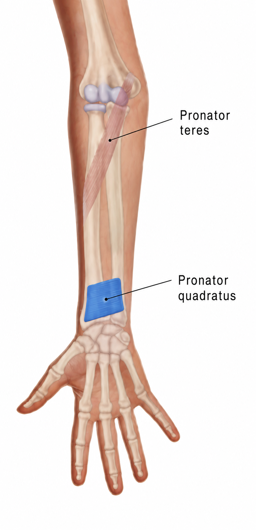
Right anterior forearm and palm, with pronator quadratus in blue and pronator teres ghosted in. Muscolino, Fig. 14-33A, p. 301. The two leader-end dots identify the named muscles; they are anatomical labels, not trigger points. No referred-pain pattern is shown.

Open full-size image
Browse related exercises (1)

Symptoms patients report

Difficulty with forearm rotation Record the direction, force and task that provoke discomfort; the distal radioulnar joint and neighboring tendons also contribute.

Weak turning or pinch True motor loss, particularly with thumb/index-tip weakness, calls for examination beyond local muscle tenderness.

Contributors to assess

Increased turning demand A change in tool use may expose a task limitation without establishing which tissue is responsible.

Wrist injury or nerve dysfunction An injury history or motor deficit changes the assessment and the choice of loading.

For clinicians and supervised training

Clinical reference

Anatomy and clinical reference; no source-supported trigger-point coordinates or referral map are supplied.

Anatomy and palpation

Anatomical relationships

A deep transverse muscle from the anterior distal ulna to the anterior distal radius. It pronates the forearm; deep fibers help bind the radius and ulna. Motor supply is the anterior interosseous branch of the median nerve.[1][2]

  • Distal ulna on the little-finger side of the forearm.
  • Distal radius on the thumb side, proximal to the wrist joint.

Nearby structures. Deep to the long flexor tendons near the interosseous membrane and distal radioulnar joint; nearby nerves and vessels limit direct pressure access.

Palpation

Patient position. Forearm supported, hand relaxed.

Find the landmarks

  • Distal radius.
  • Distal ulna and wrist joint line.

Examination sequence

  1. Observe rotation and identify surface landmarks with light contact.
  2. Record the broad location of tenderness without claiming to isolate the deep muscle.

Document the findings

  • Local tenderness is nonspecific and does not establish a trigger point or nerve entrapment.

Limits and precautions

  • Do not use forceful deep compression at the volar wrist or over an acute injury.
[1][3]

Examination and tests

2

Forearm pronation performance[1][4]

Compare comfortable rotation and motor performance.

Position
Elbow bent, forearm supported and initially palm up.
Technique
Observe active palm-down rotation; if injury has been excluded, a clinician can compare gentle resistance while limiting shoulder substitution.
Positive finding
Repeatable weakness, restricted motion or pain warrants regional assessment.
Interpretation
Helps distinguish a motion limitation from a motor complaint.
Limitations
Pronator teres also contributes. This does not isolate PQ, diagnose AIN palsy or identify a trigger point.

Anterior interosseous motor pattern[3][4]

Compare thumb-tip, index-tip and pronation function when motor weakness is reported.

Position
Seated with the forearm supported.
Technique
Observe thumb IP and index DIP flexion, including a gentle tip-to-tip pinch. Compare forearm pronation and examine other median/ulnar functions and sensation as indicated.
Positive finding
A reproducible motor deficit needs a tendon and neurological differential.
Interpretation
The combined pattern can guide further assessment.
Limitations
Pain, tendon injury and effort can alter performance. An OK-sign difficulty alone neither diagnoses AIN palsy nor a trigger point; sensory symptoms suggest a broader assessment.

Distinguishing other causes

Distal radioulnar or other wrist injury[1][6]

Findings that matter. Rotation pain after trauma, swelling or mechanical restriction.

Next step. Assess the joint and injury history before resisted exercise.

Anterior interosseous or broader median motor disorder[3]

Findings that matter. Rotation weakness combined with thumb/index-tip flexion loss.

Next step. Perform a neurological/tendon examination; electrodiagnostic or imaging assessment may be indicated.

Regional flexor tendon or forearm load-related pain[2][6]

Findings that matter. Symptoms vary with gripping or wrist/finger movement rather than rotation alone.

Next step. Compare the relevant tasks and adjacent structures; do not infer a PQ lesion from pain location.

Treatment selection and progression

Self-care and activity

Adjust the task that provokes symptoms[5][6]

Consider when. Mild task-related symptoms after significant injury and new motor loss have been excluded.

Reduce the force or uninterrupted time needed for turning a handle or tool; try assistance or a different handle and keep comfortable daily use.

Dose and review
Match exposure to the current response; no muscle-specific schedule is established.
Progression
Restore one demand at a time.
Stop or reassess
Seek review for persistent functional loss or worsening symptoms.
Evidence and limits
Regional hand guidance supports activity adaptation; this is not a treatment for a proven isolated muscle lesion.

Exercise and rehabilitation

Maintain comfortable active movement[5]

Consider when. Assessed mild stiffness without a suspected tendon tear, unstable injury or postoperative restriction.

With the elbow comfortably bent, turn the palm up and down. Stay within an easy range without forcing the movement.

Dose and review
Use brief practice adjusted to symptoms; a treating clinician sets any injury-specific dose.
Progression
Increase comfortable range before resistance.
Stop or reassess
Stop for sharp pain, increasing swelling or new weakness.
Evidence and limits
A regional mobility option, not a validated isolated-muscle exercise protocol.

Clinician-delivered care

Match rehabilitation to the examination[5][6]

Consider when. Persistent pain, stiffness or weakness that limits a useful hand task.

A clinician distinguishes tendon, joint and nerve findings, then selects mobility, support or graded loading. Follow the surgical team’s plan after repair.

Dose and review
Set from the diagnosis and measured deficit; no universal strengthening dose.
Progression
Retest the same movement and functional task before increasing load.
Stop or reassess
New loss of active movement changes the assessment rather than calling for harder exercise.
Evidence and limits
Diagnosis determines care. Regional self-management does not establish muscle-specific treatment efficacy.
Reassessment and follow-up

  • Compare the same rotation task and range.
  • Document motor weakness separately from pain-limited effort.
  • If pinch or neurological findings change, reassess the diagnosis before loading further.

Evidence and sources

  1. University of Washington Radiology: Pronator quadratus

    2026 · University anatomy atlas

    Undated page accessed 11 October 2026. Distal ulnar/radial attachments, pronation and anterior interosseous innervation. Copyrighted artwork is not reused.

  2. Texas Tech University Health Sciences Center El Paso: Muscles of the upper limb

    2026 · University anatomy teaching reference

    Undated teaching page accessed 11 October 2026. Cross-checks finger versus thumb joint actions and extrinsic versus intrinsic muscles. Does not validate palpation diagnosis or treatment.

  3. StatPearls: Anterior Interosseous Syndrome

    2025 · Clinical reference

    July 2025 update, publisher-indexed text checked. Motor pattern includes FPL, radial FDP and PQ; pure AIN palsy lacks cutaneous sensory loss. Neuritis and other causes require assessment; not all cases are local compression.

  4. Anatomy, Shoulder and Upper Limb, Forearm Anterior Interosseous Nerve

    2026 · Clinical anatomy reference

    Publisher-indexed clinical-significance section checked 11 October 2026; year is access year. Describes thumb IP, index/middle DIP and pronation motor examination, not trigger-point tests.

  5. Cambridgeshire Community Services NHS: Wrist, hand and thumb pain

    2026 · NHS regional self-management guidance

    Undated page accessed 11 October 2026. Activity adaptation, gradual movement and graded return. Used for regional care, not efficacy or a fixed dose for any one named muscle.

  6. NHS: Wrist pain

    2026 · National patient guidance

    Accessed 11 October 2026; year is access year. Injury, joint/tendon and nerve differential context and when symptoms need assessment.