Skip to main content
Atlas · Neck

Longus Colli

Longus colli lies in front of the cervical vertebrae and helps coordinate neck flexion. It is relevant to deep-flexor examination and exercise, while an uncommon calcific tendon condition can produce abrupt neck pain and painful swallowing that needs medical assessment.
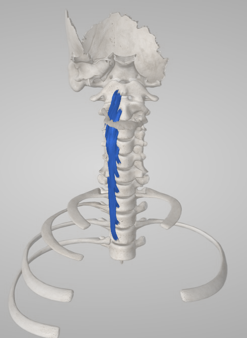
Longus Colli
Body region
Neck
Trigger points
0
documented in this muscle
Common symptoms
3
patterns cataloged
Common causes
3
contributory factors

Muscle overview

Symptoms and first steps

Practical first steps

  • Use comfortable support and brief position changes during sustained tasks.
  • Practise a small relaxed nod only when it is comfortable and appropriate to the assessed presentation.
  • Avoid deep pressure into the throat or beside the cervical airway; acute swallowing symptoms need medical assessment.

When to seek assessment

  • Sudden unusual severe neck pain or headache with double vision, speech difficulty, imbalance, facial symptoms or weakness requires emergency assessment.
  • Progressive arm weakness, hand clumsiness, gait change or new bowel/bladder dysfunction needs prompt medical assessment.
  • Acute neck pain with painful or difficult swallowing, fever, swelling or recent infection needs medical assessment before massage or stretching; breathing difficulty is an emergency.

Separate a low-load neck coordination problem from acute longus-colli calcific tendinitis. Abrupt stiffness with painful swallowing is a medical differential, not a reason to press deeper into the neck.

Examination, palpation and treatment evidence

Anatomy reference; no mapped trigger points yet.

Symptoms patients report

Neck discomfort during sustained tasks

Reading or prolonged head positioning may be uncomfortable in regional neck pain; this does not localize the complaint to longus colli.

Fatigue with a small controlled nod

A clinician can measure coordination and endurance with the CCFT rather than infer weakness from posture.

Abrupt painful neck stiffness

When accompanied by painful swallowing, this needs medical assessment for calcific tendinitis, infection and other anterior-neck causes.

Common causes

Sustained task demand

A recent increase in reading, screen time or other fixed head tasks can exceed current tolerance; vary exposure according to symptoms.

Regional neck pain with altered coordination

Deep and superficial flexors may use a different activation pattern during neck pain. This is an association and a rehabilitation target, not proof of a damaged stabilizer.

A separate inflammatory tendon presentation

Calcific tendinitis is a distinct condition near the upper longus-colli attachment; it should not be relabeled as a chronic trigger point.

Treatment & self-care

Everyday adjustments

Make sustained reading and screen tasks more comfortable

Assessed mechanical neck discomfort associated with sustained tasks, without acute swallowing or neurological symptoms. Support the forearms and bring reading material into a comfortable view. Alternate positions and take a brief movement break before the usual fatigue builds. Choose head support during rest by comfort rather than trying to hold one ideal posture all day. Build a useful task duration gradually while retaining comfortable position changes. Regional neck guidance supports education and active self-management. A screen position does not diagnose or cure a deep-flexor lesion.

Duration
Adjust task bouts to the current symptom threshold; no muscle-specific break interval is established.

Exercise and movement

Practice a comfortable small head nod

An assessed low-load coordination difficulty when a clinician finds that gentle nodding is tolerated. Lie with the head supported and jaw relaxed. Make a very small yes movement without lifting the head or pressing the neck forcefully down. Breathe normally and return to the starting position. A clinician can add feedback when technique is difficult. Build controlled repetitions or a short comfortable hold, then practise relevant upright tasks; strength work may be added separately. A 46-person chronic-neck-pain study found that six weeks of craniocervical-flexion training changed deep/superficial muscle activation compared with strengthening. Coordination findings do not establish isolated-muscle pain relief or long-term recovery.

Duration
Pragmatic starting option: 5 slow comfortable repetitions, resting between attempts. This is not the six-week trial protocol or a compulsory daily dose.

Professional care

Build a tailored neck and shoulder-girdle program

Persistent neck pain or fatigue with measured movement, coordination or endurance limitations. A physiotherapist combines the relevant low-load coordination work with neck/shoulder strength, mobility and gradual task exposure. Comfortable regional mobilization may be tried for short-term relief and followed by a functional retest; direct deep-anterior pressure is unnecessary. Use better task tolerance to progress loading; taper passive care when it does not add useful activity gains. Neck guidelines support combined exercise and regional care. The activation study concerns flexor coordination; evidence for isolated longus-colli or longus-capitis pain treatment remains limited.

Duration
Set the initial course around the measured movement or endurance deficit. Review the same head-turning or reading task and the selected examination measure before extending treatment.

Professional care

Conservative medical care for confirmed calcific tendinitis

A medically assessed acute calcific-tendinitis presentation after infection and other urgent diagnoses have been addressed. A treating clinician may use anti-inflammatory analgesia when appropriate to the medical history and provide short-term comfort measures. Restore comfortable movement as the acute episode settles. Antibiotics, drainage or muscle procedures are not automatically indicated for noninfectious calcific tendinitis. Recheck swallowing, pain and neck motion as directed; failure to improve prompts diagnostic review. The 2017 report included five local cases and a review of 242 published cases. It describes a usually self-limiting condition managed conservatively, with NSAIDs for discomfort; this is case-series evidence, not a comparative medication trial.

Duration
Drug choice and duration require review of contraindications and clinical response; no universal medication dose or immobilization schedule is provided.

Differential diagnosis

  • Acute calcific tendinitis of longus colli
  • Retropharyngeal infection or another acute throat/neck disorder
  • Regional mechanical neck pain
  • Cervical nerve-root, cord or vascular disorder

For clinicians and supervised training

Clinical reference

Anatomy and examination reference. A trigger-point map is not yet documented.

Anatomy and palpation

Anatomical relationships

Longus colli is a paired prevertebral muscle extending from the upper thoracic vertebrae toward the atlas. Vertical and oblique fascicles connect anterior vertebral bodies and cervical transverse processes. It contributes to coordinated cervical flexion; small modeled force capacity cautions against describing it as a powerful structural stabilizer.[1][2][7]

Right longus colli against the cervical vertebrae and upper thoracic attachments.
Right longus colli against the cervical vertebrae and upper thoracic attachments. Source model: Essential Anatomy 5.
  • C1 and the lower cervical/upper thoracic vertebrae orient its deep extent on anatomical imaging.
  • The upper oblique portion approaches the anterior atlas; calcific deposits in tendinitis are often identified anterior to C1–C2.
  • The muscle lies behind the retropharyngeal tissues and cannot be isolated through a superficial front-neck palpation landmark.

Nearby structures. Prevertebral fascia, retropharyngeal tissues, pharynx/esophagus, carotid sheath and sympathetic trunk are closely related. These relationships require medical or imaging-based localization when relevant; they are not manual access corridors.

Palpation

Patient position. Seated or supine with the head supported and neck comfortable; begin with history and visible movement.

Find the landmarks

  • Use the midline neck, jaw and clavicles only for regional orientation.
  • Direct identification of prevertebral fibers requires anatomical imaging or specialized examination, not increasing surface pressure.

Examination sequence

  1. Ask the patient to indicate the painful area without probing the throat.
  2. Observe a small nod and relaxed breathing; note visible SCM substitution without claiming direct palpation of longus colli.
  3. If the complaint includes acute painful swallowing or marked stiffness, stop the musculoskeletal examination and arrange medical assessment.

Document the findings

  • Record symptom location, comfortable movement and low-load performance separately.
  • A deep neck sensation or superficial tender band cannot be assigned to longus colli by touch alone.

Limits and precautions

  • Do not compress the airway, carotid region or retropharyngeal tissues.
  • Do not attempt intraoral access, deep self-release or a surface-coordinate needle approach.
[1][3][7]

Examination and tests

3

Active neck motion and symptom response[5][6]

Measure the neck movement relevant to reading, driving or looking up and identify symptoms requiring another assessment.

Position
Seated with the trunk supported; compare with the forearms comfortably supported if that changes the complaint.
Technique
Ask for slow head rotation, a small nod, flexion and extension within tolerance. Record range with a consistent method, the site of familiar pain and any headache or arm symptoms. Do not add forced end-range pressure.
Positive finding
A repeatable limitation or familiar symptom response is a regional finding; there is no positive cutoff for either deep flexor.
Interpretation
Provides a movement baseline and helps select comfortable exercise. New neurological or unusual head/neck symptoms change the examination pathway.
Limitations
Several muscles, joints and pain sensitivity affect motion. Movement restriction does not establish a longus-colli/capitis trigger point or spinal instability.

Craniocervical flexion test (CCFT)[3][2]

Assess low-load deep-neck-flexor coordination and endurance, including interaction with superficial flexors.

Position
Supine with the head comfortably supported, neck near neutral and a pressure-feedback cuff positioned behind the cervical curve.
Technique
Set the cuff baseline to 20 mmHg. Ask for a small nod through targets of 22, 24, 26, 28 and 30 mmHg, initially holding each for 2–3 seconds. Keep the head supported and watch for retraction, head lifting or superficial-flexor substitution. Once the movement is controlled, assess endurance from 22 mmHg: three 10-second holds without substitution before advancing to the next target. Record the highest controlled pressure, successful holds and symptoms; stop progression when pressure drifts or technique deteriorates.
Positive finding
Difficulty maintaining a stage, rapid fatigue or excess superficial activity is a performance finding; tenderness is not required.
Interpretation
Can guide a low-load coordination exercise and provide a repeatable regional outcome measure.
Limitations
Pressure is an indirect measure, not isolated longus-colli/capitis force. Pain, effort, breathing and learning affect performance. It neither diagnoses trigger points nor proves a need for invasive treatment.

Cervical neurological and symptom screen[5][6]

Check whether arm symptoms, hand dysfunction or gait changes indicate nerve-root or spinal-cord involvement.

Position
Seated for upper-limb testing; gait assessed with appropriate support if indicated.
Technique
Compare myotomal strength, sensation and reflexes, and ask about hand dexterity, balance and symptom progression. Add clinician-selected nerve-root tests only when indicated by the history. Sudden unusual headache with neurological symptoms requires urgent medical assessment.
Positive finding
Concordant objective neurological findings or progressive functional change warrants investigation beyond a local-muscle explanation.
Interpretation
Directs referral and regional diagnosis before exercise progression or manual treatment.
Limitations
A normal isolated finding does not exclude all disease; this screen cannot identify a painful deep flexor. It is not a self-test or a vascular clearance maneuver.

Distinguishing other causes

Acute calcific tendinitis of longus colli[7]

Findings that matter. Abrupt neck pain, marked movement restriction and painful swallowing; imaging may show prevertebral fluid and upper-cervical calcification.

Next step. Medical assessment with appropriately selected CT can distinguish it from infection and other causes. Do not diagnose from symptoms alone or treat as a trigger point.

Retropharyngeal infection or another acute throat/neck disorder[7][1]

Findings that matter. Fever, systemic illness, swelling, worsening swallowing or breathing symptoms can overlap with calcific tendinitis.

Next step. Urgent medical/ENT assessment; breathing compromise requires emergency care. Imaging and laboratory findings guide management.

Regional mechanical neck pain[5]

Findings that matter. Pain varies with movement and tasks without acute swallowing or systemic features; several muscles and joints may contribute.

Next step. Use regional classification, movement measures and active care rather than label an inaccessible tender point.

Cervical nerve-root, cord or vascular disorder[5][6]

Findings that matter. Arm neurological findings, hand/gait dysfunction, or sudden unusual head/neck pain with neurological symptoms.

Next step. Perform an appropriate neurological assessment and escalate urgently when symptoms suggest cord or vascular disease.

Treatment selection and progression

Self-care and activity

Make sustained reading and screen tasks more comfortable[5]

Consider when. Assessed mechanical neck discomfort associated with sustained tasks, without acute swallowing or neurological symptoms.

Support the forearms and bring reading material into a comfortable view. Alternate positions and take a brief movement break before the usual fatigue builds. Choose head support during rest by comfort rather than trying to hold one ideal posture all day.

Dose and review
Adjust task bouts to the current symptom threshold; no muscle-specific break interval is established.
Progression
Build a useful task duration gradually while retaining comfortable position changes.
Stop or reassess
Reassess new arm weakness, unusual headache or swallowing difficulty instead of continuing posture experiments.
Evidence and limits
Regional neck guidance supports education and active self-management. A screen position does not diagnose or cure a deep-flexor lesion.

Exercise and rehabilitation

Practice a comfortable small head nod[4][3][5]

Consider when. An assessed low-load coordination difficulty when a clinician finds that gentle nodding is tolerated.

Lie with the head supported and jaw relaxed. Make a very small yes movement without lifting the head or pressing the neck forcefully down. Breathe normally and return to the starting position. A clinician can add feedback when technique is difficult.

Dose and review
Pragmatic starting option: 5 slow comfortable repetitions, resting between attempts. This is not the six-week trial protocol or a compulsory daily dose.
Progression
Build controlled repetitions or a short comfortable hold, then practise relevant upright tasks; strength work may be added separately.
Stop or reassess
Stop for dizziness, visual symptoms, headache escalation, arm symptoms or a lasting increase in pain.
Evidence and limits
A 46-person chronic-neck-pain study found that six weeks of craniocervical-flexion training changed deep/superficial muscle activation compared with strengthening. Coordination findings do not establish isolated-muscle pain relief or long-term recovery.

Clinician-delivered care

Build a tailored neck and shoulder-girdle program[5][4]

Consider when. Persistent neck pain or fatigue with measured movement, coordination or endurance limitations.

A physiotherapist combines the relevant low-load coordination work with neck/shoulder strength, mobility and gradual task exposure. Comfortable regional mobilization may be tried for short-term relief and followed by a functional retest; direct deep-anterior pressure is unnecessary.

Dose and review
Set the initial course around the measured movement or endurance deficit. Review the same head-turning or reading task and the selected examination measure before extending treatment.
Progression
Use better task tolerance to progress loading; taper passive care when it does not add useful activity gains.
Stop or reassess
Reassess failure to improve, new swallowing symptoms or neurological change before more treatment.
Evidence and limits
Neck guidelines support combined exercise and regional care. The activation study concerns flexor coordination; evidence for isolated longus-colli or longus-capitis pain treatment remains limited.

Clinician-delivered care

Conservative medical care for confirmed calcific tendinitis[7]

Consider when. A medically assessed acute calcific-tendinitis presentation after infection and other urgent diagnoses have been addressed.

A treating clinician may use anti-inflammatory analgesia when appropriate to the medical history and provide short-term comfort measures. Restore comfortable movement as the acute episode settles. Antibiotics, drainage or muscle procedures are not automatically indicated for noninfectious calcific tendinitis.

Dose and review
Drug choice and duration require review of contraindications and clinical response; no universal medication dose or immobilization schedule is provided.
Progression
Recheck swallowing, pain and neck motion as directed; failure to improve prompts diagnostic review.
Stop or reassess
Worsening fever, swelling, swallowing or breathing symptoms requires urgent reassessment.
Evidence and limits
The 2017 report included five local cases and a review of 242 published cases. It describes a usually self-limiting condition managed conservatively, with NSAIDs for discomfort; this is case-series evidence, not a comparative medication trial.

Reassessment and follow-up

  • Compare a relevant reading or head-turning task and the same comfortable motion measure.
  • For coordination care, repeat the CCFT under the same conditions; track task improvement separately from cuff pressure.
  • In acute calcific tendinitis, follow swallowing and systemic symptoms as well as neck pain; an atypical course requires medical review.

Evidence and sources

  1. McDavid and Khan. Anatomy, Head and Neck, Prevertebral Muscles

    2023 · Clinical anatomy reference

    Verified deep position, attachments and anterior neck relationships. The chapter contains inconsistent nerve-summary wording; no dorsal-ramus innervation claim is adopted.

  2. Do longus capitis and colli really stabilise the cervical spine? A study of their fascicular anatomy and peak force capabilities

    2017 · Primary anatomy and biomechanical modeling study

    Seven cadaver dissections, MRI in six healthy volunteers and force modeling. Complex fascicles and small modeled force capacity challenge simple claims that these muscles act as powerful segmental stabilizers; no patient treatment endpoint.

  3. Jull et al. Clinical assessment of the deep cervical flexor muscles: the craniocervical flexion test

    2008 · Original clinical assessment paper

    Five progressive supine stages with pressure feedback; indirect clinical measure of deep/superficial flexor coordination and low-load endurance, not isolated longus-colli/capitis strength or a trigger-point test.

  4. Jull et al. The effect of therapeutic exercise on activation of the deep cervical flexor muscles in people with chronic neck pain

    2009 · Randomized exercise study

    46 chronic-neck-pain participants, six weeks of low-load craniocervical flexion versus flexor strengthening. Coordination/EMG outcomes differed; this study does not establish isolated-muscle pain relief or long-term clinical recovery.

  5. Blanpied et al. Neck Pain: Revision 2017

    2017 · Clinical practice guideline

    Regional classification, neurological assessment and multimodal active care. Exercise examples below are starting options, not validated doses for isolated deep-flexor pain.

  6. Rushton et al. International Framework for Examination of the Cervical Region for Potential of Vascular Pathologies of the Neck Prior to Musculoskeletal Intervention

    2023 · International clinical framework

    Published online 2022, 2023 issue. History-led vascular differential and adapted examination; positional testing cannot guarantee vascular safety.

  7. Shawky et al. Longus colli tendinitis. A review of literature and case series

    2017 · Primary case series with literature review

    Five local cases and 242 reported cases; acute neck pain, restricted motion and swallowing symptoms, with CT central to differential diagnosis. Supports conservative medical care after diagnosis, not a trigger-point release protocol.