Skip to main content
Atlas · Forearm

Flexor Pollicis Longus

Flexor pollicis longus bends the tip of the thumb for precise pinch and handling small objects. Its muscle belly lies in the forearm, distinct from the short muscles at the thumb base.

Flexor Pollicis Longus
Body region
Forearm · Hand
Reference type
Anatomy & examination
Trigger-point map not yet documented
Common symptoms
2
patterns cataloged
Contributors
2
Factors to assess

Muscle overview

Symptoms and first steps

Practical first steps

  • Use a larger grip or assistance for forceful pinch.
  • Maintain easy thumb movement if no injury or repair restriction applies.
  • Avoid repeatedly testing maximum pinch when the thumb is painful or weak.

When to seek assessment

  • New loss of thumb-tip flexion after injury needs prompt assessment.
  • Thumb and index weakness without an obvious injury also warrants a clinical examination.

Check thumb-tip bending rather than thumb opposition alone. Weakness may reflect tendon or nerve dysfunction even when the thumb can still move across the palm.

Examination, palpation and treatment evidence

Illustrated overview

Flexor Pollicis Longus · Anatomy

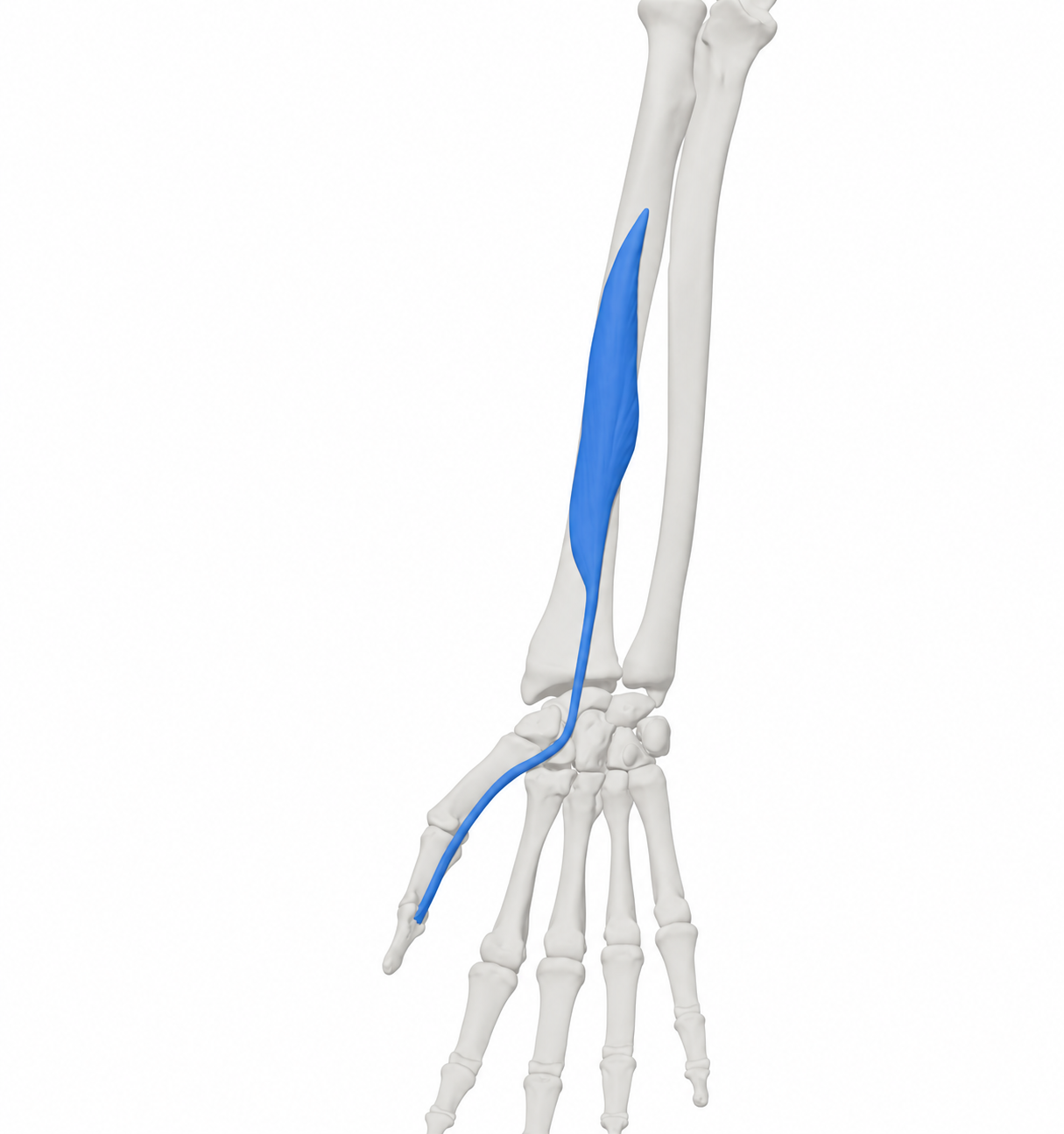
Right flexor pollicis longus with the source skeletal context. Source model rendered and material restyled; blue identifies the model structure, including its tendon-like continuations.

BodyParts3D, © The Database Center for Life Science. Adapted by MyofascialPain: source model rendered and material restyled. · CC BY-SA 2.1 Japan

Open full-size image
Browse related exercises (1)

Symptoms patients report

Difficulty bending the thumb tip Record whether this began suddenly, followed an injury or accompanies index-tip weakness.

Painful pinch Compare thumb-tip movement with base-joint movement and the actual pinch task.

Contributors to assess

Increased pinch demand Small tools or repeated fine handling can expose symptoms; change force and duration before assuming a muscle lesion.

Tendon or motor nerve dysfunction A tendon injury and AIN dysfunction require different management from ordinary task fatigue.

For clinicians and supervised training

Clinical reference

Anatomical and examination reference; no isolated FPL pain diagnosis or referral map follows from a pinch complaint.

Anatomy and palpation

Anatomical relationships

FPL arises from the anterior radius and adjacent interosseous membrane and inserts into the base of the distal thumb phalanx. It flexes the thumb IP joint; motor supply is the anterior interosseous branch of the median nerve.[1][2]

  • Deep anterior radial forearm.
  • Thumb IP joint and distal phalanx.

Nearby structures. Its tendon travels through the carpal tunnel and the palmar thumb. FPL is anatomically distinct from flexor pollicis brevis and the thenar intrinsic muscles.

Palpation

Patient position. Forearm supported, palm up and thumb relaxed.

Find the landmarks

  • Anterior radial forearm.
  • Palmar thumb tendon path.

Examination sequence

  1. Observe comfortable thumb-tip flexion and record where symptoms occur.
  2. Use gentle superficial contact if helpful; do not claim to isolate the deep forearm belly by tenderness.

Document the findings

  • A sensitive tendon path is not evidence of a muscle trigger point.

Limits and precautions

  • Avoid forceful pressure, stretch or resistance after a cut, sudden motor loss or repair.
[1][5]

Examination and tests

2

Active thumb IP flexion[1][4][5]

Check the movement produced by the long thumb flexor.

Position
Hand supported; examiner stabilizes the thumb proximal phalanx.
Technique
Ask for gentle bending of the thumb tip, comparing sides and available assisted range as appropriate.
Positive finding
Absent or reduced active flexion needs further assessment.
Interpretation
Separates end-joint movement from thenar opposition.
Limitations
Pain, joint stiffness and tendon injury can mimic nerve-related weakness; the maneuver does not identify a painful muscle focus.

Anterior interosseous motor pattern[3][4]

Compare thumb-tip, index-tip and pronation function when motor weakness is reported.

Position
Seated with the forearm supported.
Technique
Observe thumb IP and index DIP flexion, including a gentle tip-to-tip pinch. Compare forearm pronation and examine other median/ulnar functions and sensation as indicated.
Positive finding
A reproducible motor deficit needs a tendon and neurological differential.
Interpretation
The combined pattern can guide further assessment.
Limitations
Pain, tendon injury and effort can alter performance. An OK-sign difficulty alone neither diagnoses AIN palsy nor a trigger point; sensory symptoms suggest a broader assessment.

Distinguishing other causes

FPL tendon injury or loss of tendon glide[5]

Findings that matter. Active thumb-tip motion is lost or reduced after injury or surgery.

Next step. Hand-service assessment; follow protection and therapy instructions rather than generic strengthening.

Anterior interosseous motor disorder[3]

Findings that matter. Thumb IP weakness with index-tip or pronation weakness.

Next step. Assess the whole motor pattern; do not label it carpal tunnel syndrome from pinch weakness alone.

Thumb joint or tendon/sheath disorder[7][2]

Findings that matter. Pain and stiffness localized to a joint or along the tendon, sometimes with swelling.

Next step. Compare active and assisted joint motion and the painful task to direct assessment.

Treatment selection and progression

Self-care and activity

Adjust the task that provokes symptoms[6][7]

Consider when. Mild task-related symptoms after significant injury and new motor loss have been excluded.

Reduce the force or uninterrupted time needed for pinching a small object; try assistance or a different handle and keep comfortable daily use.

Dose and review
Match exposure to the current response; no muscle-specific schedule is established.
Progression
Restore one demand at a time.
Stop or reassess
Seek review for persistent functional loss or worsening symptoms.
Evidence and limits
Regional hand guidance supports activity adaptation; this is not a treatment for a proven isolated muscle lesion.

Exercise and rehabilitation

Maintain comfortable active movement[6]

Consider when. Assessed mild stiffness without a suspected tendon tear, unstable injury or postoperative restriction.

Bend and straighten the thumb tip gently with the hand supported. Stay within an easy range without forcing the movement.

Dose and review
Use brief practice adjusted to symptoms; a treating clinician sets any injury-specific dose.
Progression
Increase comfortable range before resistance.
Stop or reassess
Stop for sharp pain, increasing swelling or new weakness.
Evidence and limits
A regional mobility option, not a validated isolated-muscle exercise protocol.

Clinician-delivered care

Match rehabilitation to the examination[6][7]

Consider when. Persistent pain, stiffness or weakness that limits a useful hand task.

A clinician distinguishes tendon, joint and nerve findings, then selects mobility, support or graded loading. Follow the surgical team’s plan after repair.

Dose and review
Set from the diagnosis and measured deficit; no universal strengthening dose.
Progression
Retest the same movement and functional task before increasing load.
Stop or reassess
New loss of active movement changes the assessment rather than calling for harder exercise.
Evidence and limits
Diagnosis determines care. Regional self-management does not establish muscle-specific treatment efficacy.
Reassessment and follow-up

  • Compare active thumb-tip flexion under the same conditions.
  • Track an ordinary pinch task such as handling a button.
  • Reassess new index weakness, sensory symptoms or a change after tendon treatment.

Evidence and sources

  1. University of Washington Radiology: Flexor pollicis longus

    2026 · University anatomy atlas

    Undated page accessed 11 October 2026. Radius/interosseous membrane to distal thumb phalanx; anterior interosseous nerve.

  2. Texas Tech University Health Sciences Center El Paso: Muscles of the upper limb

    2026 · University anatomy teaching reference

    Undated teaching page accessed 11 October 2026. Cross-checks finger versus thumb joint actions and extrinsic versus intrinsic muscles. Does not validate palpation diagnosis or treatment.

  3. StatPearls: Anterior Interosseous Syndrome

    2025 · Clinical reference

    July 2025 update, publisher-indexed text checked. Motor pattern includes FPL, radial FDP and PQ; pure AIN palsy lacks cutaneous sensory loss. Neuritis and other causes require assessment; not all cases are local compression.

  4. Anatomy, Shoulder and Upper Limb, Forearm Anterior Interosseous Nerve

    2026 · Clinical anatomy reference

    Publisher-indexed clinical-significance section checked 11 October 2026; year is access year. Describes thumb IP, index/middle DIP and pronation motor examination, not trigger-point tests.

  5. British Society for Surgery of the Hand: Flexor tendon injury

    2026 · Specialist society patient guidance

    Undated page accessed 11 October 2026. Loss of active flexion after injury and protected hand therapy following repair; no generic exercise program should replace a repair protocol.

  6. Cambridgeshire Community Services NHS: Wrist, hand and thumb pain

    2026 · NHS regional self-management guidance

    Undated page accessed 11 October 2026. Activity adaptation, gradual movement and graded return. Used for regional care, not efficacy or a fixed dose for any one named muscle.

  7. NHS: Wrist pain

    2026 · National patient guidance

    Accessed 11 October 2026; year is access year. Injury, joint/tendon and nerve differential context and when symptoms need assessment.