Skip to main content
Atlas · Lower Leg

Fibularis longus (peroneus longus)

Fibularis longus, also called peroneus longus, runs from the outer lower leg behind the lateral ankle and under the foot to the first metatarsal and medial cuneiform. It helps turn the foot outward and control the first ray. Outer-leg muscle discomfort must be separated from tendon pain, instability and lateral-foot bone disease.
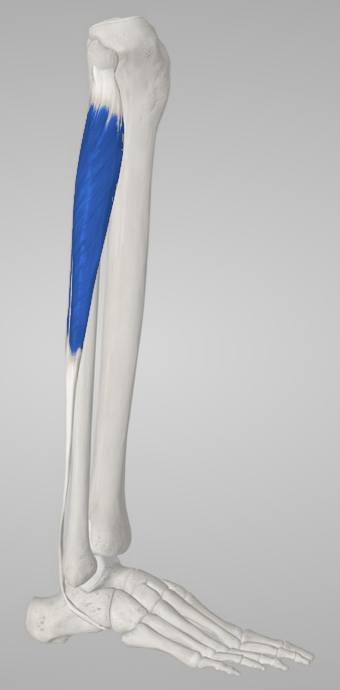
Fibularis longus (peroneus longus)
Body region
Lower Leg · Foot
Trigger points
0
documented in this muscle
Common symptoms
3
patterns cataloged
Common causes
3
contributory factors

Muscle overview

Symptoms and first steps

Practical first steps

  • Reduce provoking uneven-ground, cutting or running exposure temporarily.
  • Use stable comfortable footwear and maintain tolerable ankle motion.
  • Seek assessment for a new snap with swelling, persistent tendon displacement or focal bone pain.

When to seek assessment

  • After trauma, inability to bear weight or focal fifth-metatarsal/fibular tenderness needs prompt assessment.
  • A sudden snap with weakness, visible tendon displacement or substantial swelling warrants ankle-specialist assessment.
  • New progressive foot weakness/numbness, a cold foot or severe tense-leg pain needs urgent evaluation.

Follow the tendon conceptually around the cuboid toward the first ray. Pain under the lateral midfoot can involve the longus tendon or os peroneum and is not a muscle belly in the sole.

Examination, palpation and treatment evidence

Anatomy reference; no mapped trigger points yet.

Symptoms patients report

Lateral-leg or retromalleolar ache

Distinguish accessible outer-leg muscle discomfort from pain and swelling behind the lateral malleolus.

Pain near the plantar-lateral midfoot near the cuboid and the first-ray tendon attachment

Examine the actual painful tendon, joint or bone region; distal pain is not proof of muscle referral.

Painful eversion, uneven-ground difficulty or snapping

Repeated snapping behind the malleolus raises concern for tendon instability, especially after an ankle injury.

Common causes

Ankle inversion injury

Lateral ligaments, peroneal tendons and the fifth-metatarsal region may be injured together.

Changed running or uneven-ground demand

A rapid increase in lateral-foot control demands may exceed current muscle-tendon capacity.

Foot alignment or established tendon pathology

Assess the whole foot and tendon excursion; alignment alone is not a diagnosis or a reason for an injection.

Treatment & self-care

Exercise and movement

Progressive eversion and functional ankle loading

An assessed stable muscle-tendon presentation without fracture, major rupture or acute tendon dislocation. Start light band eversion through a comfortable range. Add supported heel rises and balance work; for longus-related distal symptoms monitor first-ray/plantar-midfoot comfort, and for brevis monitor the fifth-metatarsal insertion. Increase resistance, single-leg demand and uneven-ground exposure separately. Consensus supports progressive tendon loading after acute symptoms permit. Exercise details are clinical examples, not a verified isolated-muscle MPS protocol.

Duration
Illustrative start: 1–2 sets of 6–10 controlled repetitions on alternate days; adjust to symptoms and the next-day walking response.

Everyday adjustments

Modify the provoking terrain and footwear

Symptoms rise with running, cutting or unstable surfaces and a stable nonurgent diagnosis has been established. Use comfortable supportive footwear and choose level ground while rebuilding capacity. Temporarily reduce cutting and cambered surfaces; a clinician can assess targeted orthotic support when needed. Restore duration before speed and unpredictable surfaces. Conservative tendon guidance supports load adjustment; orthotic choices depend on the particular tendon and foot presentation.

Duration
Use a recoverable amount of activity and review fit/comfort after changes.

Exercise and movement

Balance and return to uneven-ground tasks

Walking and initial strength are tolerable, with any ankle instability being managed. Begin supported single-leg stance on firm ground and controlled step tasks. Add reach, uneven surfaces and direction changes only when the previous stage is reliable. Include the real work or sport surface before unrestricted return. A functional rehabilitation progression; persistent tendon subluxation requires reassessment rather than balance training alone.

Duration
Short quality-controlled practice with support available; stop before repeated loss of balance.

Professional care

Discuss shockwave for persistent diagnosed tendon pain

Persistent fibularis/peroneal tendinopathy after initial conservative care, once tear or instability requiring a different approach has been assessed. A clinician can discuss shockwave as a noninvasive adjunct and identify the actual tendon target, with a walking/activity outcome agreed in advance. Review the chosen function after the initial course and stop extending treatment without meaningful benefit. ESSKA-AFAS supports consideration using indirect lower-limb tendon evidence. This is not a direct longus/brevis MPS trial, and the same statement does not establish routine PRP or steroid-injection benefit.

Duration
The consensus discusses symptoms persisting beyond three months; it does not establish an optimal peroneal-specific device course or dose.

Differential diagnosis

  • Fibularis/peroneal tendon tear, tendinopathy or instability
  • Os-peroneum/longus tendon disorder or cuboid-region disease
  • Lateral ankle ligament injury or joint disease
  • Fibular/sural nerve or compartment disorder

For clinicians and supervised training

Clinical reference

Anatomy and examination reference. A trigger-point map is not yet documented.

Anatomy and palpation

Anatomical relationships

The belly arises from the fibular head and proximal lateral fibular shaft. Its tendon passes behind the lateral malleolus, along the lateral calcaneus and through the cuboid groove, then crosses the sole to the first-metatarsal base and medial cuneiform. It everts the foot, assists plantar flexion and influences first-ray loading.[1][2]

Right fibularis longus, lateral view, with its tendon course behind the ankle and beneath the foot.
Right fibularis longus, lateral view, with its tendon course behind the ankle and beneath the foot. Source model: Essential Anatomy 5.
  • Fibular head and proximal lateral-leg belly
  • Retromalleolar tendon course
  • Cuboid groove and first-ray insertion, reached by tendon rather than a plantar muscle belly

Nearby structures. The two fibularis tendons share the retromalleolar sheath and retinacular constraints. The common fibular nerve wraps around the fibular neck; superficial fibular and sural nerve territories, lateral ligaments and fibular/fifth-metatarsal bone require attention.

Palpation

Patient position. Seated or side lying with the knee slightly bent, ankle supported and lateral leg/foot accessible.

Find the landmarks

  • Lateral fibular shaft
  • Posterior edge of lateral malleolus
  • Lateral calcaneal/cuboid tendon region

Examination sequence

  1. Inspect swelling and any tendon snapping before palpation; review injury mechanism.
  2. Identify accessible lateral-leg contraction with light eversion, then relax and compare belly tenderness with tendon-course sensitivity.
  3. Examine symptoms at the plantar-lateral midfoot near the cuboid and the first-ray tendon attachment separately. Avoid repeatedly provoking a snapping tendon or pressing directly over the fibular-neck nerve.

Document the findings

  • Record the exact painful tissue region, tendon swelling and familiar pain during loading.
  • Neither a surface tender spot nor pain during eversion distinguishes longus and brevis with certainty.

Limits and precautions

  • Do not massage suspected fracture or acute tense-compartment pain.
  • A displaced tendon or major acute weakness needs assessment before resisted progression.
[1][2][3]

Examination and tests

2

Resisted eversion with symptom localization[1][2]

Assess lateral-compartment muscle-tendon loading tolerance.

Position
Seated with the lower leg supported and ankle near neutral.
Technique
Turn the sole outward against gradually applied resistance without rotating the whole hip. Compare force and record whether symptoms occur in the leg belly, behind the malleolus or at the distal attachment.
Positive finding
Familiar lateral-leg/tendon pain, a reproducible strength deficit or snapping.
Interpretation
Use the response to plan initial loading and assess tendon pathology or instability when indicated.
Limitations
Both fibularis muscles and other evertors contribute; there is no validated muscle-specific trigger-point cutoff.

First-ray loading and supported heel rise[1][2]

Assess a functional task involving longus and its distal tendon course.

Position
Seated for gentle first-ray loading, then supported standing if weight bearing is safe.
Technique
With the lesser metatarsals supported, gently press the first-metatarsal head downward while keeping the toes relaxed. Compare with a controlled heel rise and record lateral-midfoot/plantar tendon pain.
Positive finding
Familiar pain along the plantar longus tendon course or impaired first-ray loading in the functional task.
Interpretation
Directs assessment toward the longus tendon/first ray when the symptom location agrees.
Limitations
Other muscles and joint mobility affect the task. This is anatomy-informed testing and cannot independently diagnose an os-peroneum lesion or isolated longus tear.

Distinguishing other causes

Fibularis/peroneal tendon tear, tendinopathy or instability[2]

Findings that matter. Retromalleolar swelling, painful eversion, snapping/displacement or a sudden functional loss.

Next step. Clinical ankle assessment with ultrasound or MRI when appropriate; dynamic ultrasound can help with intermittent instability.

Os-peroneum/longus tendon disorder or cuboid-region disease[2][4]

Findings that matter. Focal pain at the plantar-lateral midfoot near the cuboid and the first-ray tendon attachment, particularly after trauma or increased impact.

Next step. Assess local bone and tendon; choose imaging from the injury history and examination rather than assigning the pain to muscle referral.

Lateral ankle ligament injury or joint disease[2]

Findings that matter. Inversion mechanism, joint-line swelling or instability can coexist with a tendon problem.

Next step. Examine lateral ligaments, joint motion and weight-bearing tolerance as part of the same assessment.

Fibular/sural nerve or compartment disorder[1][3]

Findings that matter. Numbness, burning, disproportionate weakness or recurrent exertional tightness.

Next step. Neurological/exertional assessment; severe acute compartment features require emergency care.

Treatment selection and progression

Exercise and rehabilitation

Progressive eversion and functional ankle loading[2][1]

Consider when. An assessed stable muscle-tendon presentation without fracture, major rupture or acute tendon dislocation.

Start light band eversion through a comfortable range. Add supported heel rises and balance work; for longus-related distal symptoms monitor first-ray/plantar-midfoot comfort, and for brevis monitor the fifth-metatarsal insertion.

Dose and review
Illustrative start: 1–2 sets of 6–10 controlled repetitions on alternate days; adjust to symptoms and the next-day walking response.
Progression
Increase resistance, single-leg demand and uneven-ground exposure separately.
Stop or reassess
Stop for snapping, new swelling, focal bone pain or loss of control.
Evidence and limits
Consensus supports progressive tendon loading after acute symptoms permit. Exercise details are clinical examples, not a verified isolated-muscle MPS protocol.

Self-care and activity

Modify the provoking terrain and footwear[2]

Consider when. Symptoms rise with running, cutting or unstable surfaces and a stable nonurgent diagnosis has been established.

Use comfortable supportive footwear and choose level ground while rebuilding capacity. Temporarily reduce cutting and cambered surfaces; a clinician can assess targeted orthotic support when needed.

Dose and review
Use a recoverable amount of activity and review fit/comfort after changes.
Progression
Restore duration before speed and unpredictable surfaces.
Stop or reassess
Reassess continued instability, worsening deformity, skin pressure or persistent focal bone pain.
Evidence and limits
Conservative tendon guidance supports load adjustment; orthotic choices depend on the particular tendon and foot presentation.

Exercise and rehabilitation

Balance and return to uneven-ground tasks[2]

Consider when. Walking and initial strength are tolerable, with any ankle instability being managed.

Begin supported single-leg stance on firm ground and controlled step tasks. Add reach, uneven surfaces and direction changes only when the previous stage is reliable.

Dose and review
Short quality-controlled practice with support available; stop before repeated loss of balance.
Progression
Include the real work or sport surface before unrestricted return.
Stop or reassess
Stop for a new snap, ankle giving way or sharp tendon pain.
Evidence and limits
A functional rehabilitation progression; persistent tendon subluxation requires reassessment rather than balance training alone.

Clinician-delivered care

Discuss shockwave for persistent diagnosed tendon pain[2]

Consider when. Persistent fibularis/peroneal tendinopathy after initial conservative care, once tear or instability requiring a different approach has been assessed.

A clinician can discuss shockwave as a noninvasive adjunct and identify the actual tendon target, with a walking/activity outcome agreed in advance.

Dose and review
The consensus discusses symptoms persisting beyond three months; it does not establish an optimal peroneal-specific device course or dose.
Progression
Review the chosen function after the initial course and stop extending treatment without meaningful benefit.
Stop or reassess
Reassess unexpected swelling, weakness, neurological symptoms or painful snapping.
Evidence and limits
ESSKA-AFAS supports consideration using indirect lower-limb tendon evidence. This is not a direct longus/brevis MPS trial, and the same statement does not establish routine PRP or steroid-injection benefit.

Reassessment and follow-up

  • Repeat eversion force, the same walking/step task and tendon swelling.
  • Check the relevant distal insertion and any snapping or instability.
  • Revisit bone, joint and nerve causes if symptoms do not improve with graded care.

Evidence and sources

  1. University of Washington Muscle Atlas: Fibularis longus

    2026 · Institutional anatomy reference

    Undated anatomy page, verified September 2026; the year records this review. Supports attachments and actions, not diagnostic accuracy of palpation or a trigger-point map. No atlas illustration reproduced.

  2. The ESSKA-AFAS international consensus statement on peroneal tendon pathologies

    2018 · International clinical consensus

    Conservative tendon care and assessment of tears/instability. Shockwave after persistent symptoms is supported by expert extrapolation from other lower-limb tendons, not a direct peroneal-muscle MPS trial. No consensus for corticosteroid injection and insufficient PRP evidence.

  3. AAOS OrthoInfo: Compartment Syndrome

    2026 · Professional society clinical information

    Undated page verified September 2026; differentiates acute emergencies from exertional presentations. Muscle palpation cannot exclude either.

  4. AAOS OrthoInfo: Stress Fractures of the Foot and Ankle

    2026 · Professional society clinical information

    Undated page verified September 2026; focal bone-load pain and the limits of early plain radiography.