Skip to main content
Atlas · Forearm

Extensor Pollicis Longus

Extensor pollicis longus straightens the end joint of the thumb and helps lift it away from a tabletop. Its tendon turns around the back of the wrist, where a tendon problem can affect thumb movement even though the muscle lies in the forearm.

Extensor Pollicis Longus
Body region
Forearm · Hand
Reference type
Anatomy & examination
Trigger-point map not yet documented
Common symptoms
2
patterns cataloged
Contributors
2
Factors to assess

Muscle overview

Symptoms and first steps

Practical first steps

  • Reduce painful repetitive thumb use while maintaining easy movement.
  • Use assistance for tasks that require forceful thumb positioning.
  • Do not try to restore a newly lost movement through hard massage or repeated resistance.

When to seek assessment

  • Arrange prompt assessment for new thumb extension loss, including after an apparently minor wrist fracture.
  • A swollen painful wrist or a significant new injury needs medical assessment.

New loss of thumb-tip extension deserves tendon assessment, particularly after a wrist fracture. It should not be explained as tightness or a trigger point without examination.

Examination, palpation and treatment evidence

Illustrated overview

Extensor Pollicis Longus · Anatomy

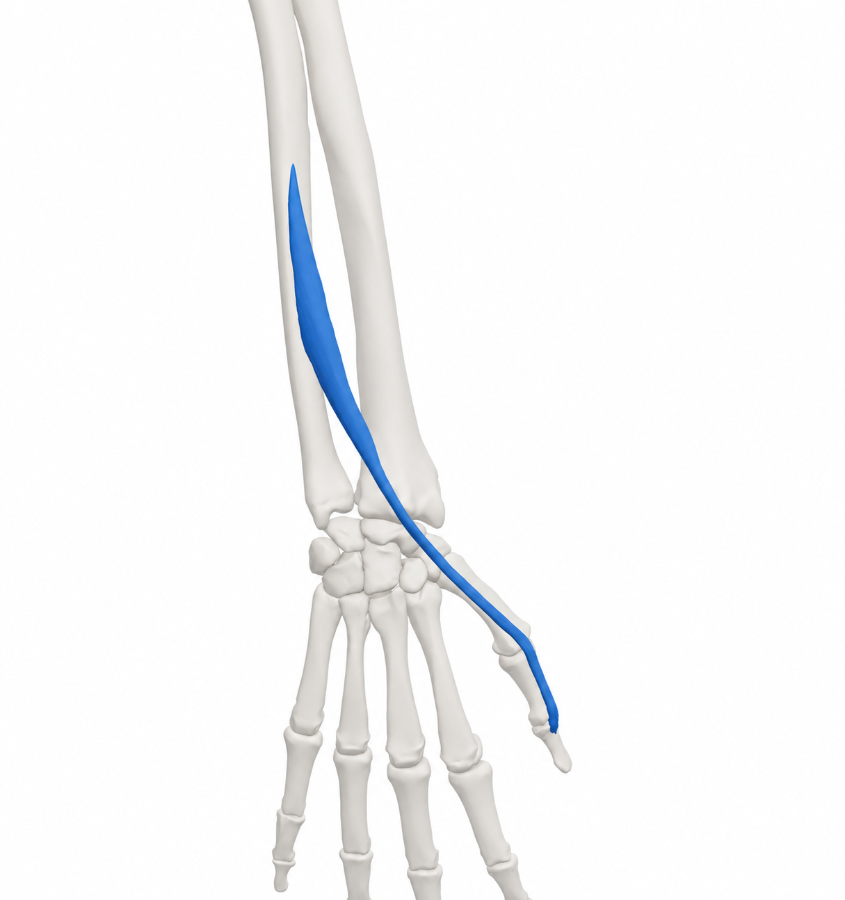
Right extensor pollicis longus with the source skeletal context. Source model rendered and material restyled; blue identifies the model structure, including its tendon-like continuations.

BodyParts3D, © The Database Center for Life Science. Adapted by MyofascialPain: source model rendered and material restyled. · CC BY-SA 2.1 Japan

Open full-size image
Browse related exercises (1)

Symptoms patients report

Difficulty straightening the thumb tip Document active motion and whether assisted straightening is still available.

Difficulty lifting the thumb from a table This functional change can accompany EPL tendon rupture and needs assessment.

Contributors to assess

Wrist fracture or inflammatory tendon disease These histories matter because the EPL tendon is vulnerable near the dorsal wrist.

Changes in thumb use Task-related discomfort without motor loss still needs comparison with nearby tendons and joints.

For clinicians and supervised training

Clinical reference

EPL is distinct from APL and EPB. An anatomical reference does not establish a trigger-point location or referral pattern.

Anatomy and palpation

Anatomical relationships

EPL arises from the posterior middle ulna and adjacent interosseous membrane and inserts into the distal thumb phalanx. It extends the thumb IP joint and assists other thumb extension movements. It is supplied by the posterior interosseous branch of the radial nerve.[1][2][3]

  • Posterior ulna in the forearm.
  • Lister’s tubercle on the dorsal radius and the distal thumb phalanx.

Nearby structures. The tendon passes through the third dorsal extensor compartment and changes direction around Lister’s tubercle. APL and EPB occupy a different compartment.

Palpation

Patient position. Forearm supported with the back of the hand upward.

Find the landmarks

  • Dorsal distal radius at Lister’s tubercle.
  • Visible dorsal thumb tendon during gentle extension.

Examination sequence

  1. Observe tendon movement and thumb posture.
  2. Use light contact to localize tenderness if there is no acute injury; record the tendon and joint location separately.

Document the findings

  • Tenderness or a poorly visible tendon alone does not confirm rupture or a trigger point.

Limits and precautions

  • Avoid repeated resistance when rupture is suspected.
[1][3]

Examination and tests

2

Active thumb-tip extension[3][1]

Compare active end-joint straightening.

Position
Hand supported; thumb proximal phalanx stabilized gently.
Technique
Ask the patient to straighten the thumb IP joint from a comfortable flexed position without forcing resistance.
Positive finding
New active extension loss requires a tendon, joint and motor differential.
Interpretation
Directly tests the movement for which EPL is important.
Limitations
Pain or stiffness can restrict movement; this is not a stand-alone diagnosis.

Thumb table lift and extensor pattern[3][2]

Assess thumb lift and whether weakness extends to other extensors.

Position
Hand resting flat on a table when comfortable.
Technique
Ask for a gentle thumb lift, then compare active finger and wrist extension. Note injury history and sensory symptoms.
Positive finding
An isolated thumb deficit or broader extensor weakness needs assessment.
Interpretation
Helps direct tendon versus neurological evaluation.
Limitations
Other muscles, pain and joint restriction affect the task; it cannot prove EPL rupture or PIN palsy alone.

Distinguishing other causes

EPL tendon rupture or injury[3]

Findings that matter. New thumb-tip extension and table-lift loss, sometimes after wrist fracture.

Next step. Arrange hand assessment; ultrasound may be selected to examine tendon continuity.

Broader radial/posterior interosseous motor disorder[2]

Findings that matter. Thumb weakness accompanied by finger or other extensor deficits.

Next step. Perform a full neurological examination rather than treating a local tender site.

Thumb joint or neighboring tendon disorder[5][2]

Findings that matter. Painful restricted movement with a different joint/tendon focus and preserved EPL function.

Next step. Assess the specific structure; radial wrist pain alone does not identify EPL or de Quervain’s disease.

Treatment selection and progression

Self-care and activity

Adjust the task that provokes symptoms[4][5]

Consider when. Mild task-related symptoms after significant injury and new motor loss have been excluded.

Reduce the force or uninterrupted time needed for repeated thumb positioning; try assistance or a different handle and keep comfortable daily use.

Dose and review
Match exposure to the current response; no muscle-specific schedule is established.
Progression
Restore one demand at a time.
Stop or reassess
Seek review for persistent functional loss or worsening symptoms.
Evidence and limits
Regional hand guidance supports activity adaptation; this is not a treatment for a proven isolated muscle lesion.

Exercise and rehabilitation

Maintain comfortable active movement[4]

Consider when. Assessed mild stiffness without a suspected tendon tear, unstable injury or postoperative restriction.

Gently bend and straighten the thumb while supporting the hand. Stay within an easy range without forcing the movement.

Dose and review
Use brief practice adjusted to symptoms; a treating clinician sets any injury-specific dose.
Progression
Increase comfortable range before resistance.
Stop or reassess
Stop for sharp pain, increasing swelling or new weakness.
Evidence and limits
A regional mobility option, not a validated isolated-muscle exercise protocol.

Clinician-delivered care

Match rehabilitation to the examination[4][5]

Consider when. Persistent pain, stiffness or weakness that limits a useful hand task.

A clinician distinguishes tendon, joint and nerve findings, then selects mobility, support or graded loading. Follow the surgical team’s plan after repair.

Dose and review
Set from the diagnosis and measured deficit; no universal strengthening dose.
Progression
Retest the same movement and functional task before increasing load.
Stop or reassess
New loss of active movement changes the assessment rather than calling for harder exercise.
Evidence and limits
Diagnosis determines care. Regional self-management does not establish muscle-specific treatment efficacy.
Reassessment and follow-up

  • Compare thumb-tip extension and comfortable table lift.
  • Record task tolerance separately from tendon continuity or nerve findings.
  • Reassess a new deficit rather than increasing exercise resistance.

Evidence and sources

  1. University of Washington Radiology: Extensor pollicis longus

    2026 · University anatomy atlas

    Undated page accessed 11 October 2026. Attachments and posterior interosseous supply; the action wording is clarified using the university table and BSSH description.

  2. Texas Tech University Health Sciences Center El Paso: Muscles of the upper limb

    2026 · University anatomy teaching reference

    Undated teaching page accessed 11 October 2026. Cross-checks finger versus thumb joint actions and extrinsic versus intrinsic muscles. Does not validate palpation diagnosis or treatment.

  3. British Society for Surgery of the Hand: Thumb extensor tendon (EPL) rupture

    2026 · Specialist society patient guidance

    Undated page accessed 11 October 2026. Thumb IP extension and table lift, Lister tubercle course, and rupture after wrist fracture or with rheumatoid arthritis. Assessment urgency is distinct from timing of surgery.

  4. Cambridgeshire Community Services NHS: Wrist, hand and thumb pain

    2026 · NHS regional self-management guidance

    Undated page accessed 11 October 2026. Activity adaptation, gradual movement and graded return. Used for regional care, not efficacy or a fixed dose for any one named muscle.

  5. NHS: Wrist pain

    2026 · National patient guidance

    Accessed 11 October 2026; year is access year. Injury, joint/tendon and nerve differential context and when symptoms need assessment.