Skip to main content
Atlas · Hip

Coccygeus

Coccygeus is a small posterior pelvic-floor muscle between the ischial spine and lower sacrum/coccyx. Tailbone or deep posterior-pelvic pain may coexist with pelvic-floor tenderness, but often requires assessment of the coccyx, surrounding ligaments and pelvic organs. This anatomical entry has no documented trigger-point map.
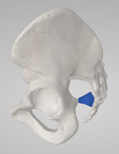
Coccygeus
Body region
Hip
Trigger points
0
documented in this muscle
Common symptoms
3
patterns cataloged
Common causes
3
contributory factors

Muscle overview

Symptoms and first steps

Practical first steps

  • Use a seat cushion or sitting position that reduces direct tailbone pressure and take comfortable movement breaks.
  • Allow relaxed unforced breathing; do not strain to urinate or prescribe yourself repeated pelvic-floor contractions for pain.
  • Keep bowel movements comfortable with clinician-guided constipation care when needed.

When to seek assessment

  • Acute severe pelvic pain, fever, significant bleeding, urinary retention or possible pregnancy with pain warrants prompt medical assessment.
  • New saddle numbness, progressive weakness or loss of bladder/bowel control requires emergency assessment.
  • Persistent unexplained night pain, a mass, weight loss, rectal bleeding or significant coccygeal trauma needs medical evaluation.

Distinguish direct tailbone pain from a pelvic-floor muscle finding. A tender coccygeal region is not proof that coccygeus is the cause, and evidence for coccydynia procedures is not isolated-muscle evidence.

Examination, palpation and treatment evidence

Anatomy reference; no mapped trigger points yet.

Symptoms patients report

Tailbone or posterior-pelvic discomfort

Record whether the main symptom is at the coccyx, a broader pelvic ache or pain during a specific activity.

Sitting or sit-to-stand pain

Direct pressure and transition-related pain may favor a coccygeal presentation and need local evaluation.

Associated bowel, bladder or sexual symptoms

These require a pelvic medical history and cannot be explained by a muscle attachment alone.

Common causes

A fall or direct coccygeal pressure

Trauma and pressure can affect bone, joints, ligaments and nearby muscles together.

Childbirth, pelvic surgery or persistent pelvic pain

These histories guide assessment without establishing an isolated coccygeus lesion.

Reduced sitting/movement tolerance

Guarding and avoidance can become part of the ongoing presentation; graded activity is tailored to comfort.

Treatment & self-care

Everyday adjustments

Reduce tailbone pressure and restore sitting

An assessed mechanical sitting-related presentation without urgent trauma or medical features. Choose a cushion/seat position that reduces direct coccygeal pressure and alternate sitting with comfortable movement. Keep a consistent task to judge benefit. Increase sitting duration gradually as the usual activity and next-day response improve. Symptom-directed conservative care for a coccygeal/pelvic presentation; it does not establish an isolated coccygeus mechanism.

Duration
Begin with a tolerable interval and adjust breaks to symptoms rather than a prescribed universal limit.

Exercise and movement

Comfortable pelvic relaxation and graded movement

An assessed pelvic-floor guarding/relaxation problem or movement avoidance related to pain. Practice unforced breathing with supported hips and allow unnecessary abdominal/gluteal contraction to settle. Add short walks and gentle hip movement; treat constipation with the appropriate clinician. Apply coordination to the relevant daily activity and add strengthening only when the examination supports it. Pelvic-pain guidance supports individualized relaxation and active care; this is not a coccygeus-specific trial protocol.

Duration
Brief comfortable practice individualized to the symptom response, without forced bearing down.

Professional care

Specialist pelvic-floor manual care

Pelvic-floor tenderness accompanying assessed bladder-pain syndrome; a pelvic-health examination determines whether local manual treatment fits the broader presentation. A pelvic-health clinician selects external pelvic, hip or abdominal soft-tissue work and, only with explicit ongoing consent, internal techniques where examined posterior pelvic-floor tenderness is relevant. Include relaxation and graded activity. Internal contact is optional; keep organ-specific assessment and treatment in place. Follow overall symptom response and a chosen goal such as sitting or wanted sexual activity. Reassess benefit at the end of the agreed course rather than treating residual tenderness indefinitely. Among 81 women with bladder pain for no longer than three years and pelvic-floor tenderness, targeted internal/external myofascial physiotherapy produced 59% global responders versus 26% with global massage at 12 weeks. Separate pain, urgency and frequency changes did not significantly differ. The regional program does not prove isolated pelvic-muscle efficacy or cure bladder disease.

Duration
The trial offered up to ten 60-minute sessions over 12 weeks. Use this as a studied reference, with dose adjusted to consent, tolerance and measurable benefit; an external-only adaptation has not been shown equivalent to the full trial program.

Professional care

Shockwave for diagnosed persistent coccydynia

A coccygeal-region pain diagnosis with persistent sitting disability after appropriate conservative care; not eligibility based on coccygeus tenderness alone. A clinician discusses shockwave directed at the assessed coccygeal pain condition and sets a sitting/function outcome. The anatomical muscle entry is not a treatment-location or device-setting guide. Review pain and sitting/function after the initial course and do not repeat without useful benefit. A 60-person trial compared radial, focused and sham shockwave for coccydynia and reported pain/disability improvement through 16 weeks with active treatment. Small groups and limited follow-up restrict confidence; this does not demonstrate a coccygeus-muscle effect.

Duration
The trial used four sessions with assessments through week 16; the clinical course is individualized.

Differential diagnosis

  • Coccydynia from coccygeal joint, ligament or injury-related disease
  • Levator-related or broader pelvic-floor pain
  • Anorectal, urological or gynecological disease
  • Pudendal, sacral or lumbar neurological pain

For clinicians and supervised training

Clinical reference

Anatomy and examination reference. Internal pelvic examination requires consent and a trained clinician.

Anatomy and palpation

Anatomical relationships

Coccygeus, or ischiococcygeus, is a triangular posterior pelvic diaphragm muscle running from the ischial spine along the sacrospinous-ligament region to the lateral lower sacrum and coccyx. It lies posterior to levator ani and contributes to pelvic support.[1]

Right coccygeus with hemipelvic and sacrococcygeal landmarks, shown in the app’s oblique view.
Right coccygeus with hemipelvic and sacrococcygeal landmarks, shown in the app’s oblique view. Source model: Essential Anatomy 5.
  • Ischial spine is the lateral attachment landmark.
  • Lower sacrum and lateral coccyx are the medial attachments.
  • The muscle is closely related to the sacrospinous ligament and cannot be isolated by surface tailbone palpation.

Nearby structures. Levator ani lies anteriorly, while the rectum, coccygeal joints/ligaments and nearby pudendal neurovascular structures influence examination and procedural safety.

Palpation

Patient position. Begin with external history, movement and coccygeal-region assessment. A trained pelvic clinician may offer internal assessment only with a separate explanation, explicit consent, stop signal and chaperone option.

Find the landmarks

  • External coccygeal region
  • Lateral posterior pelvic-floor region only within a consented specialist examination
  • Ischial-spine/sacrospinous-ligament relationship as anatomical context

Examination sequence

  1. Ask the patient to identify the main pain location and compare sitting with standing.
  2. Examine external skin/bone-region tenderness gently and note trauma or inflammatory findings.
  3. If internal assessment is indicated and accepted, document posterior pelvic-floor tenderness and relaxation without claiming surface isolation of coccygeus; stop on request or intolerance.

Document the findings

  • Distinguish direct coccygeal tenderness from broader pelvic-floor sensitivity and familiar pain.
  • A muscle finding cannot exclude joint, ligament, bowel or other pelvic disease.

Limits and precautions

  • Do not prescribe internal self-palpation, forceful coccygeal manipulation or needle trajectories.
  • An internal examination can be declined or deferred without losing access to external care.
[1][2]

Examination and tests

2

Sitting and sit-to-stand symptom localization[1][5]

Identify a repeatable coccygeal-pressure task and distinguish it from diffuse pelvic symptoms.

Position
A stable chair with a comfortable alternative cushion/seat available.
Technique
Record the usual sitting pain site and a tolerable sitting interval. Observe a controlled sit-to-stand and compare with a pressure-reducing seat position; a clinician assesses coccygeal findings when indicated.
Positive finding
Reproducible focal tailbone pain with pressure or transition, or a different deep pelvic complaint.
Interpretation
Provides a functional baseline and directs coccygeal versus broader pelvic assessment.
Limitations
The response does not isolate coccygeus or diagnose fracture/instability; medical examination and selective imaging may be needed.

Pelvic-floor relaxation and familiar-pain assessment[2][3]

Assess whether a posterior pelvic-floor coordination or tenderness finding is relevant.

Position
Supported supine or side lying; external assessment first, with internal examination only if clinically indicated and explicitly consented.
Technique
Observe comfortable breathing and ask for a gentle contraction followed by full release. A trained clinician may assess accepted posterior pelvic-floor contact for familiar pain and relaxation, while recording bladder/bowel/sexual symptoms separately.
Positive finding
Painful contraction, delayed relaxation or region-specific familiar tenderness.
Interpretation
May support pelvic-floor physiotherapy within a broader diagnosis rather than automatic strengthening.
Limitations
There is no isolated coccygeus clinical gold-standard test; tenderness does not exclude endometriosis, bladder pain or anorectal disease.

Distinguishing other causes

Coccydynia from coccygeal joint, ligament or injury-related disease[1][5]

Findings that matter. Focal tailbone tenderness, sitting-pressure pain, trauma or a painful sit-to-stand transition.

Next step. Assess the coccyx and consider targeted imaging for trauma, instability or atypical persistent symptoms.

Levator-related or broader pelvic-floor pain[2]

Findings that matter. Rectal/perineal ache, tenderness and impaired relaxation may involve several muscles.

Next step. Pelvic-health assessment with consented examination choices and a tailored care plan.

Anorectal, urological or gynecological disease[2]

Findings that matter. Bleeding, bowel change, urinary symptoms, cyclical pain or systemic illness.

Next step. Coordinate appropriate medical evaluation; muscle treatment must not serve as a rule-out test.

Pudendal, sacral or lumbar neurological pain[2]

Findings that matter. Burning, sensory change, leg weakness or bladder/bowel neurological signs.

Next step. Neurological assessment, urgently for progressive deficits or cauda equina features.

Treatment selection and progression

Self-care and activity

Reduce tailbone pressure and restore sitting[3][1]

Consider when. An assessed mechanical sitting-related presentation without urgent trauma or medical features.

Choose a cushion/seat position that reduces direct coccygeal pressure and alternate sitting with comfortable movement. Keep a consistent task to judge benefit.

Dose and review
Begin with a tolerable interval and adjust breaks to symptoms rather than a prescribed universal limit.
Progression
Increase sitting duration gradually as the usual activity and next-day response improve.
Stop or reassess
Reassess new severe pain, bleeding, a mass or persistent unexplained night symptoms.
Evidence and limits
Symptom-directed conservative care for a coccygeal/pelvic presentation; it does not establish an isolated coccygeus mechanism.

Exercise and rehabilitation

Comfortable pelvic relaxation and graded movement[3]

Consider when. An assessed pelvic-floor guarding/relaxation problem or movement avoidance related to pain.

Practice unforced breathing with supported hips and allow unnecessary abdominal/gluteal contraction to settle. Add short walks and gentle hip movement; treat constipation with the appropriate clinician.

Dose and review
Brief comfortable practice individualized to the symptom response, without forced bearing down.
Progression
Apply coordination to the relevant daily activity and add strengthening only when the examination supports it.
Stop or reassess
Stop any maneuver that increases pressure or urinary difficulty.
Evidence and limits
Pelvic-pain guidance supports individualized relaxation and active care; this is not a coccygeus-specific trial protocol.

Clinician-delivered care

Specialist pelvic-floor manual care[4]

Consider when. Pelvic-floor tenderness accompanying assessed bladder-pain syndrome; a pelvic-health examination determines whether local manual treatment fits the broader presentation.

A pelvic-health clinician selects external pelvic, hip or abdominal soft-tissue work and, only with explicit ongoing consent, internal techniques where examined posterior pelvic-floor tenderness is relevant. Include relaxation and graded activity. Internal contact is optional; keep organ-specific assessment and treatment in place.

Dose and review
The trial offered up to ten 60-minute sessions over 12 weeks. Use this as a studied reference, with dose adjusted to consent, tolerance and measurable benefit; an external-only adaptation has not been shown equivalent to the full trial program.
Progression
Follow overall symptom response and a chosen goal such as sitting or wanted sexual activity. Reassess benefit at the end of the agreed course rather than treating residual tenderness indefinitely.
Stop or reassess
Stop any unwanted or intolerable technique immediately. New unexplained bleeding, infection symptoms or sustained symptom worsening require clinical reassessment.
Evidence and limits
Among 81 women with bladder pain for no longer than three years and pelvic-floor tenderness, targeted internal/external myofascial physiotherapy produced 59% global responders versus 26% with global massage at 12 weeks. Separate pain, urgency and frequency changes did not significantly differ. The regional program does not prove isolated pelvic-muscle efficacy or cure bladder disease.

Clinician-delivered care

Shockwave for diagnosed persistent coccydynia[5]

Consider when. A coccygeal-region pain diagnosis with persistent sitting disability after appropriate conservative care; not eligibility based on coccygeus tenderness alone.

A clinician discusses shockwave directed at the assessed coccygeal pain condition and sets a sitting/function outcome. The anatomical muscle entry is not a treatment-location or device-setting guide.

Dose and review
The trial used four sessions with assessments through week 16; the clinical course is individualized.
Progression
Review pain and sitting/function after the initial course and do not repeat without useful benefit.
Stop or reassess
Reassess unexpected worsening, neurological symptoms or signs of a new medical cause.
Evidence and limits
A 60-person trial compared radial, focused and sham shockwave for coccydynia and reported pain/disability improvement through 16 weeks with active treatment. Small groups and limited follow-up restrict confidence; this does not demonstrate a coccygeus-muscle effect.

Reassessment and follow-up

  • Track sitting duration, sit-to-stand pain and the patient-selected pelvic function.
  • Distinguish improved daily activity from a less-tender internal examination alone.
  • Reassess organ, coccygeal and neurological alternatives when symptoms remain unexplained.

Evidence and sources

  1. Anatomy, Abdomen and Pelvis, Pelvis

    2023 · Anatomy reference

    Updated July 2023; verified September 2026. Supports coccygeus attachments and relation to the sacrospinous ligament. No trigger-point coordinates or isolated palpation accuracy established.

  2. EAU Guidelines on Chronic Pelvic Pain: Diagnostic Evaluation

    2026 · Clinical guideline

    Current online chapter verified September 2026; coordinated organ, musculoskeletal and neurological assessment, including pelvic-floor contraction/relaxation. Tenderness cannot exclude organ disease.

  3. EAU Guidelines on Chronic Pelvic Pain: Management

    2026 · Clinical guideline

    Individualized multidisciplinary care and specialist pelvic-floor treatment. Manual/needle evidence is heterogeneous; prostate-pain shockwave evidence cannot establish efficacy for a levator or obturator trigger point.

  4. Randomized multicenter clinical trial of myofascial physical therapy in women with interstitial cystitis/painful bladder syndrome and pelvic floor tenderness

    2012 · Randomized trial

    81 women with bladder-pain symptoms for no longer than three years and pelvic-floor tenderness; up to ten 60-minute sessions over 12 weeks. Global response was 59% with targeted internal/external pelvic, hip and abdominal treatment versus 26% with global massage. Separate pain, urgency and frequency changes did not differ significantly; no isolated pelvic-muscle or durable follow-up effect established.

  5. Comparative effects of radial and focused extracorporeal shock wave therapies in coccydynia

    2023 · Sham-controlled randomized trial

    60 adults with coccydynia; radial/focused/sham groups, four treatment sessions, outcomes at 4, 8 and 16 weeks. This is coccygeal-region pain evidence, not isolated coccygeus-muscle efficacy.