Skip to main content
Atlas · Foot

Adductor hallucis

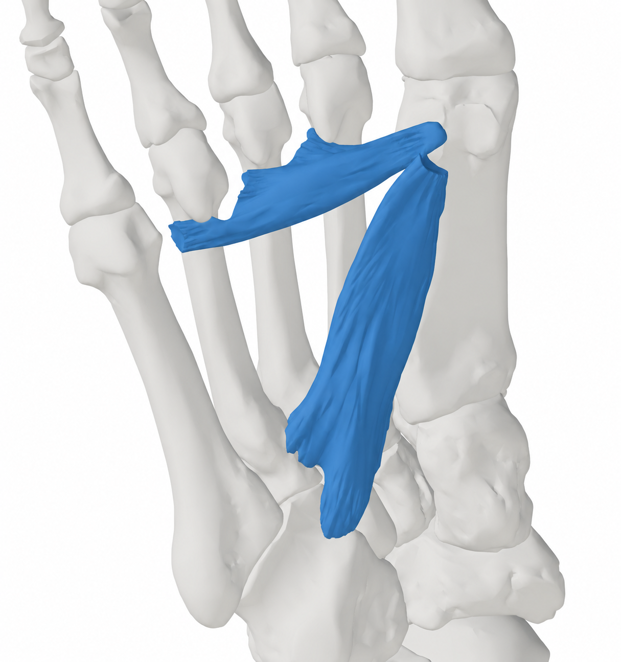
Adductor hallucis has oblique and transverse heads in the deep sole. It draws the great toe toward the second toe and contributes to forefoot control. Great-toe or forefoot pain may instead involve the first MTP joint, sesamoids, plantar plate or nerves; a bunion does not demonstrate muscle spasm.

Adductor hallucis
Body region
Foot
Reference type
Anatomy & examination
Trigger-point map not yet documented
Common symptoms
3
patterns cataloged
Contributors
2
Factors to assess

Muscle overview

Symptoms and first steps

Practical first steps

  • Keep the toes moving comfortably without forcing the painful direction.
  • Choose tolerable activity and shoes that do not compress the painful area.
  • Record the task and exact location that provoke symptoms to guide assessment.

When to seek assessment

  • Persistent focal bone pain or inability to bear weight after injury needs prompt assessment.
  • Progressive numbness, weakness or toe deformity warrants clinical review.

Adduction of the hallux is toward the second toe. Do not label it abduction or assume that strengthening or releasing this muscle corrects hallux valgus.

Examination, palpation and treatment evidence

Illustrated overview

Adductor Hallucis · Anatomy

Right adductor hallucis with the source skeletal context. Source model rendered and material restyled; blue identifies the model structure, including its tendon-like continuations.

BodyParts3D, © The Database Center for Life Science. Adapted by MyofascialPain: source model rendered and material restyled. · CC BY-SA 2.1 Japan

Open full-size image
Browse related exercises (2)

Symptoms patients report

Discomfort around the great-toe base and forefoot This is a regional complaint for assessment, not a characteristic referred-pain pattern from this muscle.

Difficulty with toe movement or push-off Pain, joint restriction and several muscles can affect the same task; note the location and activity rather than inferring a single-muscle cause.

Burning, numbness or focal swelling These findings direct attention to nerve or structural causes and should not be relabeled as muscle referral.

Contributors to assess

Changed activity or footwear demand Record recent changes in walking, running and shoe pressure; this history does not establish a muscle lesion.

Coexisting foot or toe disorder Joint alignment, plantar fascia, nerve and bone findings can alter loading and need their own assessment.

For clinicians and supervised training

Clinical reference

Anatomy and clinical examination reference. No muscle-specific trigger-point coordinates or referred-pain map have been verified for this entry.

Anatomy and palpation

Anatomical relationships

The oblique head arises around metatarsal bases 2–4 and the fibularis-longus tendon sheath. The transverse head arises from the plantar MTP ligaments of toes 3–5 and deep transverse metatarsal ligament. Both reach the lateral base of the hallux proximal phalanx. They adduct the hallux; motor supply is the deep lateral plantar nerve.[1][2][3]

  • Oblique head across the deep mid-sole
  • Transverse head near the lesser metatarsal heads
  • Lateral base of the great-toe proximal phalanx

Nearby structures. Flexor hallucis brevis and its sesamoids, the first MTP joint, plantar plates and the plantar arterial arch are nearby. Deep contact cannot reliably distinguish the two heads.

Palpation

Patient position. Seated or lying with the foot supported and the plantar surface accessible.

Find the landmarks

  • Oblique head across the deep mid-sole
  • Transverse head near the lesser metatarsal heads
  • Lateral base of the great-toe proximal phalanx

Examination sequence

  1. Start with the history, skin inspection and the person pointing to the painful region.
  2. Use light regional contact and compare with comfortable toe movement; record familiar pain without trying to force a deep structure into isolation.

Document the findings

  • Describe the actual tender region and any associated joint, skin or sensory finding.
  • Surface tenderness alone cannot confirm an individual intrinsic muscle as the pain source.

Limits and precautions

  • Avoid sustained deep pressure over painful bone, swollen joints or an area producing tingling.
  • Deep muscle layers and nearby plantar nerves limit surface localization; an anatomical diagram is not a procedural guide.
[1][2][3][7][8][5]

Examination and tests

2

Gentle hallux adduction[1][2][3][4]

Observe toe control and locate familiar discomfort during a low-load movement.

Position
Seated, with the heel supported and the foot relaxed; compare with the other side.
Technique
Starting from a comfortable great-toe position, draw it toward the second toe without twisting the whole foot or forcing a deformed or painful joint. Observe movement and symptom location.
Positive finding
Record restricted movement, a side-to-side difference or familiar pain; there is no diagnostic positive cutoff.
Interpretation
The result helps choose tolerable movement practice and directs a broader foot assessment.
Limitations
Joint alignment and motion influence performance, and the two heads are not independently tested. This is an anatomy-informed task, not a validated trigger-point test.

Regional foot examination and symptom localization[7][8][5]

Distinguish muscle-region sensitivity from nerve, bone and joint findings around the great-toe base and forefoot.

Position
Seated for inspection and gentle joint movement; assess walking only if weight bearing is comfortable.
Technique
Ask the person to indicate the painful area. Inspect skin and toe alignment, compare toe-joint motion and sensation, and identify whether tenderness is over bone, a joint, a web space or broader soft tissue.
Positive finding
Focal bone pain, sensory changes, joint swelling, altered toe alignment or a reproducible functional limitation.
Interpretation
Use the pattern to select the appropriate differential assessment; imaging depends on the history and findings.
Limitations
A tender region cannot identify one deep intrinsic muscle or exclude a structural injury. Do not repeatedly provoke painful compression.

Distinguishing other causes

Hallux valgus / first MTP-joint disorder[6]

Findings that matter. Altered great-toe alignment, a tender prominence or painful joint motion.

Next step. Assess the first ray and joint; imaging may clarify alignment or associated bone disease.

Sesamoid stress injury[5]

Findings that matter. Focal plantar pain under the first metatarsal head with loading.

Next step. Off-load the painful region pending clinical assessment and appropriate imaging.

Interdigital nerve irritation / Morton's neuroma[7]

Findings that matter. Web-space burning or tingling into adjacent toes, often aggravated by shoe pressure.

Next step. Examine sensation and the web space and consider ultrasound when indicated.

Plantar plate or lesser MTP-joint injury[8]

Findings that matter. Pain beneath a toe joint with swelling or increasing toe malalignment.

Next step. Assess joint stability and provide diagnosis-specific off-loading rather than deep muscle pressure.

Treatment selection and progression

Self-care and activity

Adjust provoking load and footwear[6][5]

Consider when. Nonurgent symptoms while the painful tissue and tolerable activity level are being assessed.

Reduce the activity or shoe pressure that consistently provokes symptoms. Keep comfortable walking and choose shoes that accommodate the foot and toes.

Dose and review
Base exposure on walking comfort and the response later that day; no isolated-muscle schedule is established.
Progression
Reintroduce the provoking activity gradually once ordinary walking is comfortable.
Stop or reassess
New swelling, persistent focal bone pain or increasing numbness calls for reassessment.
Evidence and limits
Regional foot-care advice. It does not show that one intrinsic muscle is the cause of pain.

Exercise and rehabilitation

Comfortable toe and foot-control practice[4][1][2][3]

Consider when. A stable, assessed foot presentation without a suspected fracture or unstable painful joint.

Practice small, comfortable toe-spreading and return movements with the forefoot supported. Avoid forcing the great toe toward a painful bunion or claiming selective activation of the transverse head.

Dose and review
Use a brief, comfortable practice period chosen for current control and symptoms. No validated isolated-muscle pain-treatment dose is available from the cited activation study.
Progression
Improve controlled movement first; add standing demand or resistance with a clinician when appropriate.
Stop or reassess
Stop for sharp pain, cramping that persists after relaxing, new tingling or worsening walking comfort.
Evidence and limits
The laboratory study demonstrates recruitment across intrinsic foot muscles. It does not demonstrate symptom relief or selective strengthening of this muscle.

Clinician-delivered care

Treat the established regional diagnosis[6][7][8][5]

Consider when. Persistent symptoms or examination findings suggesting a coexisting fascia, joint, nerve or bone disorder.

For symptomatic hallux valgus, a clinician can select shoe accommodation, padding or orthotic support and assess persistent joint symptoms. Examine focal plantar great-toe pain for a sesamoid or joint disorder before escalating exercise.

Dose and review
Match treatment and review timing to the actual diagnosis and functional goal; no muscle-specific procedure course is established here.
Progression
Recheck the original walking or toe task before adding treatments.
Stop or reassess
Reassess progressive deformity, neurological change or failure to regain comfortable everyday loading.
Evidence and limits
Diagnosis-specific foot guidance; this entry does not extrapolate its benefits to an unconfirmed intrinsic-muscle trigger point.
Reassessment and follow-up

  • Repeat the same comfortable toe movement and walking task.
  • Track pain location, sensation and any swelling or change in toe alignment.
  • Revisit the diagnosis if the response does not fit the planned regional care.

Evidence and sources

  1. Anatomy, Bony Pelvis and Lower Limb, Foot Muscles

    2025 · Anatomy reference

    Card and Bordoni; updated December 9, 2025, checked October 11, 2026. Used for named muscles, attachments, actions and innervation. It does not validate isolated palpation or a trigger-point map. No illustration reproduced.

  2. Texas Tech University Health Sciences Center El Paso: Anatomy Tables — Leg and Foot

    2026 · University anatomy teaching reference

    Undated teaching table; year records October 2026 verification. Corroborates adductor hallucis and dorsal/plantar interosseous attachments, the second-toe movement axis and plantar nerve supply. No atlas illustration reproduced.

  3. Ultrasound of the plantar foot: a guide for the assessment of plantar intrinsic muscles

    2023 · Peer-reviewed anatomical imaging guide

    Zaottini and colleagues. Describes the layered anatomy and ultrasound identification of plantar intrinsic muscles, including the two heads of quadratus plantae. Imaging anatomy is not proof of a painful muscle or a validated surface palpation test.

  4. Intrinsic Foot Muscle Activation During Specific Exercises: A T2 Time Magnetic Resonance Imaging Study

    2016 · Descriptive laboratory study

    Eight collegiate athletes performed foot-control exercises. MRI assessed several muscles, including FDB, quadratus plantae and the oblique adductor hallucis; interossei and lumbricals were analyzed together. Activation does not establish pain relief, isolation of a muscle or a clinical dose.

  5. AAOS OrthoInfo: Stress Fractures of the Foot and Ankle

    2026 · Professional society clinical information

    Online page checked October 2026; year records verification. Load-related focal bone pain, reduced impact and diagnostic assessment; early radiographs may be unrevealing.

  6. AOFAS FootCareMD: Bunions (Hallux Valgus)

    2026 · Professional society clinical information

    Online page checked October 2026; year records verification. Structural first-ray assessment, comfortable footwear and symptom-directed care. A bunion is not proof of adductor hallucis spasm; exercises are not presented as reversing deformity.

  7. AAOS OrthoInfo: Morton's Neuroma

    2026 · Professional society clinical information

    Online page checked October 2026; year records verification. Supports web-space, sensation and joint assessment and accommodation of forefoot pressure. No isolated interosseous or adductor-hallucis test is established.

  8. AOFAS FootCareMD: Plantar Plate Tear

    2026 · Professional society clinical information

    Online page checked October 2026; year records verification. A plantar plate injury may cause pain below a toe joint and progressive toe malalignment. This is a ligament/joint presentation, not a muscle referral map.

  9. Gautschi: Manuelle Triggerpunkt-Therapie — publisher table of contents

    2016 · Publisher contents excerpt hosted by IMTT

    Section 7.7.10 names adductor hallucis, lumbricals and interossei in the deep plantar group. Only chapter coverage was verified; this excerpt supplies no patient-specific diagnosis, point coordinates or referral contour.