Understanding Spondylolisthesis & Myofascial Overlap
Spondylolisthesis involves the anterior translation of one vertebral body over the adjacent vertebra below it, most frequently occurring at the L4-L5 or L5-S1 levels. Epidemiological data suggest a prevalence of roughly 5 to 8 percent for isthmic spondylolisthesis in the general population, while degenerative spondylolisthesis is more common in adults over 50 (reported rates vary widely depending on imaging modality and population studied). Differentiating the structural pain of a vertebral slip from secondary myofascial pain syndrome (MPS) is clinically significant. The two conditions frequently coexist, presenting with overlapping symptoms of axial low back pain that can complicate diagnosis and treatment planning.
The relationship between spondylolisthesis and MPS is characterized by complex biomechanical compensations. Segmental instability often provokes protective muscle splinting, leading to the development of myofascial trigger points in the lumbar multifidus and erector spinae. Furthermore, clinical observations suggest that hypertonicity and trigger points in the iliopsoas can exacerbate anterior shear forces on the lumbar spine, potentially aggravating the mechanical stress at the level of the slip. Hamstring tightness, a classic clinical sign of spondylolisthesis, may also involve secondary trigger point formation as the posterior chain attempts to retrovert the pelvis and stabilize the region.
While imaging confirms the structural slip, the patient's functional pain often arises from the secondary myofascial compensation surrounding the unstable segment.
Diagnostic differentiation requires a comprehensive clinical evaluation correlating physical findings with imaging studies. While standing lateral radiographs are standard for grading the slip via the Meyerding classification, the presence of a radiological slip does not inherently confirm it as the primary pain generator. Clinicians generally recommend assessing for radicular signs, neurogenic claudication, and localized step-off deformities, while simultaneously palpating for reproducible trigger point referral patterns. A trial of targeted myofascial therapy and stabilization exercises may help delineate which portion of the patient's pain is structural versus muscular.
How vertebral slip severity is classified
1–25% slip
26–50% slip
51–75% slip
76–100% slip
Spondyloptosis (>100%)
Key Muscular Compensations
Vertebral instability forces surrounding musculature to adapt, often leading to chronic overload. Understanding these compensatory patterns is essential for differentiating structural pain from myofascial pain.
The multifidus provides critical segmental stability to the lumbar spine. In the presence of spondylolisthesis, MRI studies frequently demonstrate multifidus atrophy and fatty infiltration at and above the level of the slip. Trigger points often develop in these compromised fibers as they attempt to stabilize the hypermobile segment, contributing to deep, localized axial pain.
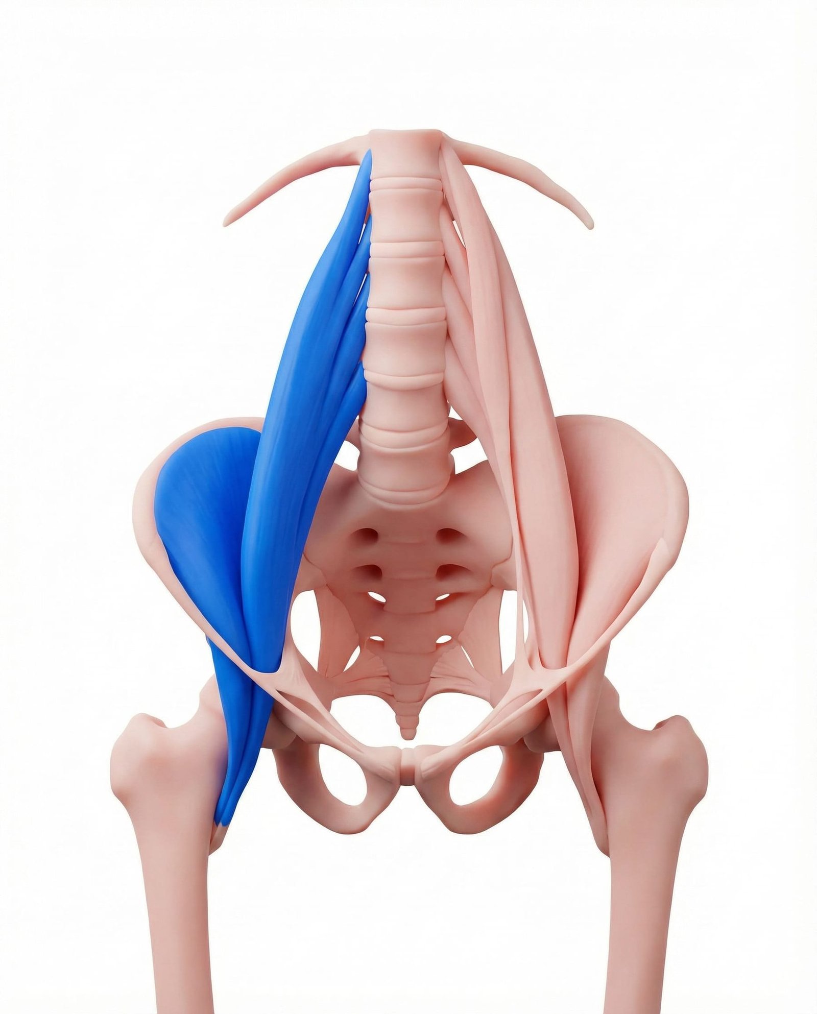
As a primary hip flexor that attaches to the lumbar vertebrae, the iliopsoas exerts an anterior and inferior pull on the lumbar spine. Hypertonicity and trigger points in the psoas major can increase anterior shear forces at the L4-L5 or L5-S1 levels. Addressing psoas tension is often a key component in conservative management to reduce mechanical stress on the pars interarticularis.
The longissimus and iliocostalis muscles frequently undergo protective splinting in response to lumbar instability. This chronic isometric contraction can lead to the formation of dense myofascial trigger points. While these muscles attempt to brace the spine, their sustained tension often exacerbates compressive loads on the posterior elements of the vertebrae.
Hamstring tightness is a hallmark clinical sign of isthmic spondylolisthesis, particularly in adolescents. Current biomechanical models suggest this tightness is a compensatory mechanism to retrovert the pelvis and reduce the lumbosacral angle, thereby decreasing anterior shear. Trigger points in the hamstrings frequently develop secondary to this chronic, altered pelvic biomechanics.
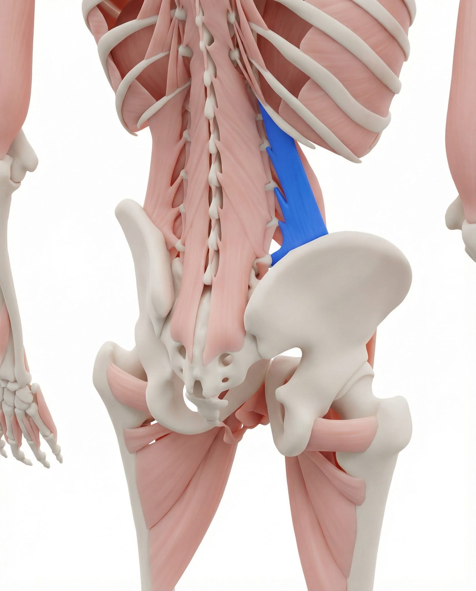
The quadratus lumborum (QL) assists in stabilizing the pelvis and lumbar spine during gait and lateral movements. In patients with degenerative spondylolisthesis, the QL often compensates for weakened deep core musculature. Trigger points here can cause severe, debilitating lower back pain that mimics the acute mechanical pain of a shifting vertebral segment.
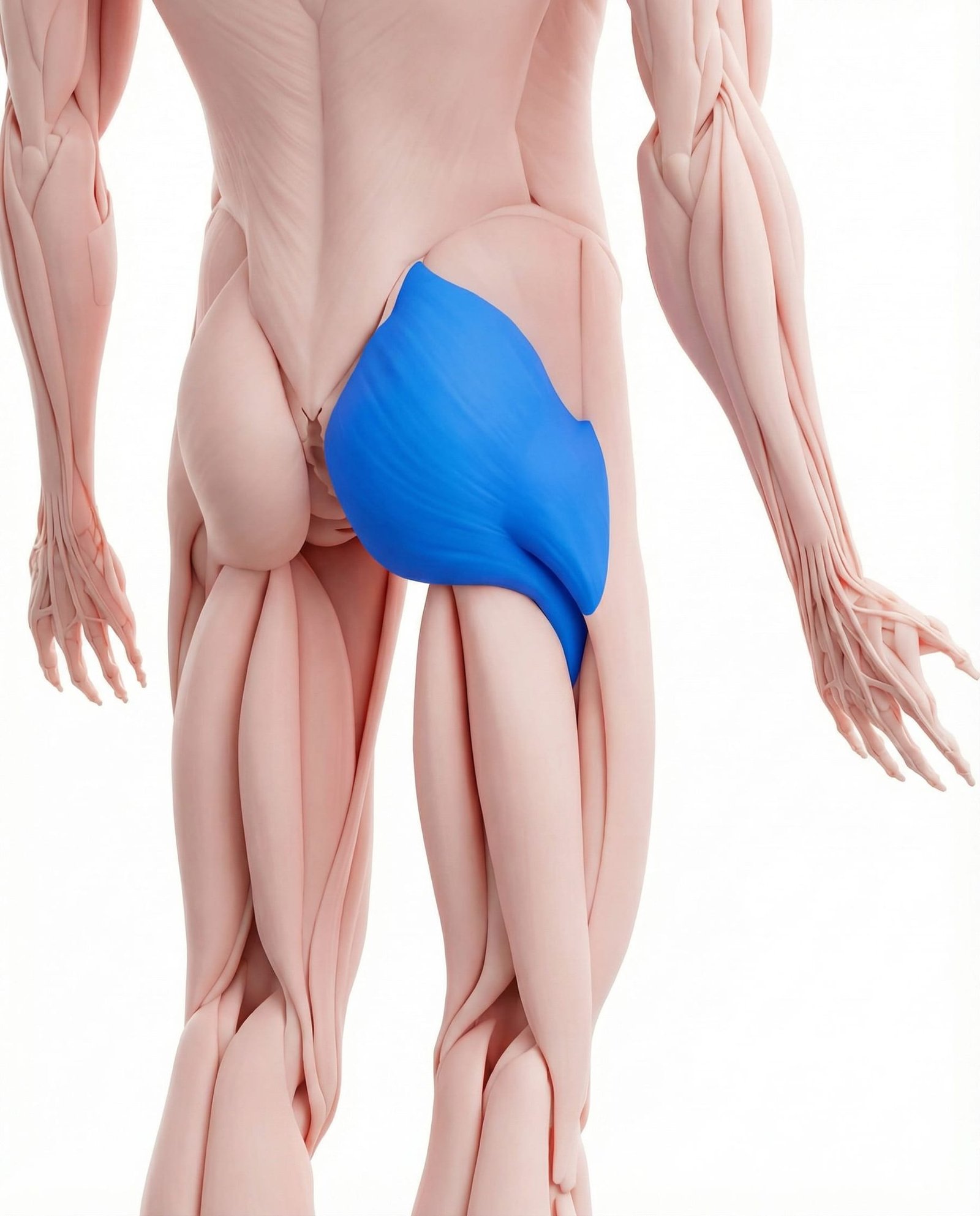
The gluteus maximus plays a vital role in controlling pelvic tilt and providing lumbopelvic stability. In chronic spondylolisthesis, pain inhibition often leads to gluteal weakness, forcing the hamstrings and lumbar erectors to overwork. Addressing gluteal trigger points and restoring strength is generally recommended to improve load transfer through the pelvis.
Anterior Shear Forces
Mechanical stress on the pars interarticularis and intervertebral disc due to gravity and altered biomechanics.
Muscular Splinting
Chronic, protective contraction of the multifidus and erector spinae leading to ischemia and trigger point formation.
Neural Compression
Narrowing of the lateral recess or neural foramen, potentially causing radicular pain or neurogenic claudication.
Segmental Instability
Abnormal motion between vertebrae during functional tasks, triggering acute mechanical pain and reflex muscle spasm.
Overlapping Pain Patterns
Trigger points in the muscles compensating for spondylolisthesis can produce referral patterns that mimic radiculopathy or discogenic pain. Recognizing these patterns aids in accurate diagnosis.
Lumbar Multifidus
Iliopsoas
Quadratus Lumborum
Conservative vs Surgical Management
Slip grade
Neurological status
Pain pattern
Typical first-line treatment
Time horizon
Clinical Assessment Techniques
Clinical evaluation of suspected spondylolisthesis involves assessing for structural deformity, segmental instability, and compensatory muscle tightness. These assessments help guide further imaging and treatment.
Palpation for Step-Off Deformity
Single-Leg Hyperextension (Stork) Test
Popliteal Angle (Hamstring Flexibility)
Prone Instability Test
Comprehensive Management Pathways
Current guidelines generally recommend a conservative approach for low-grade spondylolisthesis, focusing on muscular stabilization, trigger point management, and activity modification before considering surgical options.
Self-Care
- Activity modification to avoid repetitive lumbar hyperextension
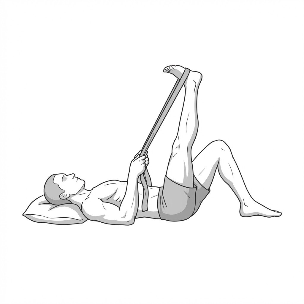
- Gentle hamstring stretching (if not exacerbating pelvic tilt)
- Use of a supportive lumbar brace during acute flare-ups (short-term)
- Application of heat for muscle spasms and ice for acute inflammation
- Pacing of daily activities to prevent muscular fatigue
Manual Therapy
- Ischemic compression of iliopsoas and QL trigger points
- Soft tissue mobilization of the erector spinae and hamstrings
- Avoidance of high-velocity posterior-to-anterior (PA) thrusts at the slip level
- Myofascial release techniques targeting the thoracolumbar fascia
- Gentle passive range of motion focusing on lumbar flexion
Rehabilitation
- Flexion-biased lumbar stabilization exercises (e.g., Williams flexion exercises)
- Deep abdominal and transverse abdominis activation
- Gluteal strengthening to improve lumbopelvic control
- Motor control training for the lumbar multifidus
- Postural re-education to manage anterior pelvic tilt
Interventional
- Epidural steroid injections for associated radicular pain
- Pars interarticularis injections for diagnostic and therapeutic purposes
- Trigger point injections for refractory myofascial pain
- Surgical decompression and fusion (for progressive neurological deficits or high-grade slips)
- Radiofrequency ablation of the medial branches (if facet arthropathy is present)
Red Flags — When to See a Doctor
While many cases of spondylolisthesis are stable and manageable with conservative care, progressive neurological symptoms or high-grade instability require prompt medical investigation.
Spondylolisthesis involves the anterior translation of a vertebra, most commonly at L4-L5 (degenerative) or L5-S1 (isthmic).
Myofascial pain syndrome frequently coexists with spondylolisthesis as muscles like the multifidus and erector spinae spasm to protect the unstable segment.
Hamstring tightness is a classic compensatory sign, particularly in isthmic spondylolisthesis, acting to reduce anterior shear forces.
Trigger points in the iliopsoas can exacerbate the mechanical stress on the lumbar spine by increasing anterior shear.
Conservative management, including targeted myofascial therapy and core stabilization, is generally recommended as the first-line treatment for Grade I and II slips.
While imaging confirms the structural diagnosis, physical examination is required to determine if the pain is primarily structural, neurological, or myofascial in origin.
Movement therapy
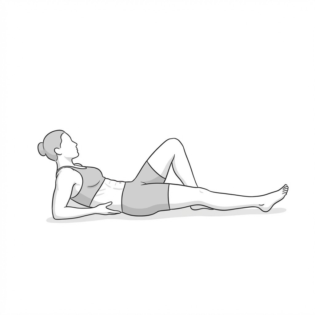
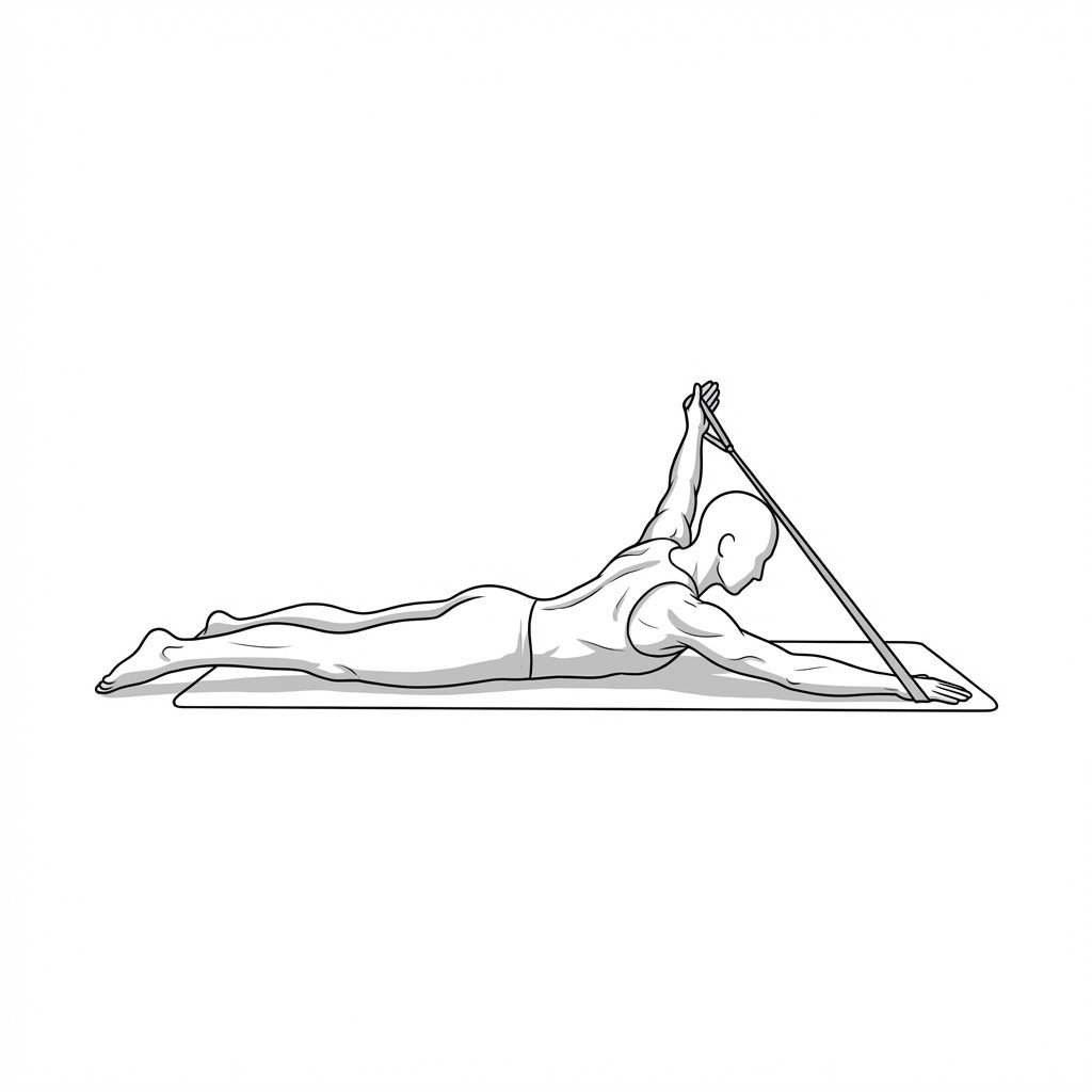
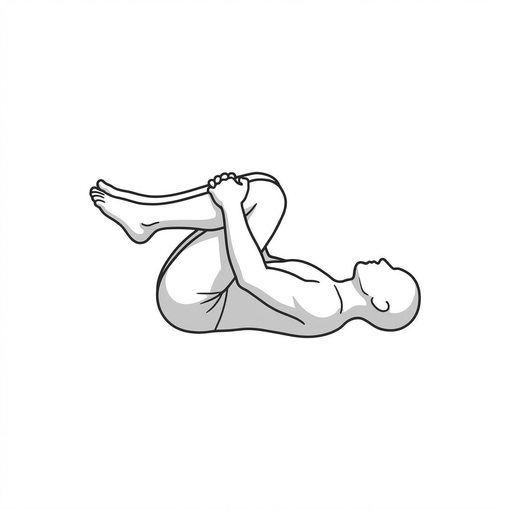
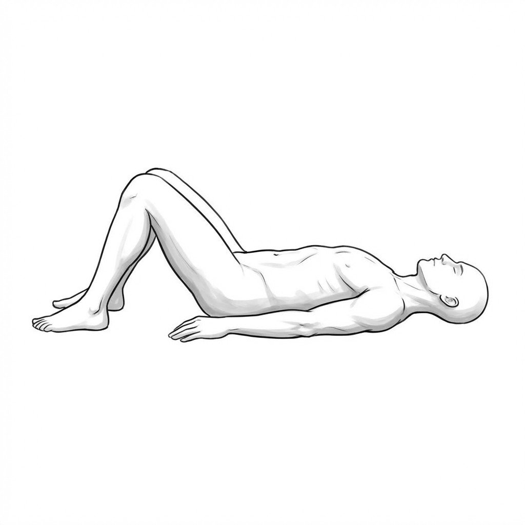
Exercises for Spondylolisthesis
These exercises focus on reducing anterior shear at the slip segment — multifidus holds and bracing rebuild segmental co-contraction, pelvic tilts train neutral lumbar position, and hamstring stretching reduces the hip flexion posture that increases shear forces. Start with 2–3 and progress as tolerated.