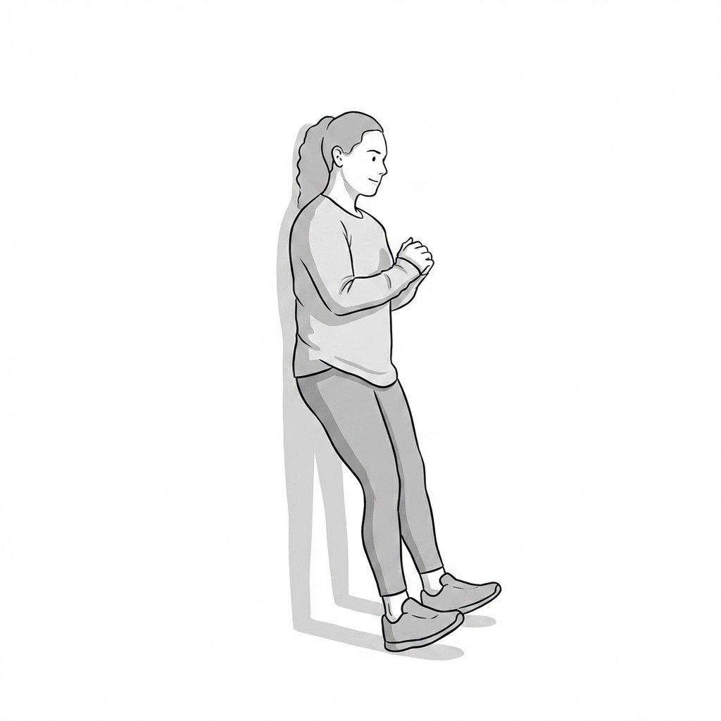
Understanding Shin Splints & Differential Diagnosis
Medial tibial stress syndrome (MTSS), commonly referred to as shin splints, presents as exercise-induced pain along the posteromedial tibial border. Current epidemiological data suggest it accounts for a significant percentage of running-related injuries, often arising from rapid increases in training volume or mechanical loading. The primary pathology is generally understood to involve periosteal traction and localized bone stress rather than pure inflammation. Differentiating MTSS from other lower extremity pathologies, particularly tibial stress fractures and chronic exertional compartment syndrome, is critical for appropriate clinical management.
Myofascial pain syndrome frequently coexists with or mimics MTSS, complicating the clinical picture. Trigger points within the deep posterior compartment musculature—specifically the tibialis posterior, flexor digitorum longus (FDL), and soleus—can refer pain directly to the posteromedial tibial border. Furthermore, excessive tension in these fascial compartments may exacerbate periosteal traction at their tibial attachments. Clinical observations suggest that addressing myofascial restrictions in these muscle groups may reduce the mechanical load on the tibia, though high-quality randomized trials isolating this effect remain limited.
Differentiating between periosteal traction, myofascial referral, and bone stress is the foundation of effective lower leg pain management.
A systematic diagnostic approach is necessary to rule out higher-risk conditions. Focal point tenderness, particularly on the anterior tibial crest, or pain that persists at rest warrants imaging to evaluate for stress fractures. Conversely, symptoms characterized by a feeling of tightness, numbness, or weakness that predictably occur at a specific exercise volume and resolve with rest may indicate chronic exertional compartment syndrome, which requires intracompartmental pressure testing for definitive diagnosis. Popliteal artery entrapment syndrome should also be considered in patients presenting with exercise-induced claudication-like symptoms.
Lower Leg Pain — Four Common Sources
Tenderness pattern
Pain timing
Hop / load test
Neurological signs
First-line workup
Anatomical Considerations
The lower leg is divided into four distinct fascial compartments. Understanding the contents and fascial attachments of these compartments is essential for diagnosing the source of exercise-induced leg pain.
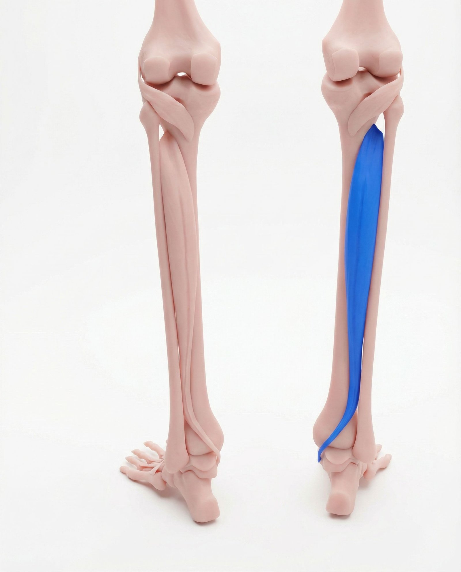
Situated deep within the posterior compartment of the lower leg, the tibialis posterior plays a critical role in plantarflexion and inversion of the foot, as well as dynamic support of the medial longitudinal arch. Excessive eccentric loading during the stance phase of running can lead to myofascial trigger points and increased fascial tension. This tension is hypothesized to contribute to the periosteal traction along the medial tibia, a primary mechanism in medial tibial stress syndrome (MTSS).
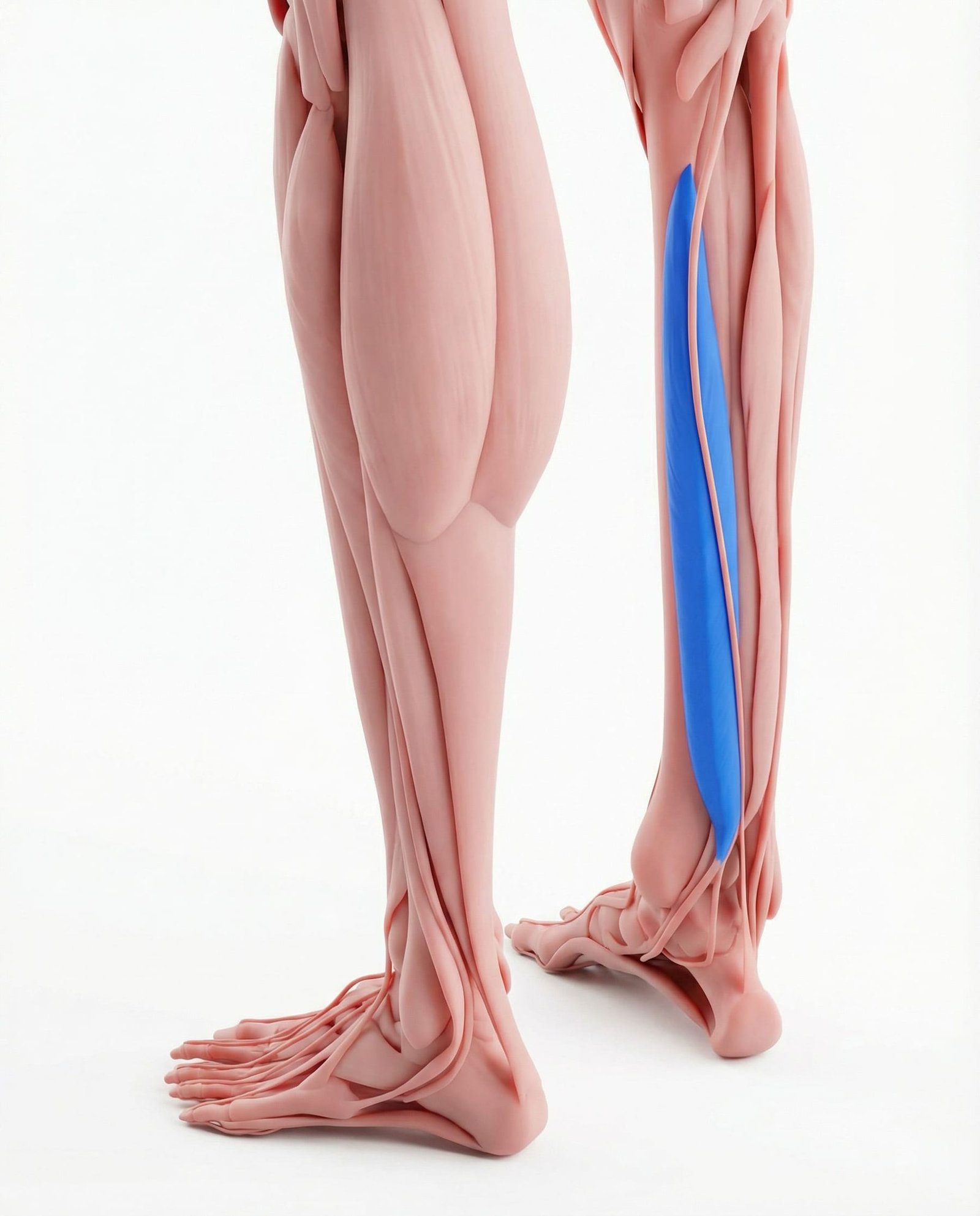
The FDL originates on the posterior surface of the tibia and is responsible for flexing the lateral four toes. Because its anatomical origin overlaps significantly with the typical pain distribution of MTSS, myofascial trigger points in the FDL can perfectly mimic shin splint symptoms. Clinical evaluation often reveals that palpation of the FDL reproduces the patient's familiar posteromedial tibial pain.
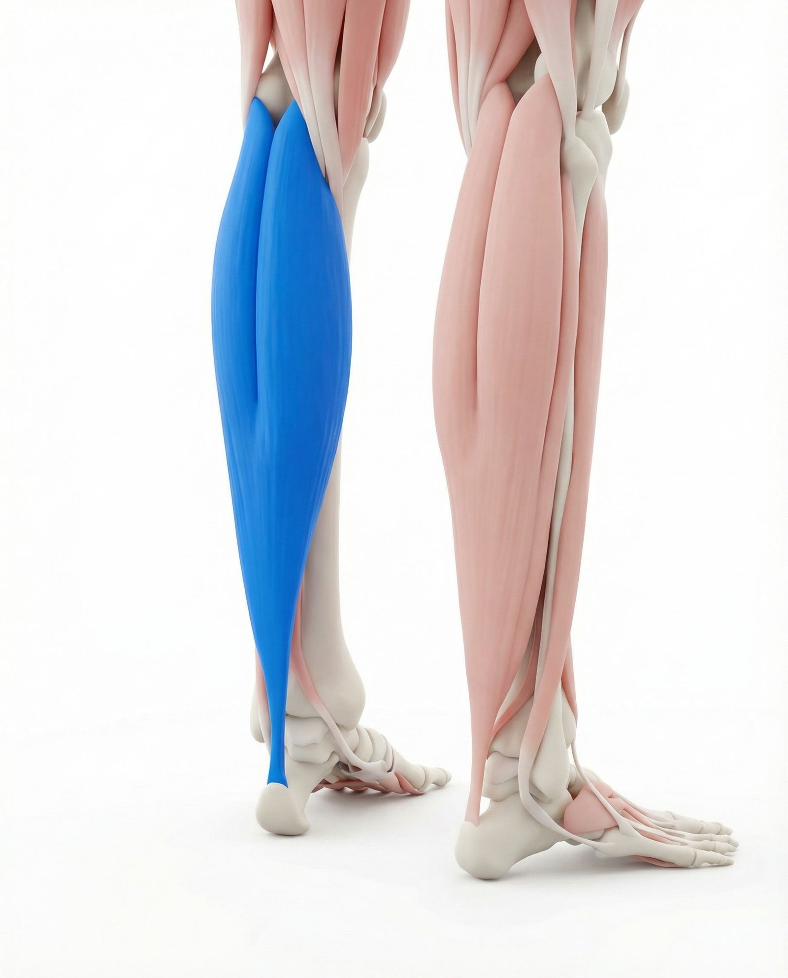
A broad, flat muscle lying deep to the gastrocnemius, the soleus is a primary plantarflexor and is heavily recruited during endurance running. The medial soleus fascial attachment on the tibia is another potential site for periosteal traction. Trigger points in the medial aspect of the soleus can contribute to a generalized aching in the lower half of the calf, complicating the clinical presentation of MTSS.
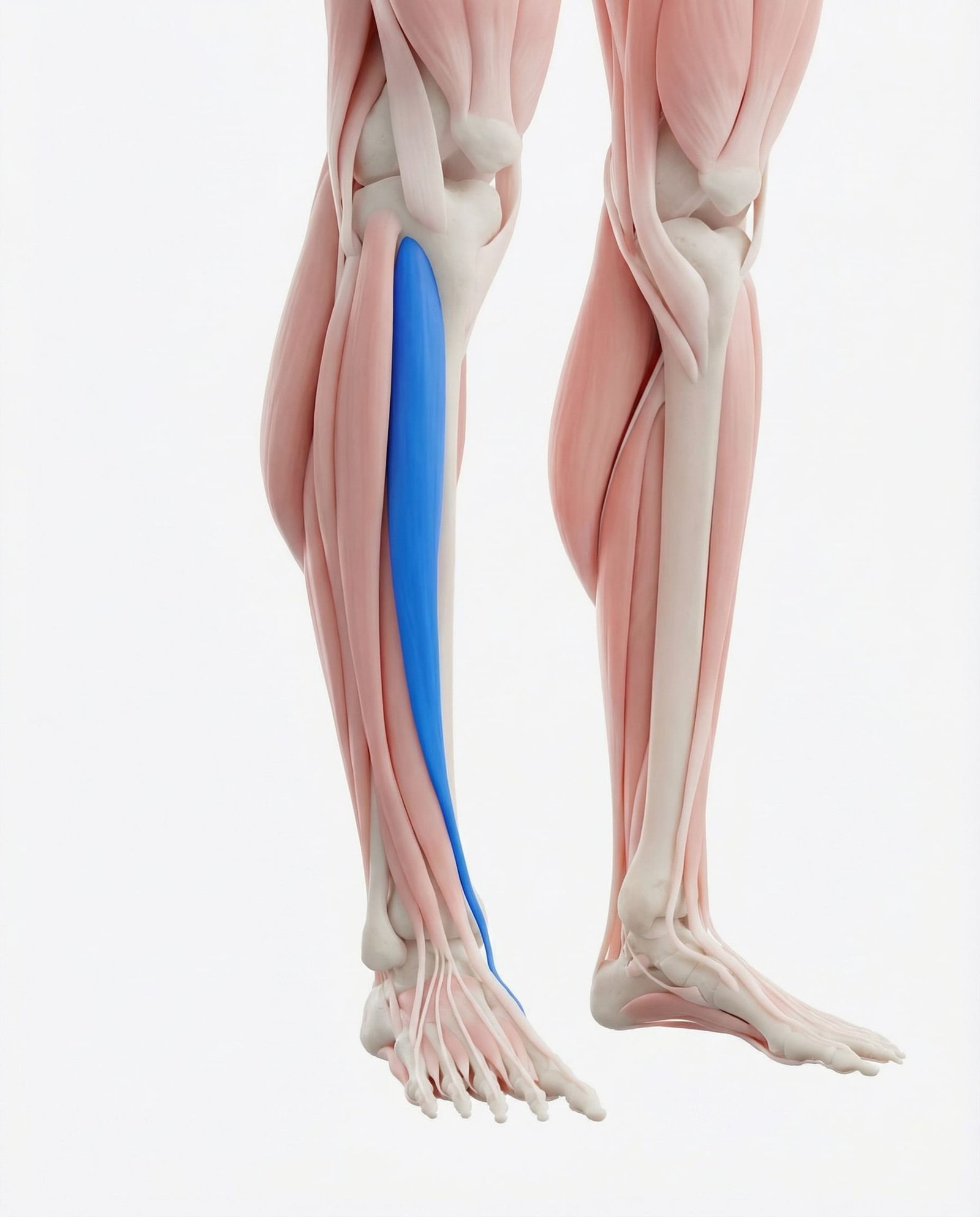
Located in the anterior compartment of the leg, this muscle is the primary dorsiflexor of the ankle. While MTSS typically presents posteromedially, "anterior shin splints" involve the tibialis anterior and are often related to eccentric overload during heel strike. Trigger points here can cause significant anterior leg pain and weakness, occasionally mimicking a mild foot drop or anterior compartment syndrome.
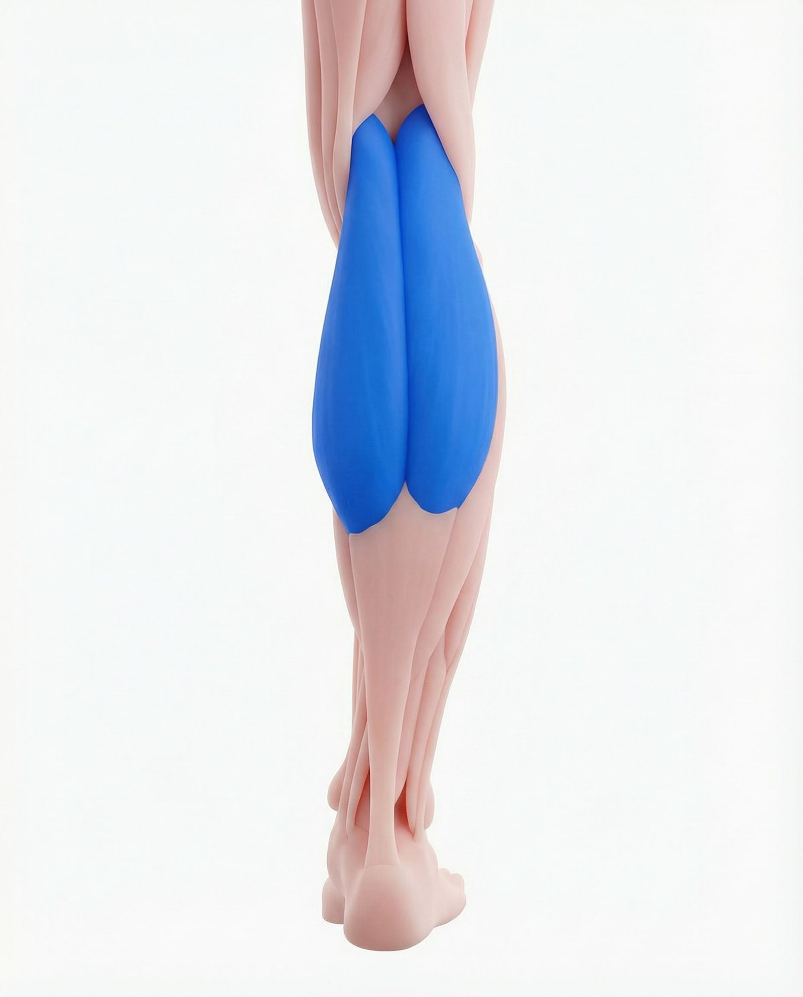
The superficial, two-headed muscle of the calf that crosses both the knee and the ankle joints. While less directly involved in the periosteal traction of MTSS than the deep posterior compartment muscles, gastrocnemius tightness alters ankle biomechanics. Reduced dorsiflexion secondary to gastrocnemius tension forces compensatory over-pronation, indirectly increasing the load on the tibialis posterior and medial tibial border.
Comprising the longus and brevis muscles in the lateral compartment, these act as the primary evertors of the foot. They provide lateral ankle stability and counterbalance the inversion forces of the tibialis posterior. Weakness or myofascial restriction in the peroneal group can lead to poor frontal-plane control of the foot during running, exacerbating the medial stresses that contribute to MTSS.
Periosteal Traction
Repetitive pulling of the deep fascial attachments on the medial tibia, leading to localized stress and pain.
Bone Stress
Accumulation of microdamage in the tibial cortex due to repetitive axial loading, potentially progressing to a stress fracture.
Myofascial Referral
Pain projected to the shin or ankle from active trigger points in the deep posterior or anterior compartment muscles.
Ischemia & Pressure
Increased volume of muscle tissue during exercise within an unyielding fascial compartment, compromising microcirculation.
Myofascial Referral Patterns
Trigger points in the lower leg musculature can project pain to the shin, ankle, and foot. Recognizing these patterns helps differentiate myofascial pain from primary bone or tendon pathology.
Tibialis Posterior & FDL
Tibialis Anterior
Soleus
Clinical Assessment & Differentiation
Clinical evaluation of lower leg pain relies heavily on palpation and load-testing. These assessments help differentiate between muscular, fascial, and osseous sources of pain.
Tibial Palpation Test
Single-Leg Hop Test
Resisted Inversion & Toe Flexion
Passive Stretch Assessment
Evidence-Based Treatment Pathways
Management of lower leg pain requires a multimodal approach centered on load management. Current guidelines generally recommend addressing both the mechanical overload and any contributing myofascial restrictions.
Self-Care
- Temporary reduction in running volume and intensity
- Transition to low-impact cross-training (cycling, swimming)
- Application of ice to the medial tibial border post-activity
- Assessment of footwear for appropriate cushioning and support
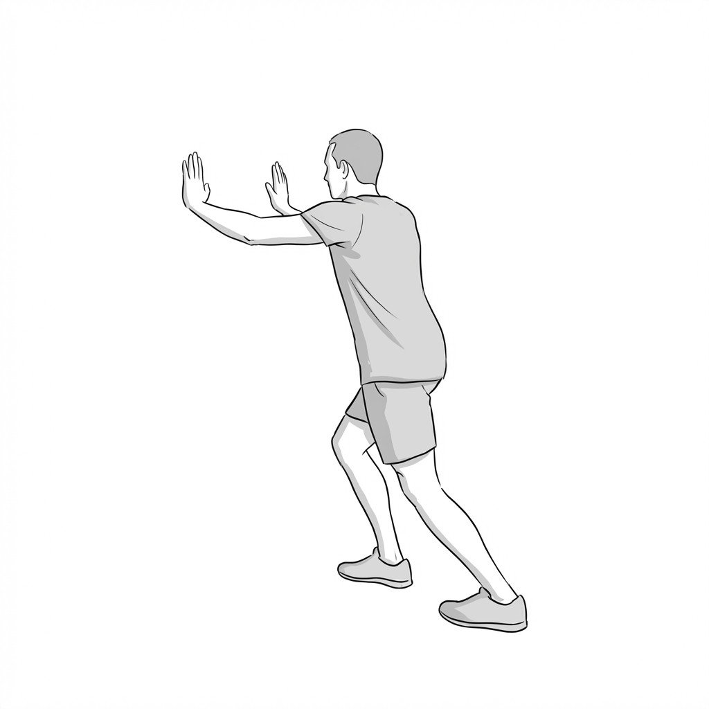
- Gentle, progressive stretching of the gastrocnemius and soleus
Manual Therapy
- Ischemic compression of tibialis posterior and FDL trigger points
- Deep longitudinal stripping of the medial soleus fascia
- Myofascial release of the anterior and lateral compartments
- Joint mobilization of the talocrural and subtalar joints
- Soft tissue mobilization to address periosteal adhesions
Interventional
- Extracorporeal shockwave therapy (ESWT) for refractory MTSS
- Dry needling of deep posterior compartment trigger points
- Intracompartmental pressure testing (if chronic compartment syndrome is suspected)
- High-resolution MRI or bone scan to rule out stress fractures
- Gait analysis using 3D kinematics or pressure mapping
Rehabilitation
- Graduated, symptom-guided return to running program
- Eccentric strengthening of the posterior calf musculature
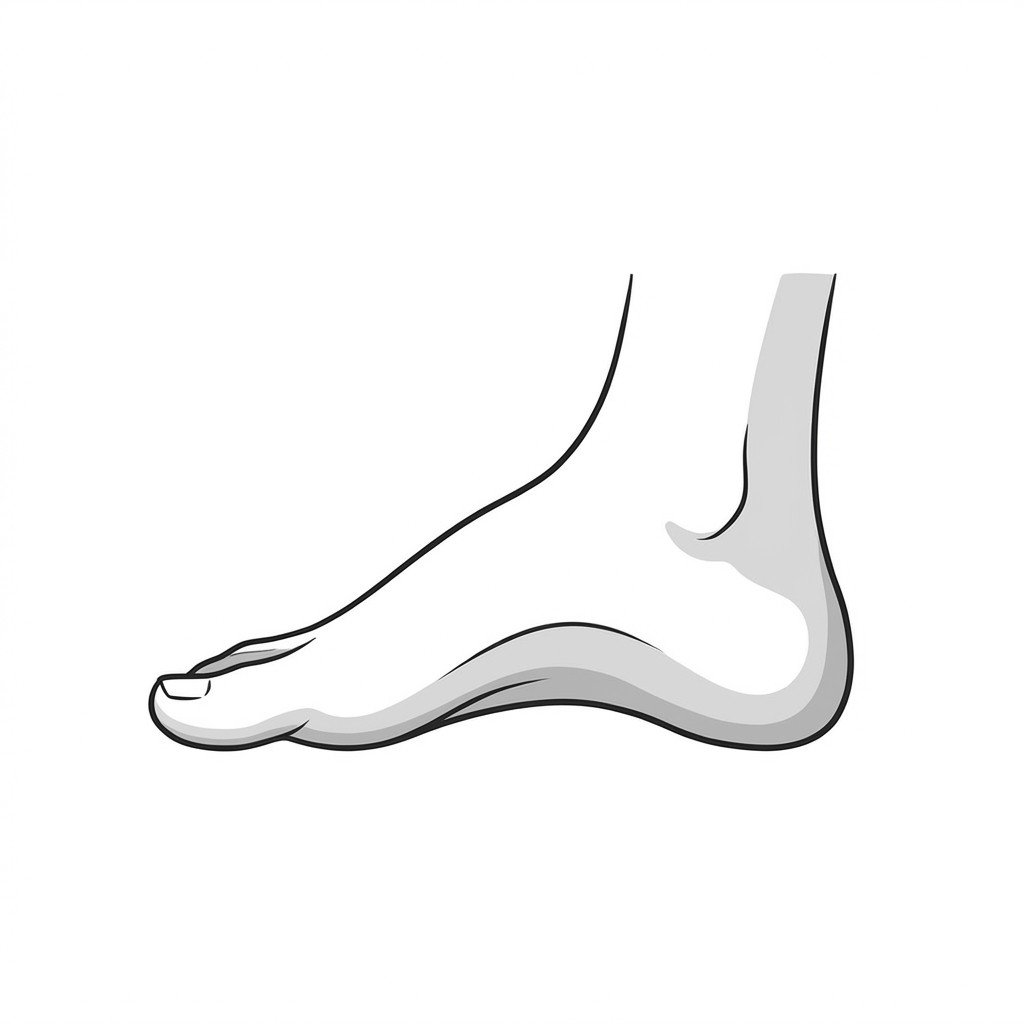
- Intrinsic foot muscle strengthening (e.g., foot doming exercises)
- Hip abductor and external rotator strengthening to control femoral internal rotation
- Cadence manipulation (increasing step rate by 5-10%) to reduce tibial shock
Red Flags & Differential Diagnoses
Lower leg pain can occasionally be a symptom of serious vascular, neurological, or osseous pathology. The following signs require prompt medical investigation.
Medial tibial stress syndrome (MTSS) is characterized by diffuse periosteal pain, whereas tibial stress fractures typically present with highly focal point tenderness.
Myofascial trigger points in the tibialis posterior and flexor digitorum longus can perfectly mimic the pain distribution of MTSS.
Load management—specifically modifying running volume, frequency, and intensity—remains the cornerstone of conservative treatment for bone stress injuries.
Symptoms of tightness, numbness, or weakness that predictably occur during exercise and resolve with rest should raise suspicion for chronic exertional compartment syndrome.
Addressing proximal biomechanical deficits, such as hip abductor weakness, is often necessary to reduce distal medial tibial loads.
Current guidelines generally recommend a trial of conservative management for 4 to 8 weeks; failure to improve warrants advanced imaging.
Movement therapy
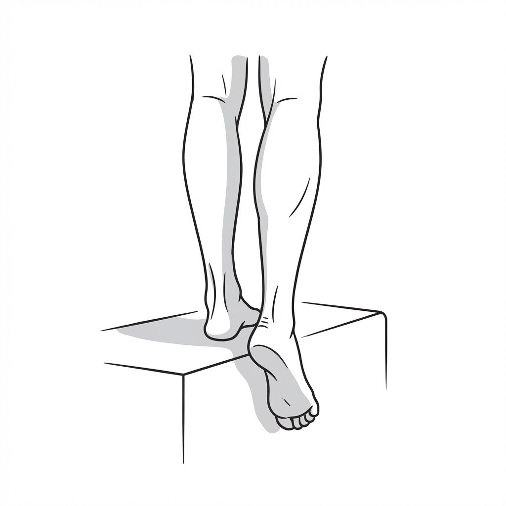
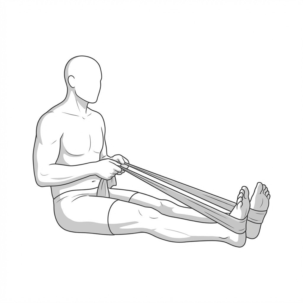
Exercises for Shin Splints
These exercises strengthen the tibialis anterior and posterior, soleus, and foot intrinsics that absorb tibial stress during running — building the lower-leg capacity and arch control that reduce periosteal traction and lower re-injury risk. Start with 2–3 and progress as tolerated.