Understanding the Overlap
Rotator cuff tendinopathy, often framed within the broader diagnosis of Subacromial Pain Syndrome (SAPS), is a leading cause of shoulder pain and disability. It represents a spectrum of tendon pathology, from reactive tendinitis to degenerative tendinosis, most commonly affecting the supraspinatus tendon. Differentiating this tendon-based pain from the referred pain of myofascial trigger points in the shoulder girdle is a critical, and often challenging, clinical task. An accurate diagnosis is paramount because effective treatment for a dysfunctional tendon (load management) differs significantly from that for a dysfunctional muscle (release and activation).
The relationship between tendinopathy and myofascial pain is complex and often bidirectional. Myofascial trigger points in muscles like the upper trapezius and serratus anterior can lead to scapular dyskinesis—abnormal movement of the shoulder blade. This altered mechanic can reduce the subacromial space, leading to impingement and secondary irritation of the rotator cuff tendons. Conversely, the persistent pain and guarding from a primary tendinopathy can lead to the development of satellite trigger points in surrounding muscles, such as the infraspinatus and deltoid, creating a layered and confusing clinical picture.
In the shoulder, the site of pain is often a poor indicator of the source of the problem.
A skilled clinician differentiates these conditions through a careful history and a detailed physical examination. The history focuses on the mechanism of injury and aggravating factors (e.g., pain with overhead motion vs. pain at rest). The physical exam uses specific orthopedic tests to stress the tendons, combined with careful palpation to distinguish tenderness directly over a tendon insertion from a taut band within a muscle belly. Often, both conditions coexist, and the clinical approach involves addressing both the "hardware" (the tendon) and the "software" (the muscular control and movement patterns) simultaneously.
Rotator Cuff Tendinopathy vs Myofascial Mimic
Pain behavior
Pain location
Palpation finding
First-line treatment
Key Muscular Anatomy
The rotator cuff is not just four individual muscles; it is a functional unit that compresses and stabilizes the humeral head. Its health is intimately linked to the position and movement of the scapula, which is controlled by a separate group of larger muscles.
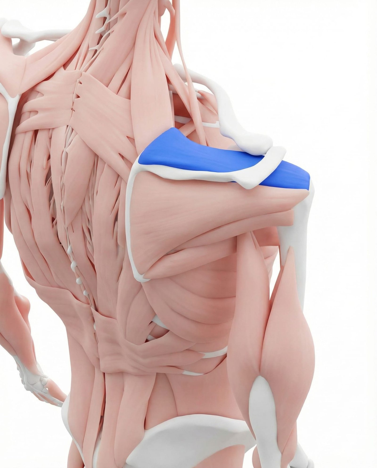
The supraspinatus is one of the four rotator cuff muscles, situated in a small fossa above the spine of the scapula. Its primary roles are to initiate abduction (lifting the arm out to the side) and to stabilize the head of the humerus within the glenoid socket. Due to its path beneath the acromion, its tendon is the most common site for impingement and degenerative tendinopathy, often leading to a characteristic painful arc of motion.
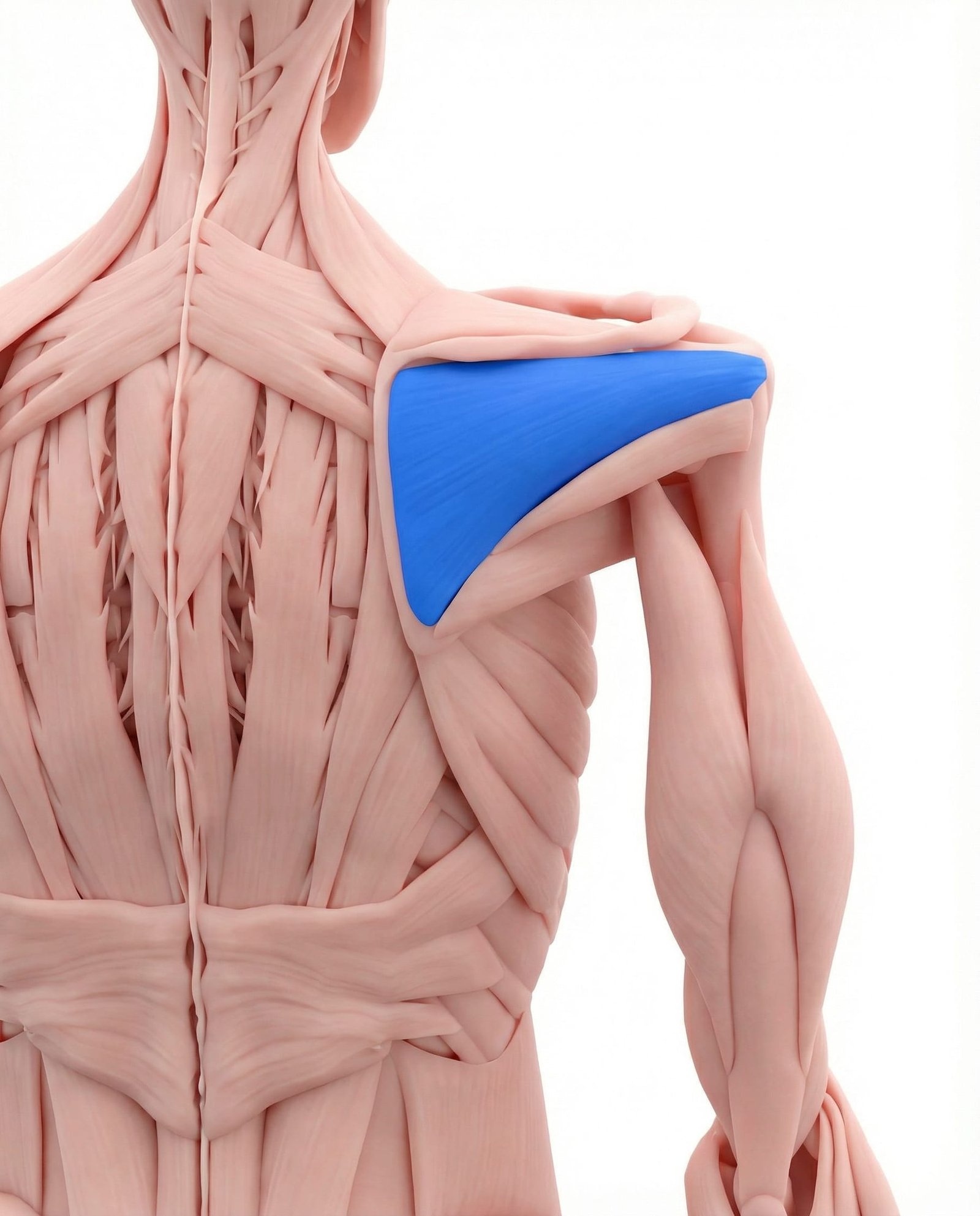
Located below the spine of the scapula, the infraspinatus is a primary external rotator of the shoulder. Trigger points within this muscle are a notorious mimic of other shoulder pathologies. They frequently refer a deep, aching pain to the front of the shoulder, bicipital groove, and down the arm, often leading to a misdiagnosis of anterior joint problems when the source is posterior.
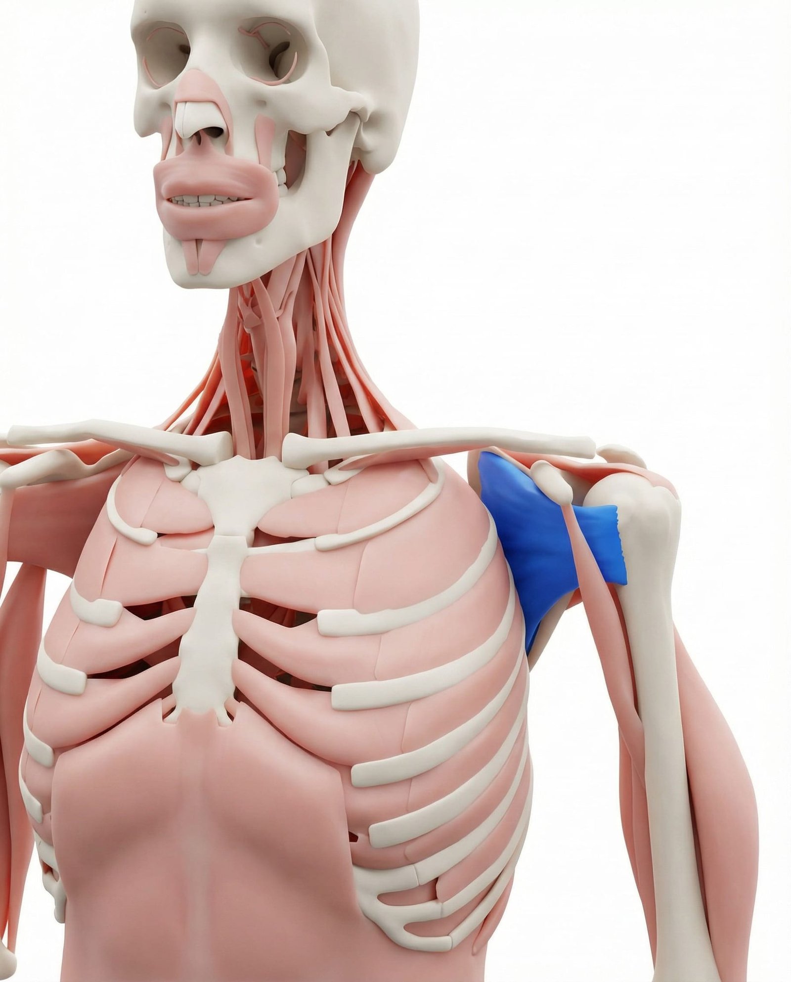
The largest of the rotator cuff muscles, the subscapularis lies on the anterior surface of the scapula and is the primary internal rotator of the shoulder. Its trigger points are less common but can produce a distinct band of pain over the posterior shoulder and a "catch" sensation during movement. Due to its deep location, it is often overlooked in clinical examinations.
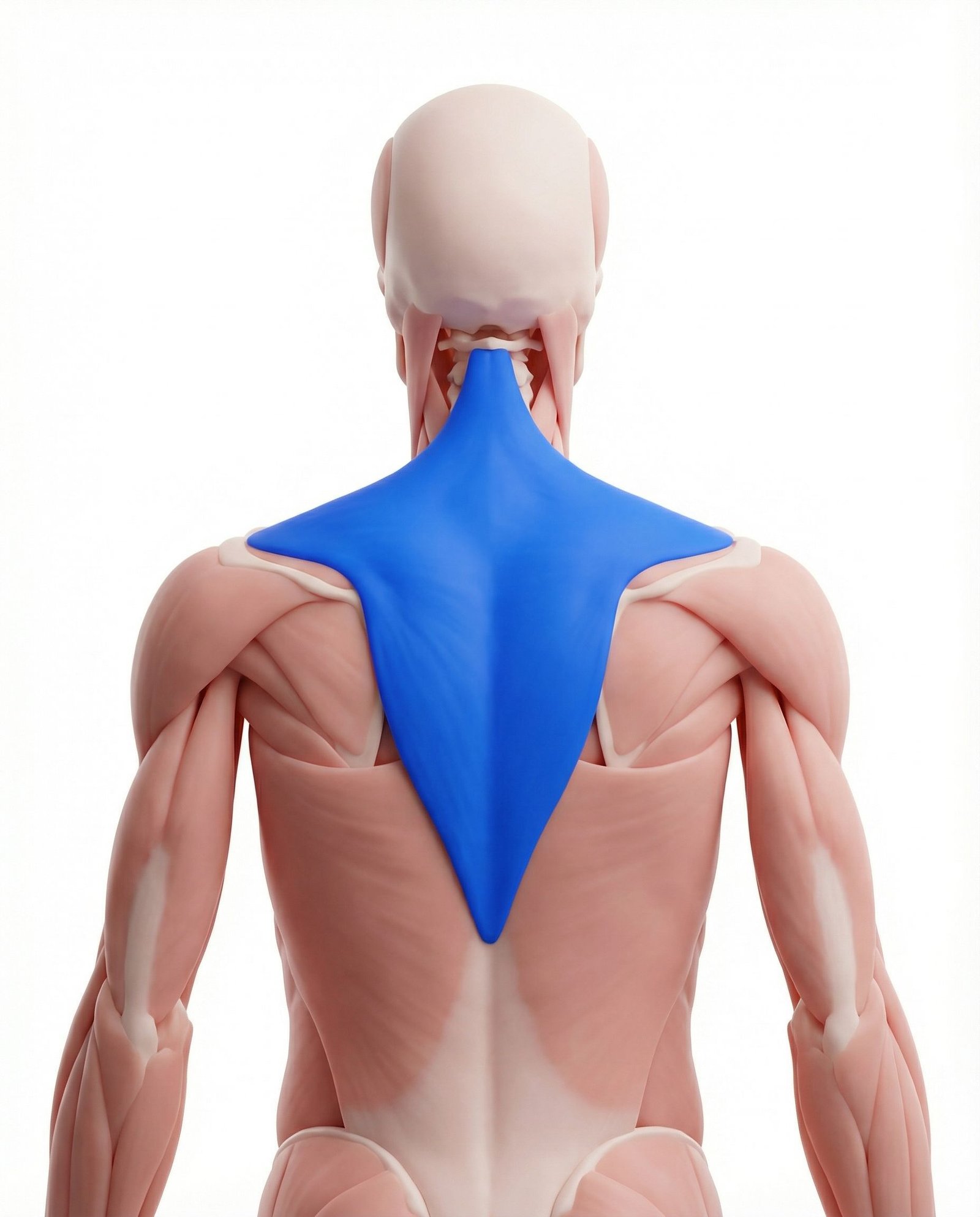
This large, superficial muscle elevates and upwardly rotates the scapula. While not part of the rotator cuff, its function is critical for proper shoulder mechanics. Myofascial trigger points in the upper trapezius are common and can contribute to scapular dyskinesis, altering the position of the acromion and potentially increasing subacromial impingement on the rotator cuff tendons.
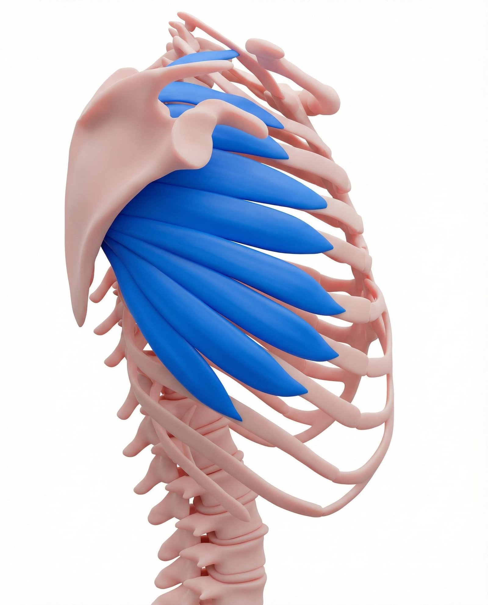
Known as the "boxer's muscle," the serratus anterior holds the scapula against the rib cage and is the primary protractor. Weakness or myofascial inhibition of this muscle leads to medial winging of the scapula. This instability alters the glenohumeral rhythm, placing excessive strain on the rotator cuff muscles during overhead activities and is a key target in rehabilitation.
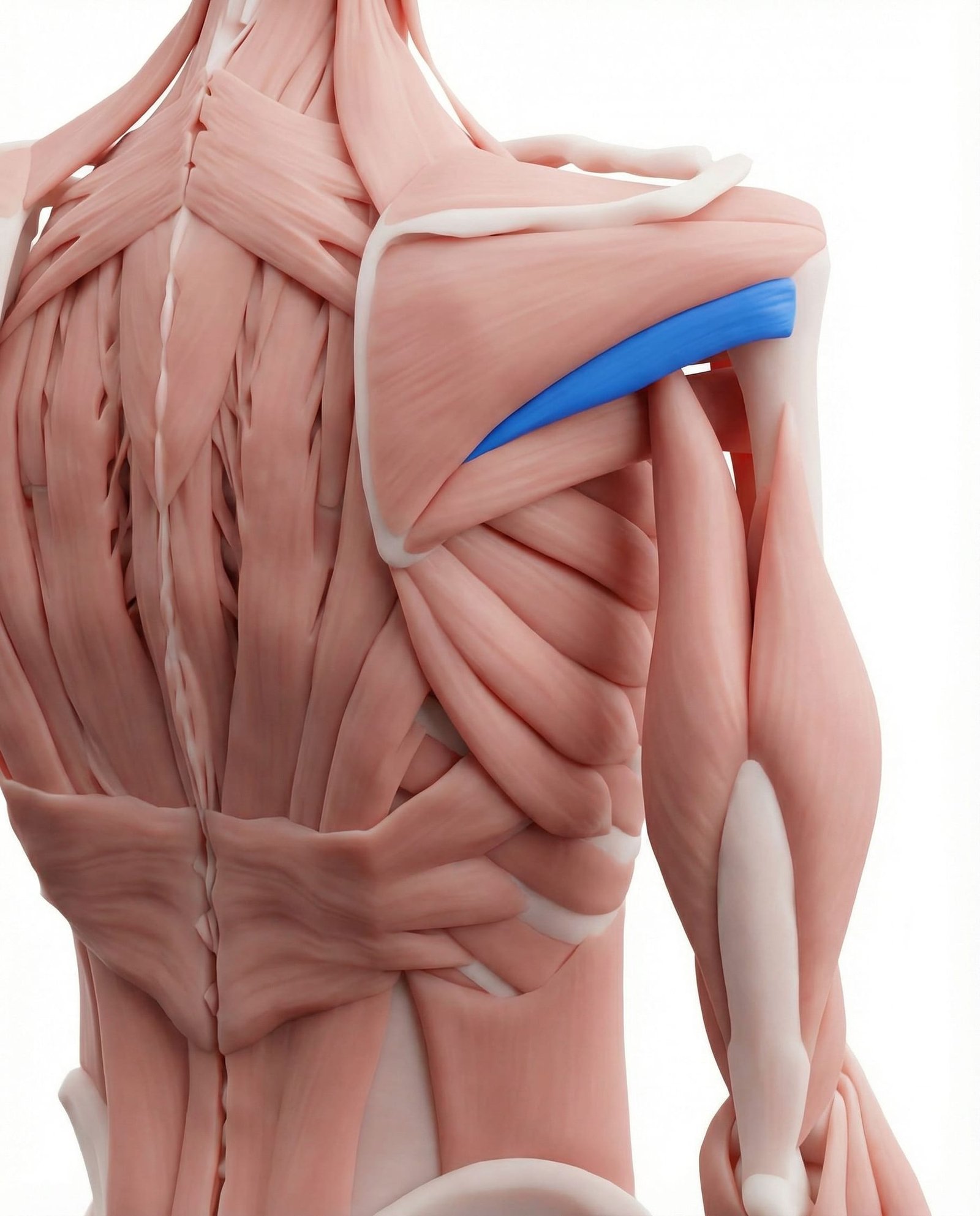
Working in concert with the infraspinatus, the teres minor assists in external rotation and shoulder stabilization. It is less frequently injured in isolation but is an integral part of the posterior cuff. Its myofascial trigger points can cause localized, deep pain in the posterior deltoid region.
Tendon Overload
Repetitive or excessive load beyond the tendon's capacity can lead to a continuum of pathology, from a reactive, inflammatory state to chronic, degenerative changes (tendinosis).
Subacromial Impingement
Poor scapular mechanics or anatomical variations can narrow the space below the acromion, leading to mechanical compression and abrasion of the supraspinatus tendon and subacromial bursa.
Myofascial Referral
Active trigger points in muscles like the infraspinatus, subscapularis, or scalenes can generate referred pain that is perceived in the shoulder joint, mimicking intra-articular pathology.
Central Sensitization
With persistent pain, the central nervous system can become hypersensitive. This can lead to pain that is disproportionate to the degree of tissue injury, spreads to wider areas, and is less responsive to local treatments.
Common Referral Patterns
Myofascial trigger points are hyperirritable spots in muscle that can refer pain and tenderness to distant, yet predictable, locations. Understanding these patterns is key to avoiding diagnostic errors and targeting treatment effectively.
Infraspinatus
Supraspinatus
Subscapularis
Scalenes
Self-Assessment Tests
These tests can help you explore the nature of your shoulder pain. They are designed to selectively stress different structures. Perform them gently and stop if you experience sharp pain.
Painful Arc Test
Hawkins-Kennedy Test
Empty Can (Jobe) Test
Infraspinatus Trigger Point Palpation
External Rotation Strength Test
Integrated Treatment Pathways
A successful treatment plan for shoulder pain typically integrates multiple approaches. The goal is to calm irritated tissues, restore normal movement patterns, and progressively build capacity and resilience in both the tendons and the supporting musculature.
Self-Care
- Activity modification to avoid overhead and painful movements
- Pendulum exercises for gentle, pain-free range of motion
- Application of ice or heat based on preference and acuity
- Postural awareness and ergonomic adjustments
- Gentle isometric exercises in non-provocative positions
Manual Therapy
- Soft tissue mobilization of periscapular muscles (trapezius, levator scapulae)
- Trigger point release for infraspinatus, subscapularis, and teres minor
- Glenohumeral and thoracic spine joint mobilization
- Myofascial release techniques for the shoulder girdle
- Cross-friction massage over the affected tendon (clinician-guided)
Interventional
- Diagnostic musculoskeletal ultrasound to assess tendon integrity
- Corticosteroid injection for subacromial bursitis (short-term relief)
- Platelet-Rich Plasma (PRP) injections (evidence is still emerging)
- Trigger point dry needling for associated myofascial pain
- In rare cases, surgical consultation for full-thickness tears or failed conservative care
Rehabilitation
- Structured, progressive loading program for the rotator cuff (eccentrics, HSR)
- Scapular stabilization exercises (serratus anterior, lower trapezius)
- Neuromuscular re-education for proper glenohumeral rhythm
- Kinetic chain strengthening (core, hips)
- Functional movement pattern retraining for sport or work activities
Red Flags — When to See a Doctor
While most shoulder pain is musculoskeletal in origin, it is important to be aware of signs and symptoms that could indicate a more serious condition. The following "red flags" warrant prompt consultation with a medical professional.
Rotator cuff tendinopathy and myofascial pain from surrounding muscles frequently coexist and can create a self-perpetuating cycle of dysfunction.
The location of shoulder pain is an unreliable guide to its source; deep anterior shoulder pain is commonly referred from trigger points in the posterior infraspinatus muscle.
A comprehensive clinical examination is essential, as it must assess not only the glenohumeral joint but also the cervical spine and scapulothoracic function.
Imaging findings such as "tendinosis" or "partial-thickness tears" are prevalent in asymptomatic individuals and must be interpreted in the context of the patient's clinical presentation.
The cornerstone of effective, long-term management for most rotator cuff conditions is a well-structured, progressive exercise program focused on load management and restoring function.
Addressing scapular dyskinesis by strengthening the serratus anterior and lower trapezius is often critical to reducing subacromial impingement and allowing the rotator cuff to heal.
Passive treatments may provide temporary relief, but an active approach involving rehabilitation is generally necessary for lasting improvement.
Movement therapy
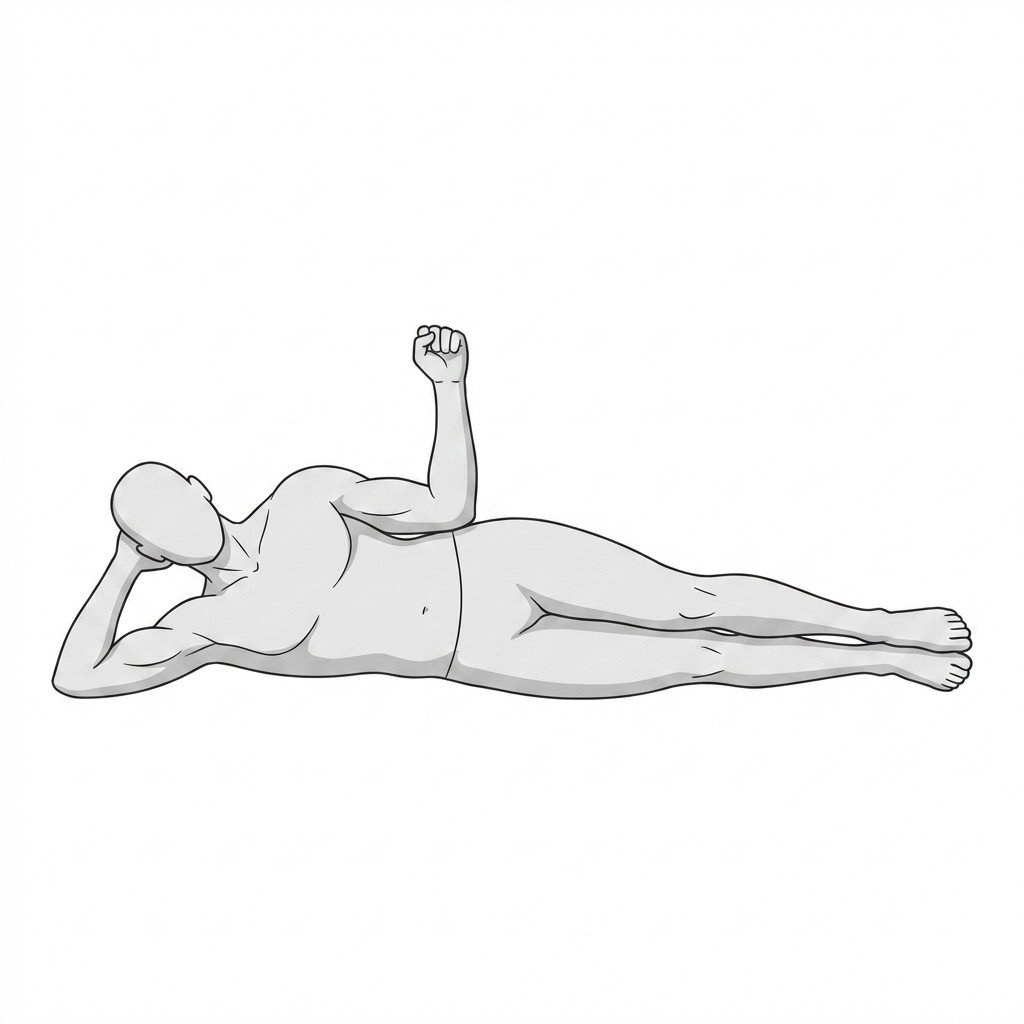
Exercises for Rotator Cuff Tendinopathy
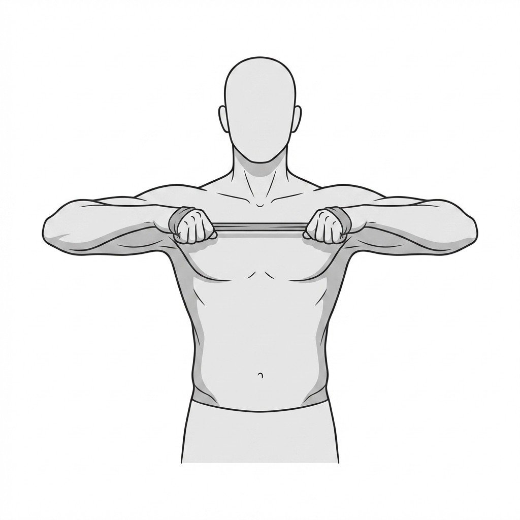
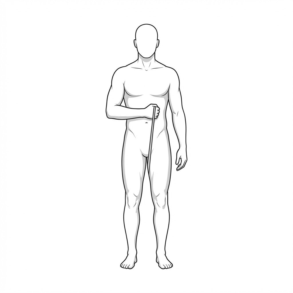
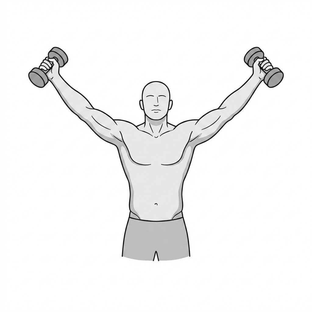
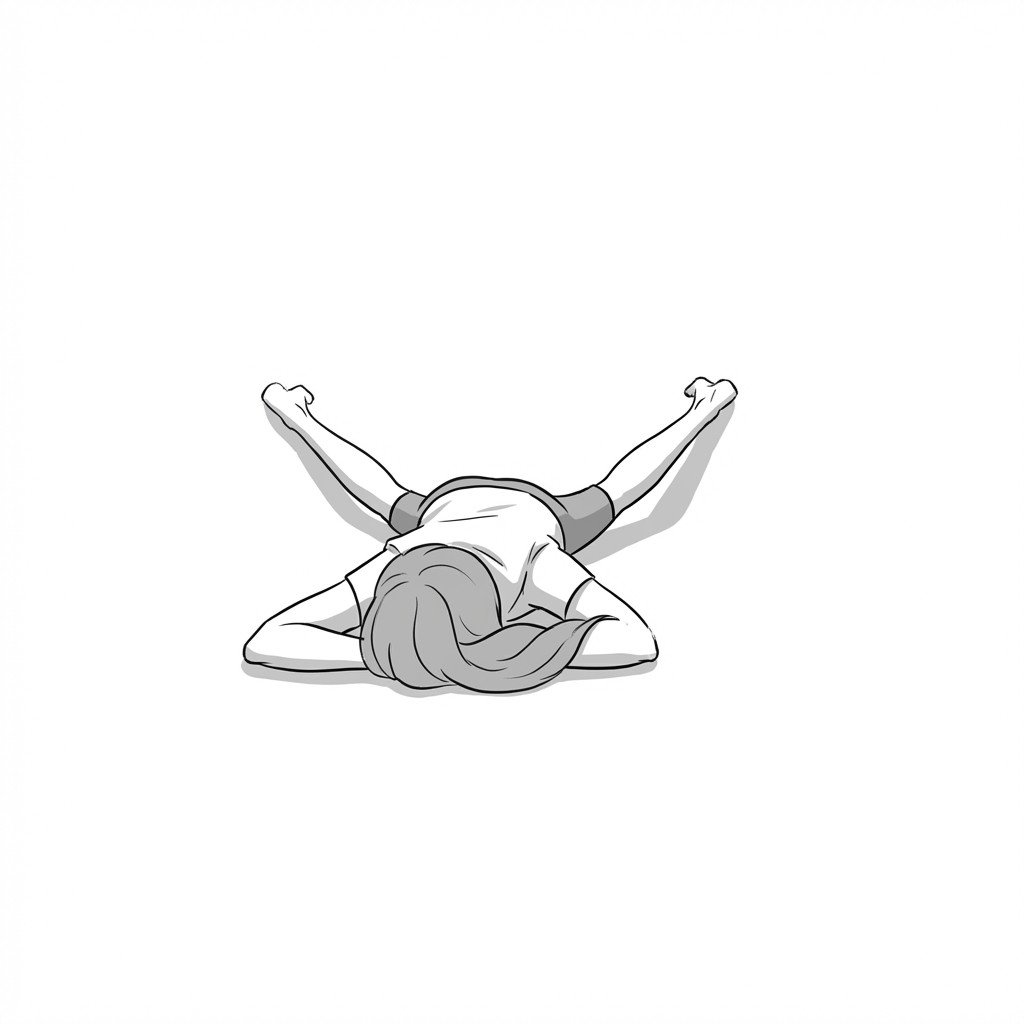
These exercises load the rotator cuff through its pain-free range — eccentric external rotation and scaption to rebuild tendon capacity, W-raises and internal rotation to rebalance the shoulder girdle. Start with 2–3 and progress as tolerated.