Understanding Anterior Knee Pain
Patellar tendinopathy, often called jumper's knee, is a clinical pain syndrome centered around the patellar tendon, usually at or near the inferior pole of the patella. It is especially common in sports that involve repeated jumping, deceleration, and forceful knee extension. Current models describe it as a load-related tendon disorder rather than a simple inflammatory condition.
Anterior knee pain often has more than one contributor. In some patients, quadriceps trigger points or proximal tissue tension amplify anterior knee symptoms or alter mechanics enough to increase tendon stress. That does not mean every tendon diagnosis is really myofascial pain, but it does mean the two often overlap.
Localized inferior pole pain points toward tendon involvement, but proximal quadriceps and hip contributors often shape how severe and persistent the problem becomes.
Clinical differentiation depends on pain location, symptom behavior, tendon-loading tests, palpation, and broader biomechanical assessment. Imaging can help in selected cases, but imaging findings must still match the patient’s actual presentation.
Patellar Tendinopathy vs Myofascial Mimic
Pain behavior
Pain location
Palpation finding
First-line treatment
Anatomical Considerations
The extensor mechanism depends on coordination among the quadriceps, patella, patellar tendon, and the way the hip and trunk control load. Problems in one part of the system often affect the rest.
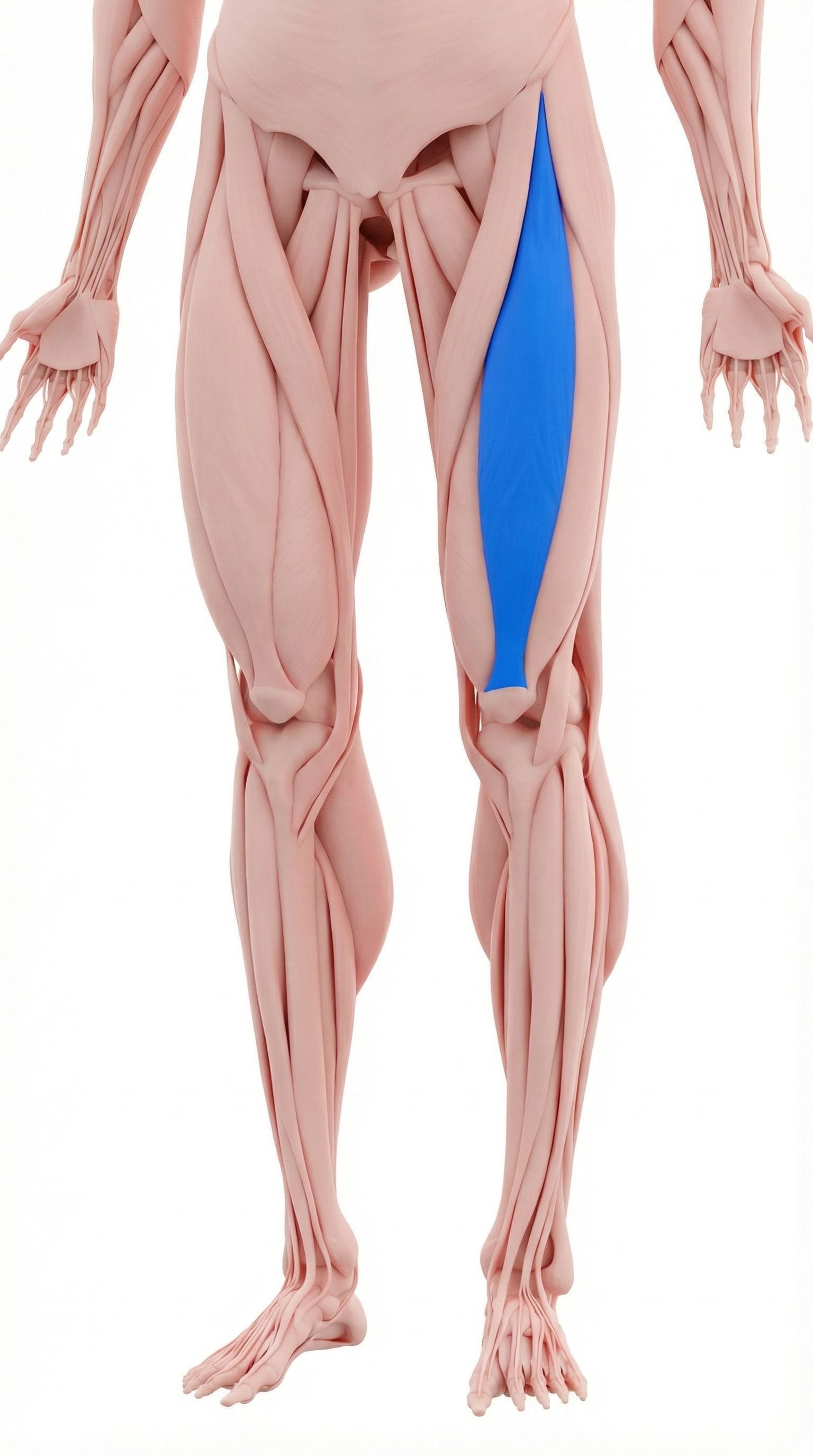
A biarticular quadriceps muscle that crosses both the hip and the knee. Trigger points or increased resting tone here can contribute to deep anterior knee discomfort and may increase extensor mechanism load during jumping, running, and stair descent.
A deep quadriceps muscle lying beneath the rectus femoris. Pain from this region may feel diffuse and deep in the anterior thigh or superior patellar region, which can complicate clinical differentiation from tendon-related pain.
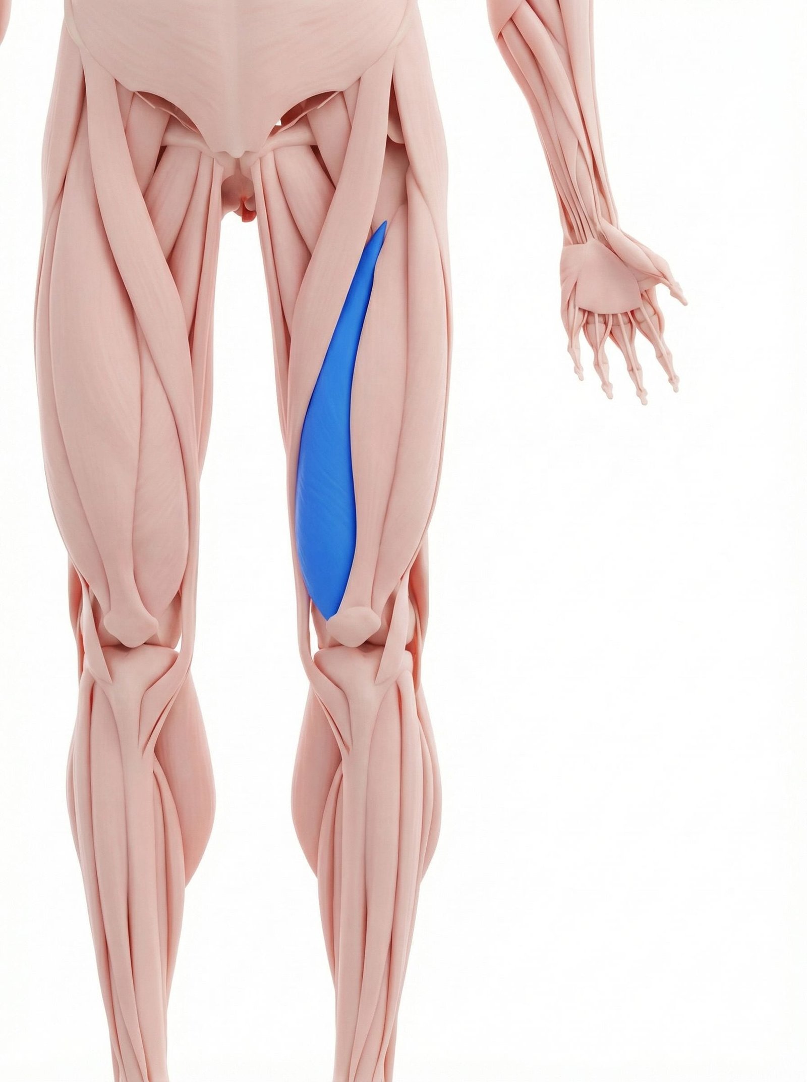
An important medial quadriceps component involved in knee extension and patellar control. Distal myofascial irritation here may contribute to medial patellar or anteromedial knee pain and may coexist with patellofemoral tracking problems.
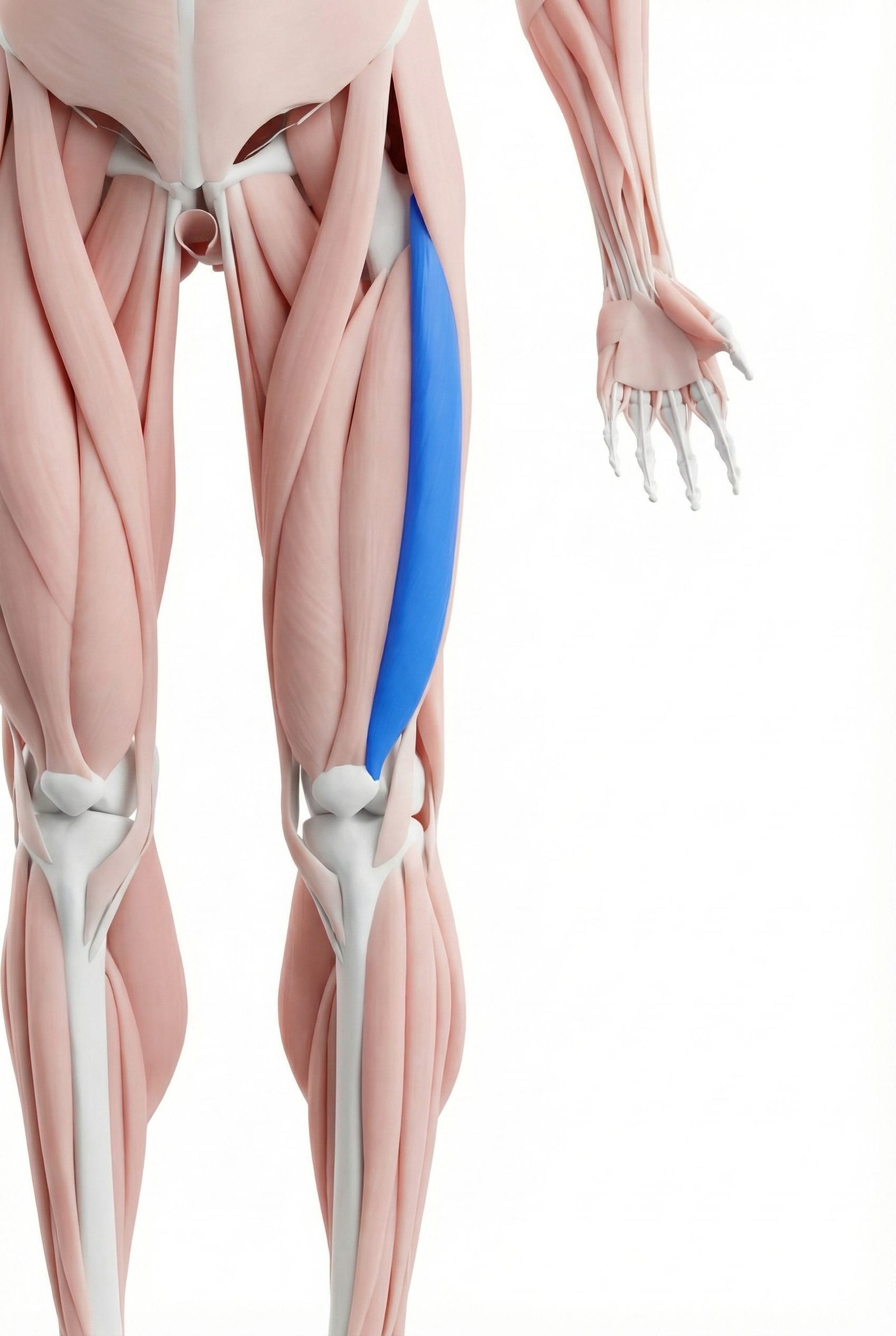
A large lateral quadriceps muscle that can contribute to lateral thigh tension and altered patellofemoral loading when stiff, overactive, or myofascially irritated.
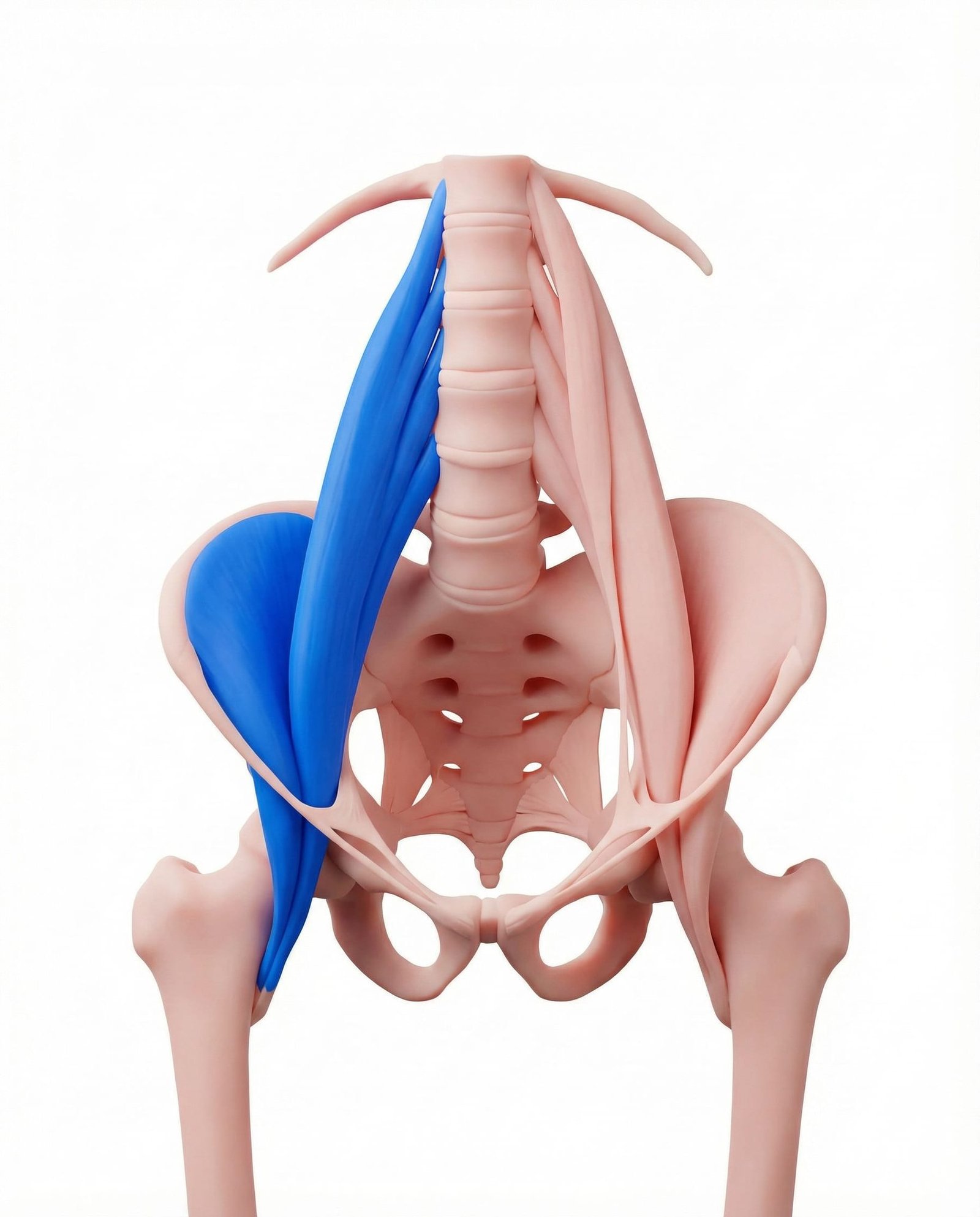
A primary hip flexor complex. Although it does not typically refer pain directly into the patellar tendon, iliopsoas restriction or poor hip extension can change landing and gait mechanics, increasing quadriceps demand and anterior knee load.

An important hip adductor involved in frontal-plane control and pelvic stability. Dysfunction here may contribute to altered femoral mechanics, which can indirectly change patellofemoral and tendon loading during sport or repetitive activity.
Tensile Load
Repeated forceful loading through the patellar tendon during jumping, landing, and deceleration.
Compressive Load
Deep knee flexion and certain movement positions may increase compressive stress around the tendon origin.
Myofascial Tension
Quadriceps and hip muscle tension may increase baseline load across the extensor mechanism.
Biomechanical Contribution
Hip, trunk, ankle, and landing mechanics can all influence how much load the tendon repeatedly absorbs.
Myofascial Referral Patterns
Quadriceps trigger points can project pain toward the knee and may complicate the clinical picture. Recognizing this helps avoid under-treating proximal contributors in a patient whose pain feels “purely patellar.”
Rectus Femoris
Vastus Intermedius
Vastus Medialis
Clinical Assessment
Provocative loading and targeted palpation are often more clinically useful than vague symptom description alone. The goal is to reproduce the familiar pattern safely, not to push through severe pain.
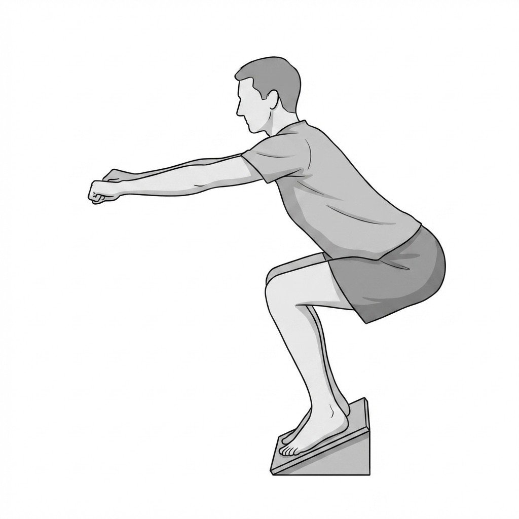
Single-Leg Decline Squat
Inferior Pole Palpation
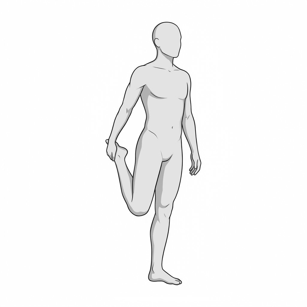
Rectus Femoris Tension Test (Modified Thomas Test)
Hoffa's Fat Pad Compression
Evidence-Based Management
Current management favors progressive loading rather than prolonged rest. When proximal myofascial tension or poor movement mechanics are also present, treatment often needs to address both the tendon and the surrounding system.
Self-Care
- Modify jumping, sprinting, or deep-loading activities during high irritability phases
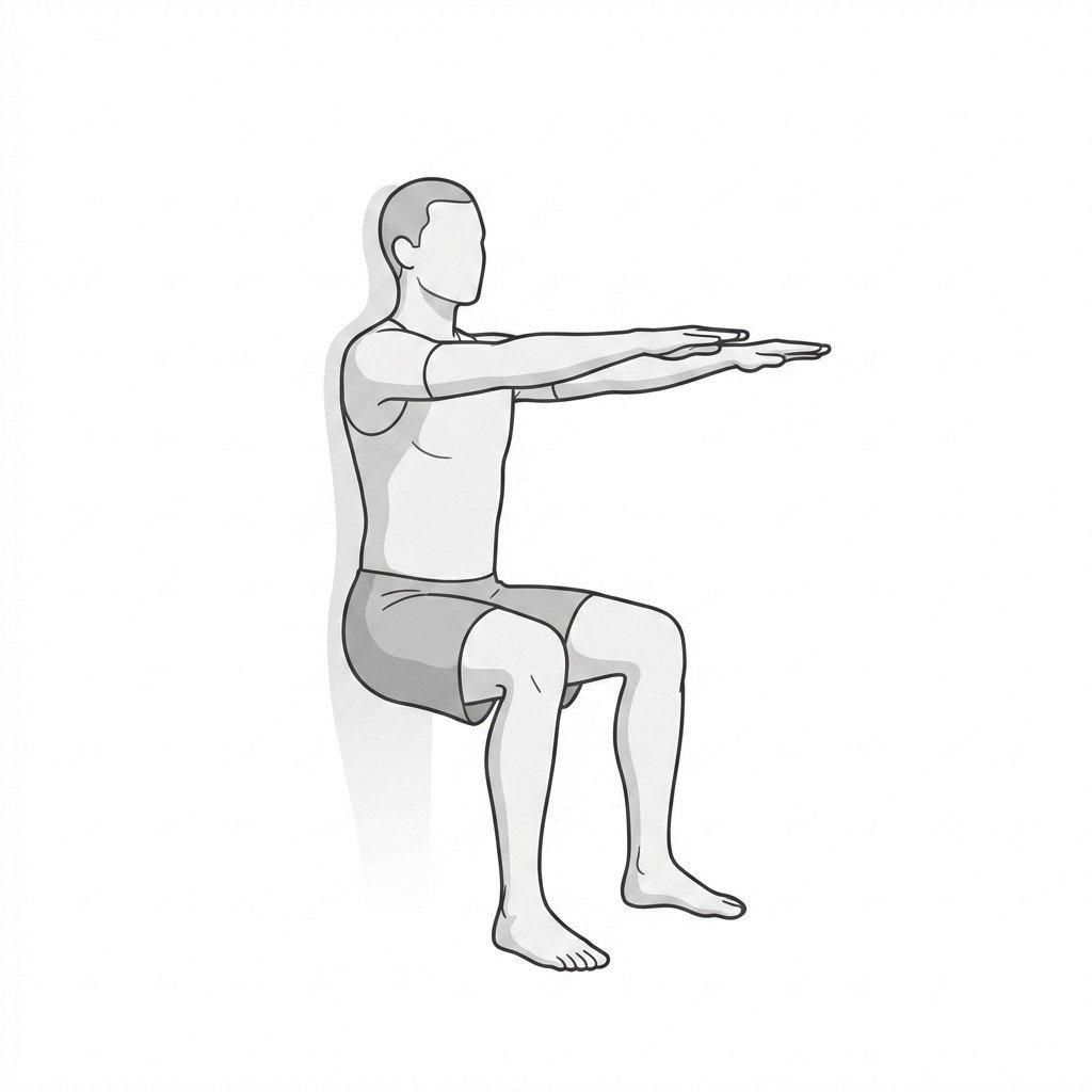
- Use isometric quadriceps work in selected cases for short-term pain modulation
- Use gentle self-treatment of proximal quadriceps tissues rather than pressing repeatedly on the painful tendon itself
- Apply ice after clearly provocative activity if it helps calm a flare
- Use a patellar strap only if it subjectively helps and does not replace load management
Manual Therapy
- Treat relevant quadriceps trigger points or myofascial restriction when present
- Use soft-tissue treatment to improve quadriceps and anterior-hip tissue tolerance
- Address patellofemoral and tibiofemoral mobility when clinically relevant
- Use friction-based tendon techniques cautiously, since evidence and tolerance vary
- Integrate manual therapy with loading progression rather than using it alone
Interventional
- Shockwave therapy may be considered in selected chronic tendon cases
- PRP may be discussed in refractory cases, though evidence remains mixed
- Dry needling or trigger point injection may help when quadriceps myofascial pain is clearly part of the picture
- Image-guided procedures may be considered in specialist settings for selected chronic cases
- Corticosteroid injection into the patellar tendon is generally avoided because of tissue-risk concerns
Rehabilitation
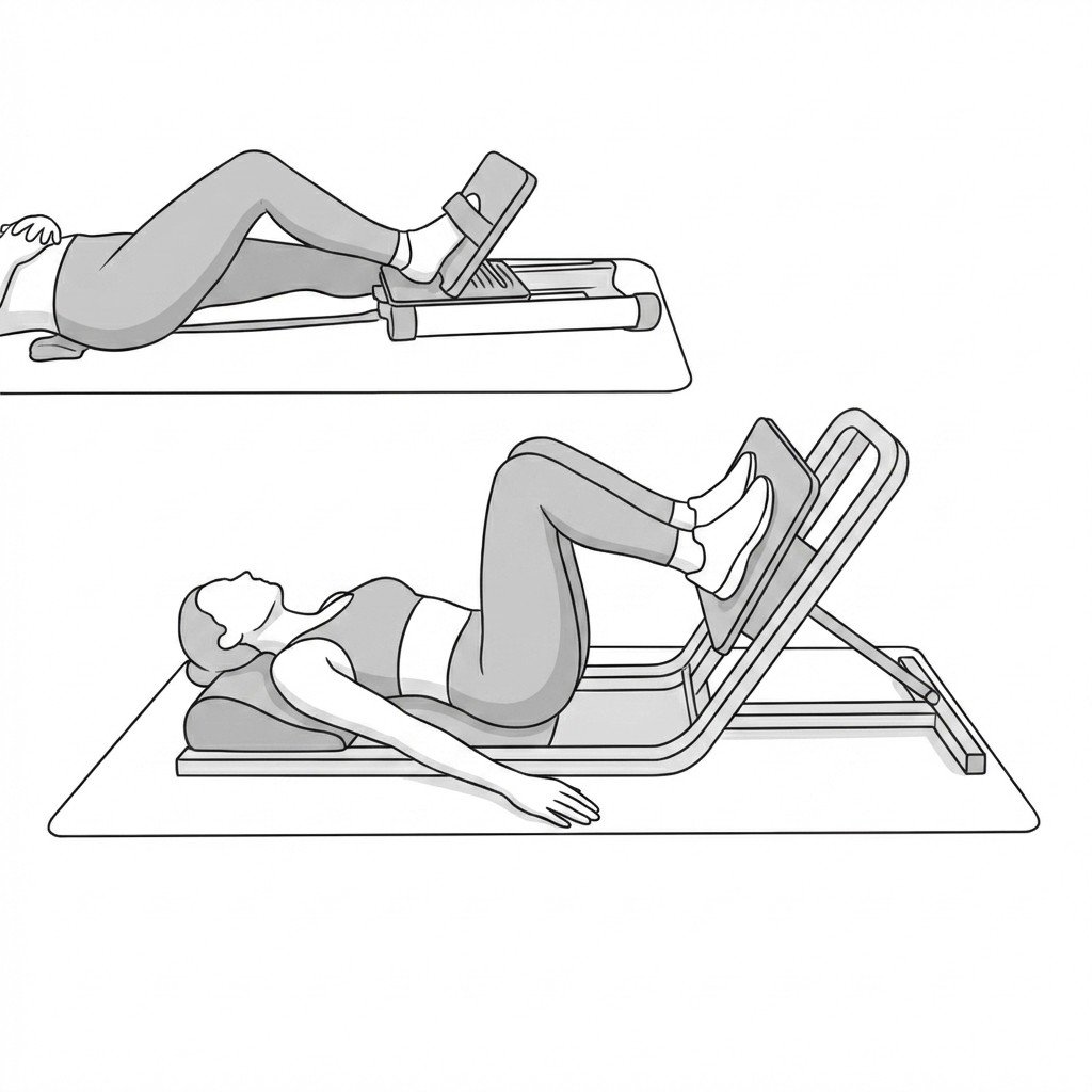
- Heavy slow resistance training is one of the most established tendon-loading strategies
- Progressive tendon loading usually matters more than passive treatment alone
- Hip and trunk strengthening often helps reduce recurring knee overload
- Plyometric and energy-storage work belong later in rehabilitation, not early
- Biomechanical review of sport or landing strategy can be useful when recurrence is persistent
Red Flags & Differential Diagnosis
Most anterior knee pain is mechanical, but some findings require urgent medical review before a tendon-based or myofascial explanation is assumed.
Patellar tendinopathy usually presents with localized pain at the inferior pole of the patella, especially with energy-storage or tendon-loading activities.
Quadriceps trigger points — especially in the rectus femoris and vastus intermedius — can mimic or coexist with patellar tendon pain.
One clinically useful feature of tendinopathy is the warm-up pattern, where pain improves during activity and worsens later or the following day.
The single-leg decline squat and localized inferior-pole palpation are helpful tendon-loading tests, but they should be interpreted alongside the rest of the examination.
Progressive tendon loading remains a first-line treatment principle in patellar tendinopathy.
Addressing proximal quadriceps and hip-related contributors often improves tendon rehabilitation outcomes.
Corticosteroid injection directly into the patellar tendon is generally avoided because of tissue-risk concerns.
Movement therapy
Exercises for Patellar Tendinopathy
These exercises target loading tolerance, quadriceps function, and supporting movement patterns. Start with 2-3 and progress based on symptom response.