Understanding the Condition
Lateral epicondylalgia (LE), commonly referred to as tennis elbow, is primarily considered a tendinopathy of the common extensor origin, most notably involving the extensor carpi radialis brevis (ECRB). Current literature suggests the underlying pathology is typically a degenerative tendinosis rather than an acute inflammatory process, characterized by angiofibroblastic hyperplasia and disorganized collagen. It is highly prevalent among manual workers, individuals engaged in repetitive gripping, and racket sport athletes. However, differentiating true insertional tendinopathy from regional myofascial pain syndromes (MPS) is critical, as misdiagnosis frequently leads to refractory symptoms and treatment failure.
Myofascial pain frequently coexists with or directly mimics LE. Trigger points within the supinator, extensor carpi radialis longus (ECRL), and ECRB muscle bellies can refer pain directly to the lateral epicondyle, creating a clinical presentation nearly indistinguishable from tendinopathy. Furthermore, proximal myofascial dysfunction, particularly within the scalenes and infraspinatus, can refer pain to the lateral elbow or contribute to a "double-crush" phenomenon. In this scenario, mild cervical nerve root irritation (often C6) sensitizes the peripheral nerves, lowering the threshold for pain and dysfunction at the common extensor tendon.
Lateral elbow pain is rarely an isolated tendon issue; it frequently represents a complex interplay of local tendinopathy, regional myofascial trigger points, and cervical sensitization.
Clinical differentiation requires precise palpation and provocative testing. True LE typically presents with point tenderness directly at or immediately anterior to the lateral epicondyle, exacerbated by resisted wrist extension (Cozen's test) or passive wrist flexion with elbow extension (Mill's test). Conversely, maximal tenderness located 2 to 4 centimeters distal to the epicondyle, particularly over the mobile wad, should raise clinical suspicion for supinator trigger points or radial tunnel syndrome (posterior interosseous nerve entrapment). Assessing grip strength with the elbow fully extended versus flexed can also help differentiate local tendon loading intolerance from neurodynamic tension.
True Tendinopathy vs Myofascial Mimic
Pain location
Provocation
Palpation finding
First-line treatment
Anatomical Context
The lateral elbow is a crowded anatomical junction where the common extensor tendon, the supinator muscle, and the radial nerve converge. Understanding this spatial relationship is vital for accurate diagnosis.
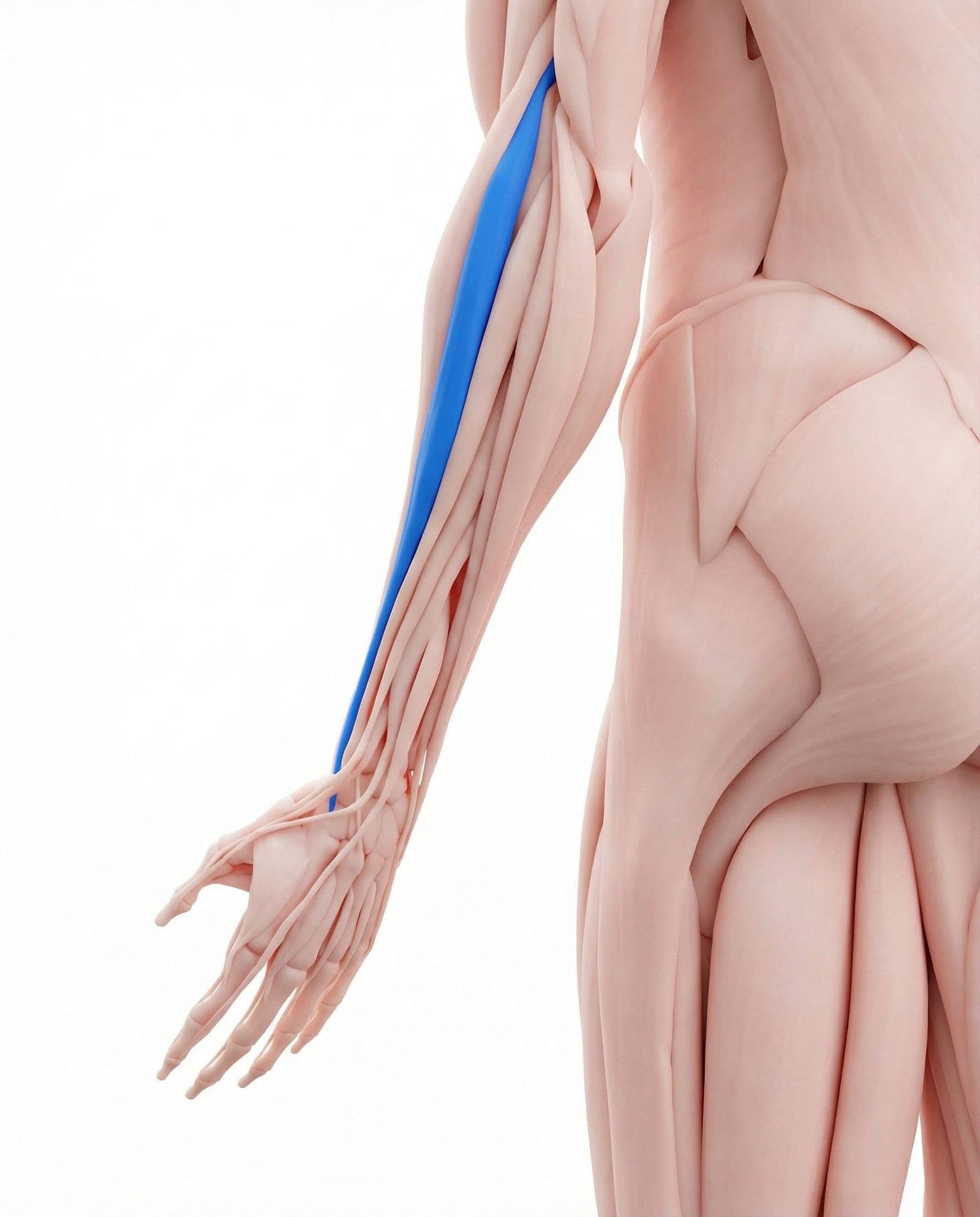
The ECRB is the primary tendon implicated in true lateral epicondylalgia. It functions to extend and abduct the wrist, stabilizing the wrist during gripping activities. Myofascial trigger points within the ECRB muscle belly frequently develop secondary to tendinopathy or repetitive overload, referring pain directly to the lateral epicondyle and the dorsum of the hand.
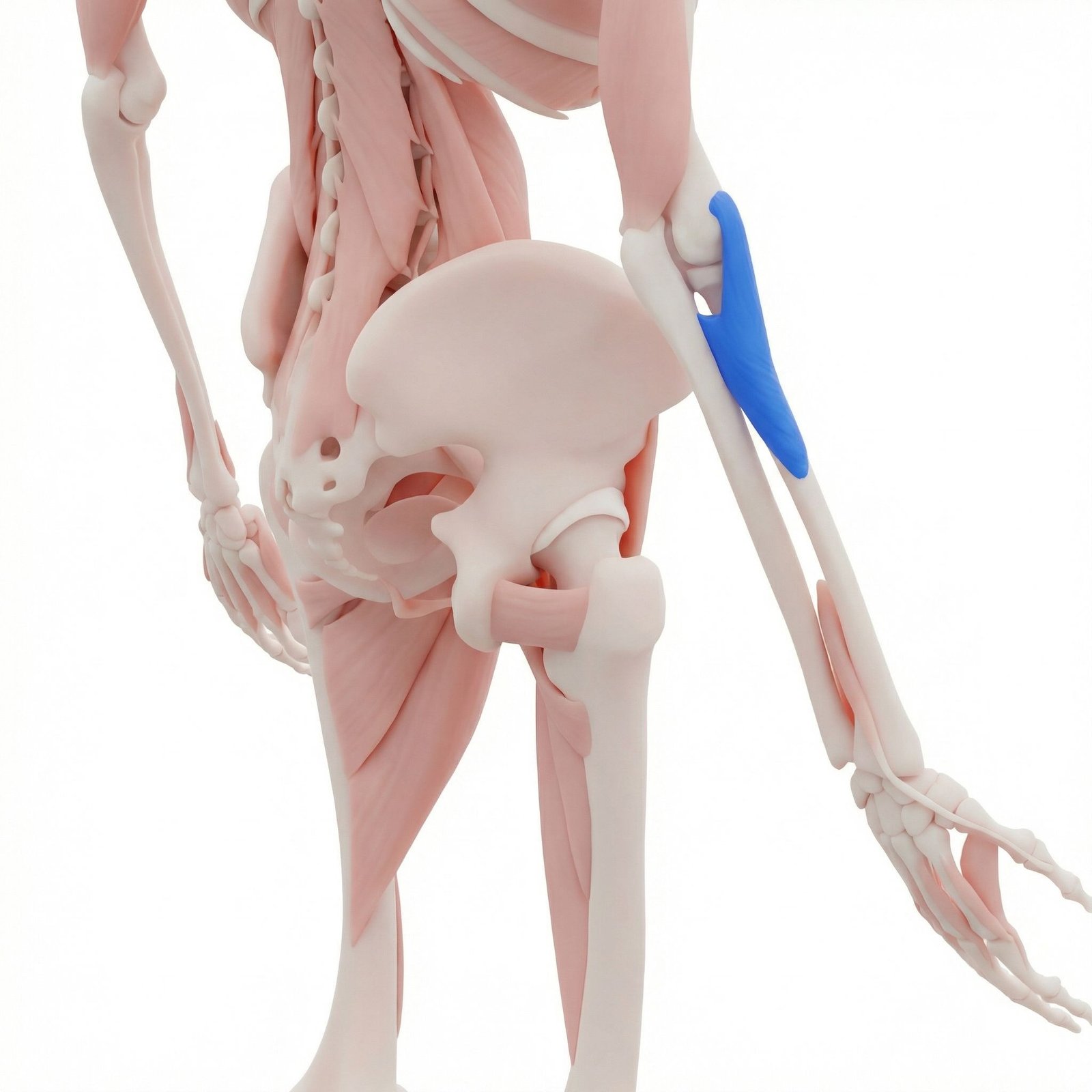
The supinator is a classic mimicker of lateral epicondylalgia. It wraps around the proximal radius and is pierced by the deep branch of the radial nerve. Trigger points in the supinator refer pain strongly to the lateral epicondyle and dorsal web space of the thumb, often complicating the clinical picture and contributing to radial nerve sensitization.
Working in tandem with the ECRB, the ECRL originates slightly higher on the lateral supracondylar ridge. While less commonly the primary site of tendinopathy, it frequently harbors trigger points in patients with chronic lateral elbow pain, particularly those involved in racket sports or heavy manual labor.
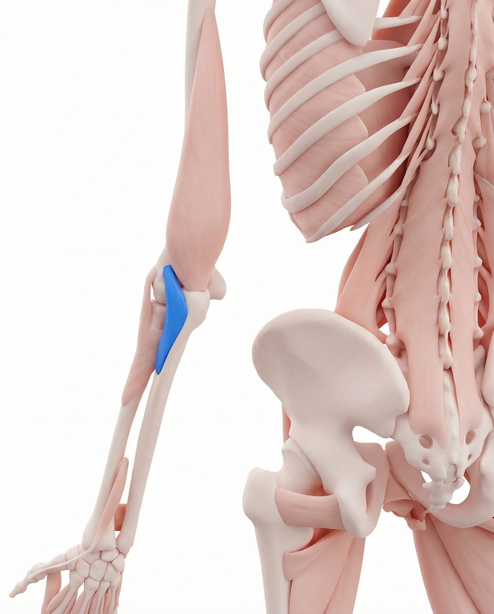
A small triangular muscle at the posterior aspect of the elbow joint that assists in elbow extension and stabilizes the ulna. Trigger points here are often overlooked but can produce localized, aching pain directly at the lateral epicondyle, mimicking insertional tendinopathy.
The anterior and middle scalenes can entrap or irritate the roots of the brachial plexus (particularly C5-C6). Trigger points in the scalenes frequently refer pain down the radial aspect of the arm to the lateral elbow. This cervical contribution can create a "double-crush" scenario, sensitizing the distal tissues and making the elbow more susceptible to tendinopathy.
The lateral head of the triceps attaches near the lateral epicondyle. Trigger points in this region can refer pain locally to the lateral epicondyle and occasionally down the dorsal forearm. It is often involved in patients who perform repetitive pushing or elbow extension under load.
ECRB Tendinopathy
Degenerative changes (tendinosis) at the insertion of the extensor carpi radialis brevis due to repetitive overload and failed healing.
Myofascial Trigger Points
Hyperirritable nodules in the supinator, ECRB, or triceps that refer aching pain directly to the lateral epicondyle.
Radial Nerve Sensitization
Irritation or entrapment of the deep branch of the radial nerve (PIN) as it passes through the Arcade of Frohse in the supinator muscle.
Cervical Referral
Radicular pain from C6 nerve root irritation or somatic referral from scalene trigger points mimicking lateral elbow pathology.
Pain Referral Patterns
Pain at the lateral epicondyle can originate from local tendon pathology, but it is frequently referred from trigger points in the forearm or the cervical spine. Recognizing these patterns helps differentiate the source.
Supinator
Extensor Carpi Radialis Brevis
Scalenes
Anconeus
Clinical Assessment
Clinical evaluation of lateral elbow pain involves stressing the extensor tendons, palpating for specific tissue tenderness, and screening the cervical spine to rule out proximal referral.
Cozen's Test (Resisted Wrist Extension)
Mill's Test (Passive Stretch)
Maudsley's Test (Resisted Middle Finger Extension)
Supinator Palpation vs. Epicondyle Palpation
Cervical Foraminal Compression (Spurling's Screen)
Treatment Pathways
A comprehensive approach to lateral epicondylalgia addresses local tendon pathology through progressive loading, while simultaneously managing regional myofascial trigger points and cervical sensitization.
Self-Care
- Activity modification to reduce repetitive gripping and wrist extension
- Use of a counterforce brace 2-3 cm distal to the epicondyle during provocative tasks
- Ergonomic assessment of workstations and tool handles
- Gentle, pain-free stretching of the wrist extensors
- Application of ice or heat based on symptom acuity and patient preference
Manual Therapy
- Ischemic compression of trigger points in the supinator and ECRB/ECRL
- Myofascial release of the cervical spine and scalenes to address proximal sensitization
- Cross-friction massage at the common extensor origin (evidence is mixed, but often utilized)
- Joint mobilization of the radiohumeral and proximal radioulnar joints
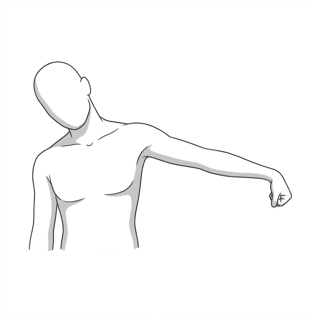
- Radial nerve neurodynamic mobilization (gliding techniques)
Interventional
- Dry needling of the supinator, wrist extensors, and relevant cervical musculature
- Platelet-rich plasma (PRP) injections (moderate evidence for intermediate-to-long-term benefit)
- Corticosteroid injections (meta-analyses suggest short-term relief but potential long-term detriment)
- Autologous blood injections for chronic, refractory tendinosis
- Acupuncture targeting local and regional meridians
Rehabilitation
- Progressive isometric loading of the wrist extensors for pain modulation
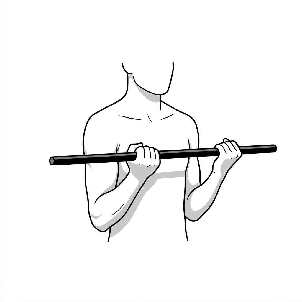
- Eccentric exercise protocols (e.g., the Tyler Twist using a flexible rubber bar)
- Heavy slow resistance (HSR) training for tendon remodeling
- Scapular and rotator cuff strengthening to improve proximal kinetic chain stability
- Gradual return-to-play or return-to-work functional conditioning
Red Flags — When to See a Doctor
While most lateral elbow pain is mechanical or myofascial in nature, certain signs indicate potentially serious neurological or structural pathology that requires prompt medical investigation.
Lateral epicondylalgia is primarily a degenerative tendinopathy, but myofascial pain frequently coexists or mimics the condition entirely.
Trigger points in the supinator muscle are the most common mimickers of lateral epicondylalgia and must be assessed in refractory cases.
Maximal tenderness 2-4 cm distal to the epicondyle suggests supinator involvement or radial tunnel syndrome, rather than isolated ECRB tendinopathy.
Cervical spine dysfunction, particularly involving the C6 nerve root and scalene muscles, can sensitize the lateral elbow via a "double-crush" mechanism.
Current guidelines generally recommend progressive tendon loading (eccentrics or heavy slow resistance) as the cornerstone of long-term rehabilitation.
Meta-analyses suggest corticosteroid injections may provide short-term relief but are associated with higher recurrence rates and potential long-term tendon detriment.
Movement therapy
Exercises for Lateral Epicondylalgia
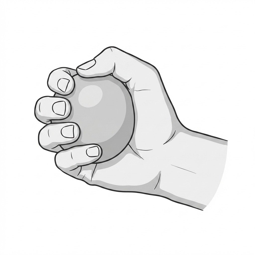
These exercises progressively load the wrist extensors and supinator through their full range — addressing the tendon overuse at the lateral epicondyle — while radial nerve glides reduce the neural component that often sustains the pain cycle. Start with 2–3 and progress as tolerated.