Understanding Myofascial Knee Pain
Knee & Patellofemoral Pain

Knee & Patellofemoral Pain

Anatomy and Pain Pattern Overview

Knee pain is one of the most common musculoskeletal complaints, and patellofemoral pain is a major part of that burden. In many patients, the pain picture is not explained by cartilage, bone, or tendon findings alone. Muscular contributors — especially in the quadriceps and lateral thigh — are often part of the clinical picture.

Trigger points in the quadriceps group can refer pain directly into and around the knee. This is one reason some patients feel deep aching “in the joint” even when the most useful palpation findings are located well above the kneecap. The goal is not to replace structural diagnosis with trigger point diagnosis, but to recognize that knee pain can be partly muscular even when the symptoms feel articular.

Knee pain is often more than a knee problem. The thigh and hip frequently influence what the patient feels at the joint.

The distinction between myofascial contributors and structural pathology still matters. Meniscal tears, ligament injuries, inflammatory joints, and acute traumatic problems have their own patterns and still need to be considered. But when the examination repeatedly reproduces knee pain from the thigh muscles and the joint signs are relatively quiet, a myofascial component deserves attention.

Anatomy & Key Trigger Points

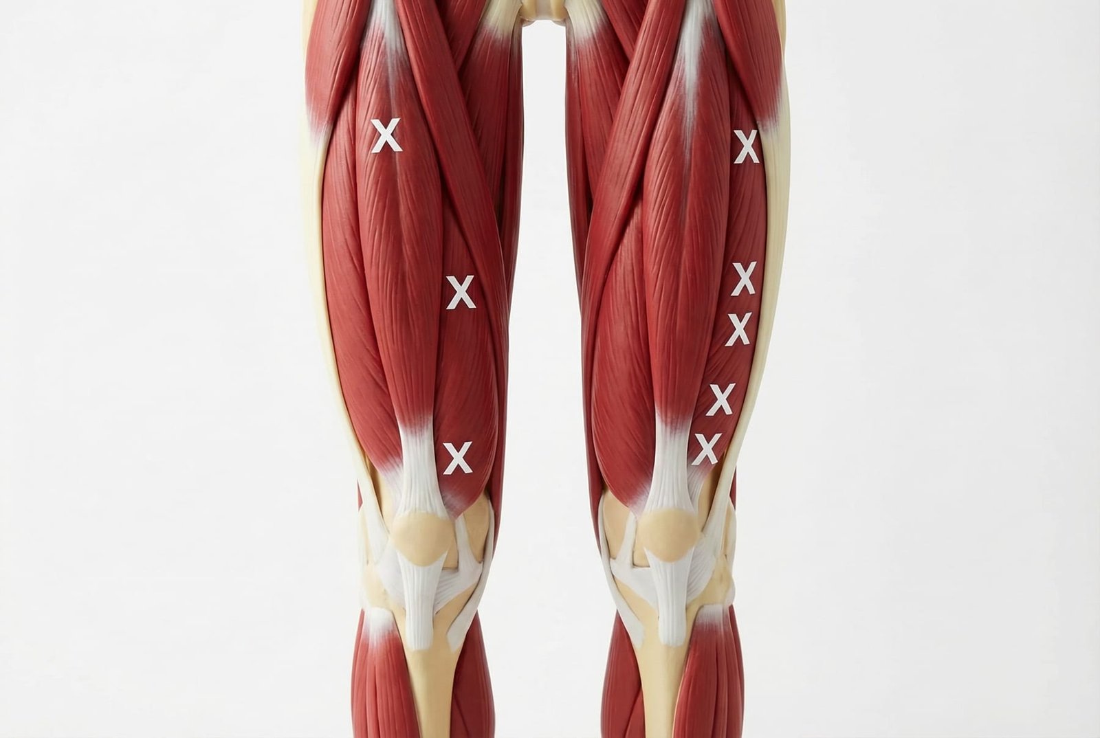
The knee is influenced by several muscle groups that cross above and below it. Trigger points in these muscles can create pain patterns that overlap with common orthopedic diagnoses, especially around the patella and lateral knee.
Knee & Patellofemoral Pain

Knee & Patellofemoral Pain

Vastus Medialis

The vastus medialis, including its oblique fibers (VMO), contributes to patellar tracking and anterior knee control. Trigger points in this muscle may refer pain to the anteromedial knee and sometimes create a deep aching sensation around the joint. In some patients, VMO dysfunction is also associated with a subjective feeling of knee weakness or giving way, even when ligament testing is normal.Anteromedial knee, peri-patellar ache, sense of instability

Rectus Femoris

The only quadriceps muscle that crosses both the hip and knee. Trigger points in the rectus femoris may refer pain to the anterior knee, over and around the patella, and sometimes into the anterior thigh. This muscle is often irritated in patients who spend long periods sitting, perform repeated hip-flexion activities, or overload the quadriceps during sport.Anterior knee and peri-patellar pain

Vastus Lateralis

The largest quadriceps muscle, the vastus lateralis can contribute to lateral thigh and lateral knee pain patterns. Trigger points here may overlap with symptoms often labeled as ITB syndrome, lateral patellar overload, or lateral knee soft-tissue irritation. Careful palpation of the lateral thigh can help determine whether a myofascial component is part of the picture.Lateral thigh and lateral knee pain

Vastus Intermedius

Located deep to the rectus femoris, the vastus intermedius can contribute to deep anterior thigh pain and anterior knee pain, especially when the quadriceps are persistently overloaded. Because of its depth, it is less accessible to manual pressure and may be harder to isolate clinically than the other quadriceps muscles.Deep anterior thigh and anterior knee pain

Tensor Fasciae Latae / IT Band

The TFL feeds into the iliotibial band, which crosses the lateral knee region. Trigger points in the TFL and surrounding lateral hip/thigh tissues can contribute to lateral knee pain patterns that overlap with classic ITB-related complaints. This does not mean the IT band is never involved; rather, the lateral knee picture is often more mixed than a simple friction model suggests.Lateral knee near the femoral epicondyle / ITB pattern

Popliteus

A small but clinically important posterior knee muscle that helps control rotation and unlock the knee from extension. Trigger points in the popliteus may refer pain to the posterior knee and can mimic other posterior knee problems, especially when symptoms are provoked by downhill walking or rotational loading.Posterior knee pain

Gastrocnemius (Proximal)

The proximal gastrocnemius heads arise just above the knee and can contribute to posterior knee and popliteal-region pain. In some patients, proximal calf trigger points overlap with posterior joint-line complaints or a feeling of tightness behind the knee.Posterior knee, popliteal fossa, and sometimes the foot arch

Sartorius

The sartorius crosses from the ASIS to the medial tibia at the pes anserinus. Trigger points in the distal sartorius may contribute to anteromedial knee pain and can overlap with pes anserine region symptoms, particularly during twisting, crossing the legs, or hip-flexion-biased movement patterns.Anteromedial knee / pes anserine region
Knee Pain Generators
Vastus Medialis (VMO)

Can contribute to anteromedial or peri-patellar pain and a sense of knee weakness.

Rectus Femoris

Can contribute to anterior knee pain, especially with prolonged sitting or repeated hip-flexion loading.

Vastus Lateralis / ITB Region

Can contribute to lateral knee pain patterns that overlap with ITB or lateral patellar complaints.

Popliteus / Gastrocnemius

Can contribute to posterior knee pain that overlaps with posterior joint-line complaints.

Referral Patterns

Trigger point referral to the knee can be clinically confusing because patients often feel the pain at the joint while the relevant muscle findings sit higher in the thigh or calf. Reproducing the familiar pain from those muscles is often one of the most useful clues.

Vastus Medialis

May refer pain to the anteromedial knee and around or behind the patella. Some patients describe a deep joint ache, even though the pain can be reproduced by examining the muscle above the knee rather than the joint itself.

Rectus Femoris

May refer pain to the anterior knee and peri-patellar region, with or without accompanying anterior thigh discomfort. Distal involvement can overlap with patellar tendon pain; proximal involvement may broaden the thigh pain picture.

Vastus Lateralis

May refer down the lateral thigh toward the lateral knee, overlapping with ITB-related and lateral patellar pain patterns. Reproduction of the familiar pain from the mid-lateral thigh can be a helpful clue.

Popliteus

May refer pain to the posterior knee and popliteal fossa. This pattern can mimic posterior meniscal or cyst-related pain and usually requires careful differential diagnosis.

Gastrocnemius (Proximal)

May refer pain into the posterior knee and calf from the proximal medial or lateral heads. Because this overlaps with other posterior knee conditions, palpation and movement testing are important.

Self-Assessment

These tests may help identify whether a myofascial component is contributing to the knee pain pattern. They do not replace a full knee examination when red flags or major structural symptoms are present.

Test · 01

VMO Palpation Test

Technique →
Sit with the knee slightly bent (about 20 degrees of flexion). Palpate the vastus medialis just above and medial to the patella. Press firmly but tolerably into the muscle belly, looking for taut bands or distinct tender spots. Sustain pressure briefly on a tender point. A useful finding is reproduction of familiar anteromedial knee pain.
Positive Sign →
Positive: Reproduction of familiar anteromedial knee pain or peri-patellar ache
Test · 02

Rectus Femoris Provocation

Technique →
Lie prone with the knee bent. Have a partner or clinician guide the heel toward the buttock while the anterior thigh is palpated. A useful finding is reproduction of familiar anterior knee pain from a clearly tender rectus femoris region, rather than only from the patellar tendon or joint itself.
Positive Sign →
Positive: Anterior knee pain reproduced during rectus femoris stretch with tender thigh findings
Test · 03

Lateral Thigh Sweep Test

Technique →
Lie on the unaffected side. Using thumb pressure or a roller, gradually explore the lateral thigh from the hip toward the knee. If pressing a tender area in the mid-lateral thigh reproduces the same lateral knee pain you usually feel, that supports a myofascial contribution.
Positive Sign →
Positive: Familiar lateral knee pain reproduced from a tender spot in the lateral thigh
Test · 04

Posterior Knee Palpation

Technique →
Lie supine with the knee slightly bent over a rolled towel. Palpate the popliteal region and the proximal gastrocnemius heads. A useful finding is reproduction of familiar posterior knee pain from muscle palpation rather than only from the joint line.
Positive Sign →
Positive: Posterior knee pain reproduced by proximal gastrocnemius or nearby soft tissue palpation
Test · 05

Movie Sign Assessment

Technique →
Sit with the knees bent for 20-30 minutes. Progressive anterior knee pain that worsens during prolonged sitting and improves when the knee is straightened is commonly called the “movie sign.” This pattern is often seen in patellofemoral pain and may coexist with quadriceps trigger points, especially rectus femoris and vastus intermedius involvement.
Positive Sign →
Positive: Progressive anterior knee pain with prolonged sitting, improved by straightening the knee

Treatment Pathways

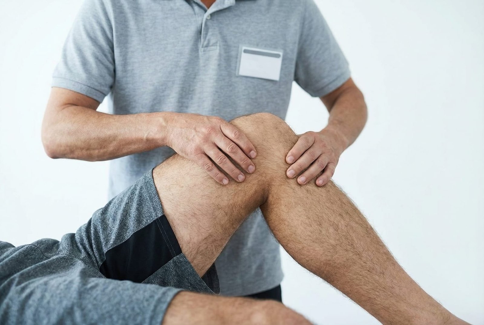
Treatment of myofascial knee pain usually works best when trigger point treatment, load modification, and progressive rehabilitation are combined. Lasting improvement usually requires better quadriceps and hip function, not just symptom relief.
Treatment Pathways

Treatment Pathways

Mechanism Diagram

Self-Care

  • Foam rolling the quadriceps group as tolerated — anterior, medial, and lateral surfaces
  • Targeted self-release of clearly tender quadriceps areas when symptoms are reproducible from the thigh
  • Quadriceps and hip-flexor stretching within tolerance
  • Reducing prolonged deep knee flexion when it clearly aggravates symptoms
  • Activity modification during flares, especially stairs, squatting, or repetitive loading

Manual Therapy

  • Sustained pressure release of trigger points in the VMO, rectus femoris, and vastus lateralis
  • Soft tissue treatment to the quadriceps and surrounding tissues based on symptom behavior
  • Patellar mobilization when patellar movement is restricted or painful
  • Myofascial treatment of the lateral thigh / ITB-adjacent region when lateral knee symptoms are present
  • Hip and ankle mobility assessment and treatment when lower-limb mechanics suggest broader contributors

Interventional

  • Dry needling of quadriceps trigger points in selected patients with persistent myofascial findings
  • Dry needling of the popliteus or proximal gastrocnemius only by appropriately trained clinicians because of local anatomy
  • Trigger point injection into accessible quadriceps trigger points when clinically appropriate
  • Ultrasound-guided needling may be considered for deeper targets such as the vastus intermedius

Rehabilitation

  • Progressive quadriceps strengthening, especially through well-tolerated ranges
  • VMO and patellofemoral loading work where clinically relevant
  • Hip abductor and external rotator strengthening to improve proximal control
  • Balance, proprioception, and single-leg control work
  • Graded return to stairs, squatting, running, and jumping with attention to load tolerance
§ Seek Immediate Evaluation

Red Flags — When to See a Doctor

Before attributing knee pain to a myofascial source, check for structural, inflammatory, and traumatic red flags that may require urgent care.

Key Takeaways
  1. Myofascial trigger points in the quadriceps and surrounding muscles can meaningfully contribute to anterior, lateral, or posterior knee pain patterns.

  2. Quadriceps trigger points often coexist with patellofemoral pain rather than replacing the diagnosis entirely.

  3. The “movie sign” is common in patellofemoral pain and can also be amplified by rectus femoris or vastus intermedius myofascial involvement.

  4. A false sense of knee instability can occur when pain inhibits the quadriceps, even when the ligaments are intact.

  5. Treatment usually works best when symptom-relieving soft tissue work is paired with progressive quadriceps and hip strengthening.

  6. Structural knee pathology and myofascial contributors can coexist, so both need to be considered in the evaluation.

Movement therapy

Exercises for Patellofemoral Pain

These exercises load the quadriceps and hip stabilizers most implicated in patellofemoral pain. Start with 2-3 and progress as tolerated.
Quadriceps stretch

Quadriceps stretch

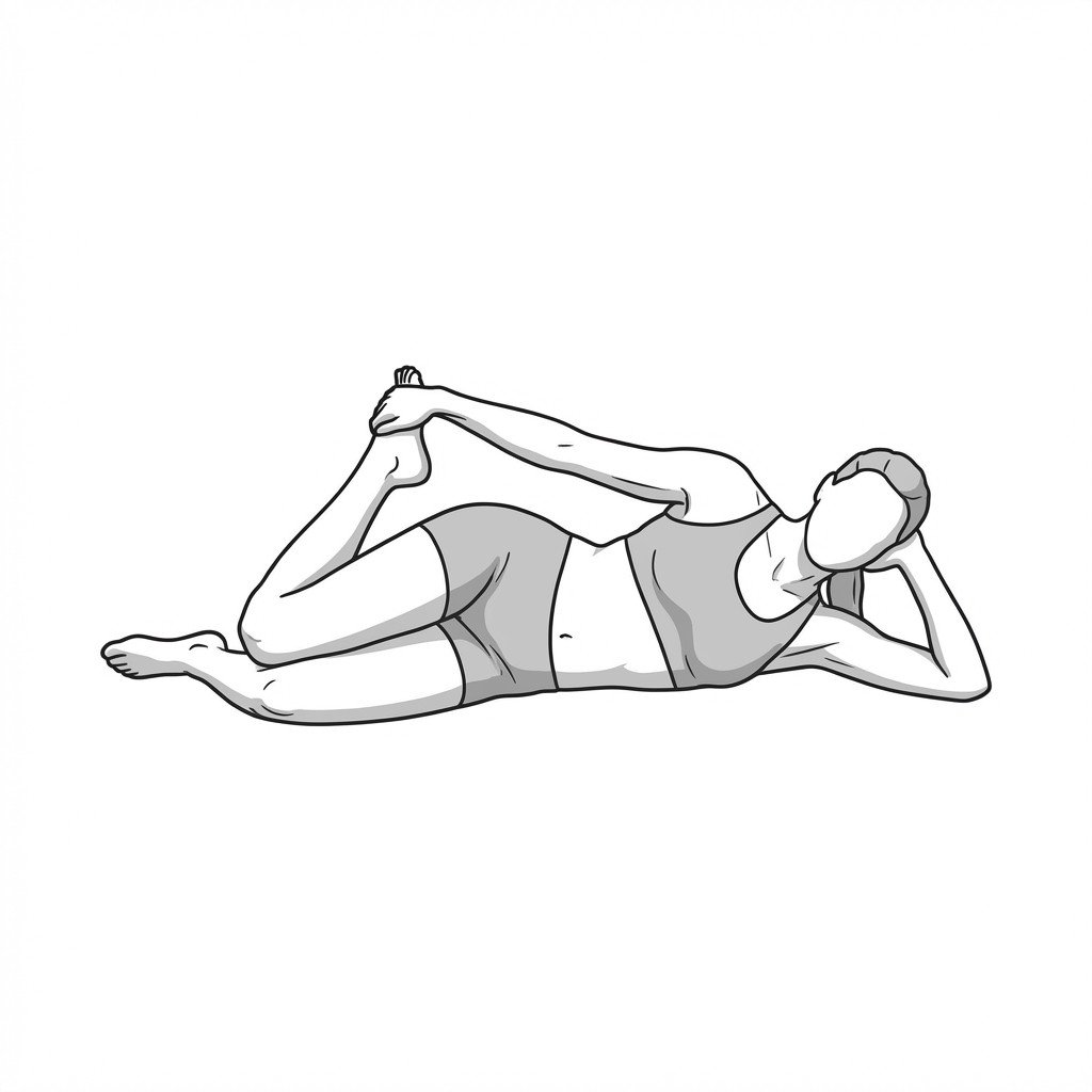
The quadriceps stretch lengthens the rectus femoris and vastus muscles, reducing passive tension on the patella. By decreasing the resting tone of these anterior thigh muscles, it mitigates compressive forces at the patellofemoral joint during movement. This improved flexibility helps normalize patellar tracking and may reduce anterior knee pain.

3 sets × 30-second hold, both sides, daily→ Start Exercise →
Step-downs

Step-downs

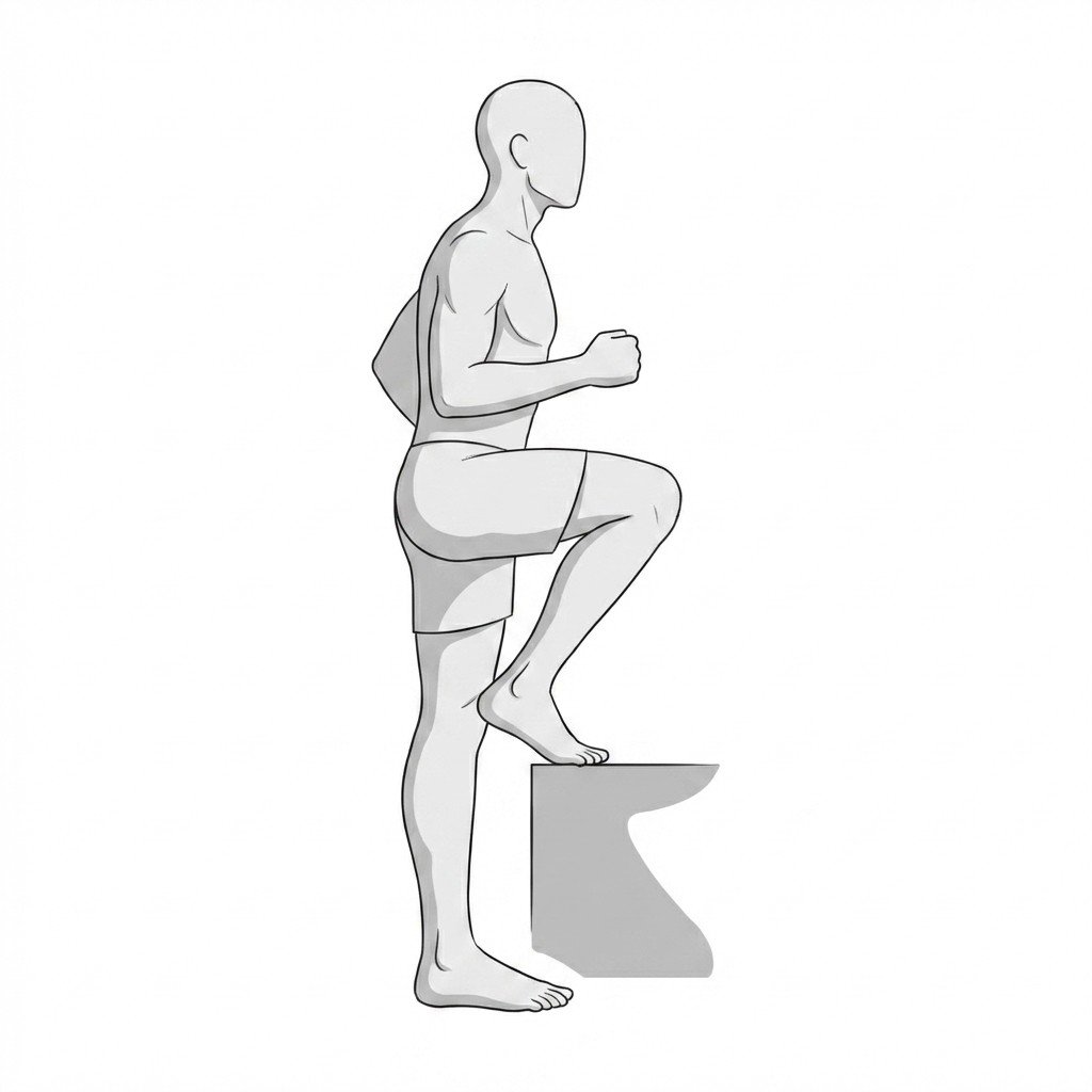
Step-downs focus on the eccentric control of the quadriceps while demanding dynamic stabilization from the hip abductors. By improving neuromuscular control of the lower extremity, this exercise helps control medial knee collapse, which may reduce abnormal contact stress on the patellofemoral joint.

3 sets × 10 reps, both sides, 3-4 times per week→ Start Exercise →
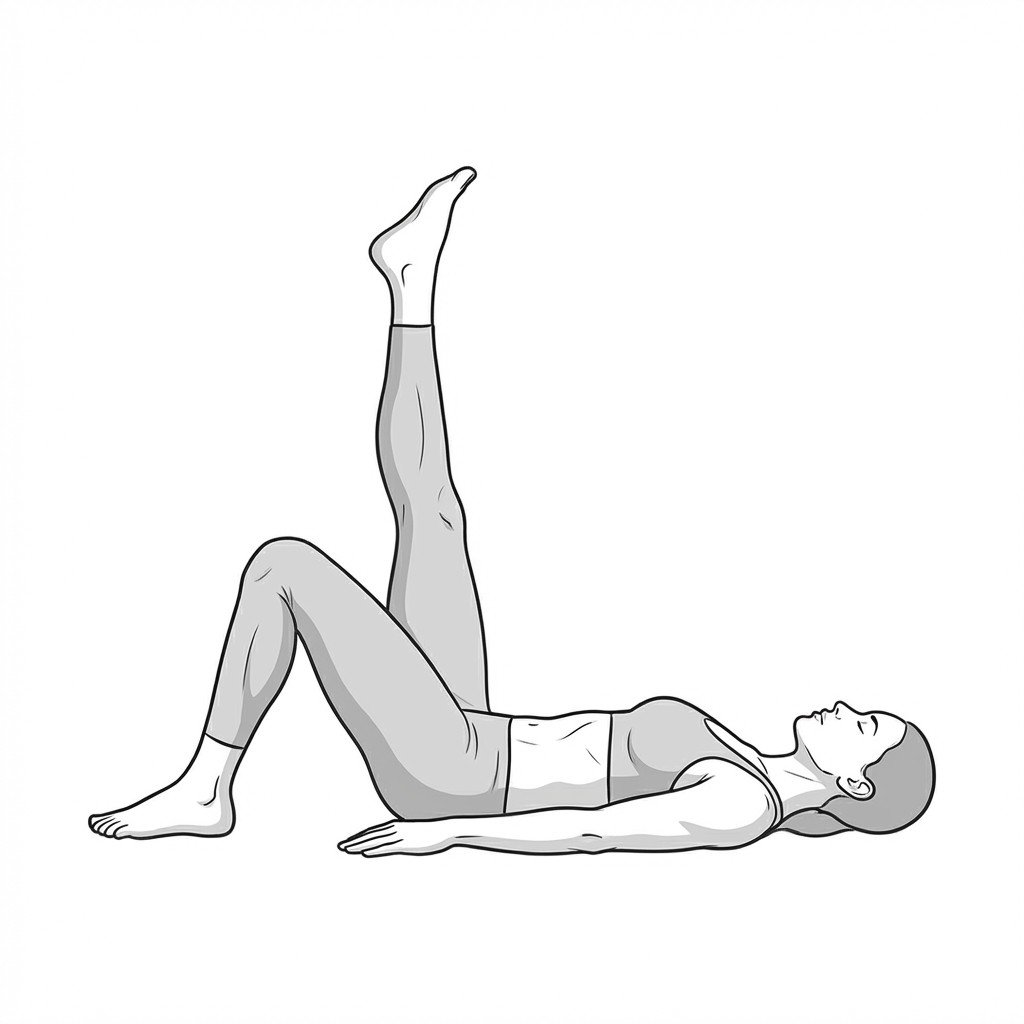
Straight leg raises

Straight leg raises

Straight leg raises activate the quadriceps complex without placing compressive load on the patellofemoral joint. By performing this exercise with a fully extended knee, it strengthens the thigh and hip flexor musculature while minimizing patellar tracking stress, making it highly tolerable for early-stage rehab.

3 sets × 10-15 reps, both sides, daily→ Start Exercise →
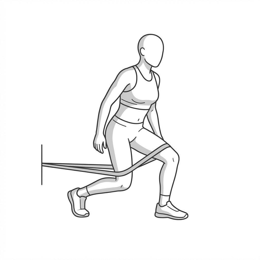
Terminal knee extensions

Terminal knee extensions

Terminal knee extensions isolate the final degrees of knee extension, preferentially engaging the quadriceps and heavily recruiting the vastus medialis. By strengthening these structures and improving motor control in the terminal range, this exercise may help improve patellar tracking and reduce patellofemoral compressive stress.

3 sets × 15 reps, both sides, daily→ Start Exercise →
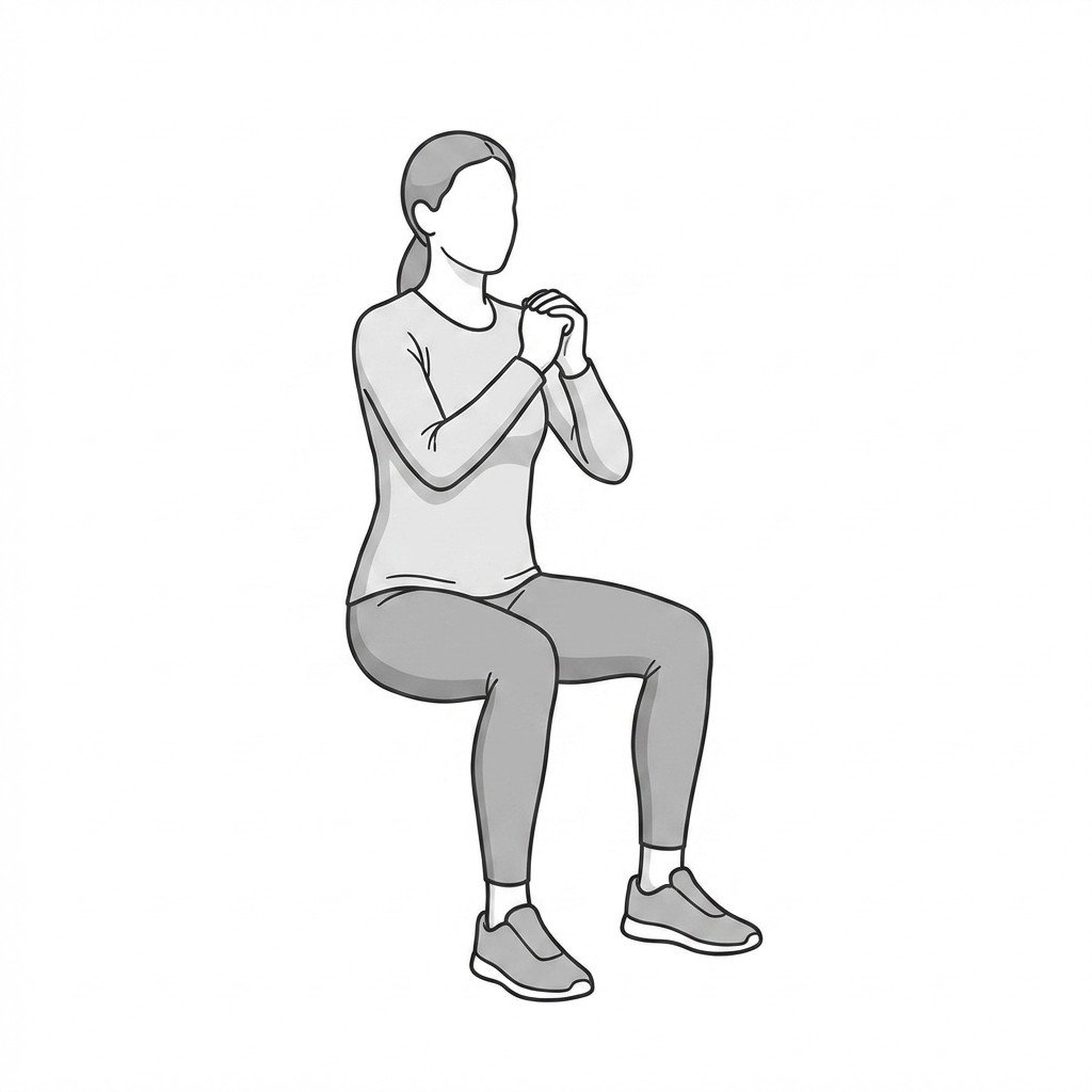
Wall sits

Wall sits

Wall sits provide a closed-kinetic chain isometric contraction of the quadriceps, targeting the vastus muscles while minimizing shear forces on the patellofemoral joint. This isometric loading may provide a short-term local analgesic effect and builds the baseline muscular endurance necessary for dynamic knee stability.

3-4 sets × 30-45 seconds hold, daily→ Start Exercise →