Understanding the Clinical Picture
Knee osteoarthritis (OA) is a highly prevalent, progressive condition characterized by the degradation of articular cartilage, subchondral bone remodeling, and osteophyte formation. It most commonly affects the medial tibiofemoral compartment due to natural varus alignment tendencies during the gait cycle, though the lateral and patellofemoral compartments are also frequently involved. Clinical guidelines consistently note a significant dissociation between radiographic severity and patient-reported pain intensity. Many patients with severe radiographic changes remain asymptomatic, while others with mild structural changes experience debilitating pain. This discrepancy highlights the complex, multifactorial nature of knee pain, where structural joint damage is only one component of the nociceptive experience.
Myofascial pain syndrome (MPS) frequently coexists with, and is often secondary to, knee osteoarthritis. As joint pain alters gait mechanics and reduces range of motion, the surrounding musculature—particularly the quadriceps, hamstrings, and gastrocnemius—is subjected to abnormal loading patterns. This chronic mechanical stress promotes the development of active trigger points. Notably, trigger points in the vastus medialis can refer pain directly to the anteromedial knee, creating a symptom profile that is virtually indistinguishable from medial compartment OA. Furthermore, chronic nociceptive input from the degenerating joint can lead to central sensitization, which lowers the threshold for trigger point activation and amplifies the overall pain experience.
Radiographic evidence of knee osteoarthritis does not preclude the presence of clinically significant, treatable myofascial pain generators.
Differentiating primary articular pain from myofascial pain requires careful clinical examination. Palpation is a critical tool; exquisite tenderness strictly localized to the joint line suggests intra-articular pathology, whereas reproduction of the patient's familiar knee pain upon palpation of the proximal muscle bellies strongly implicates a myofascial source. Diagnostic intra-articular injections (using local anesthetic under fluoroscopic or ultrasound guidance) can be highly informative. If a patient experiences minimal relief following a confirmed intra-articular block, extra-articular pain generators, such as myofascial trigger points or anserine bursitis, must be thoroughly investigated. Current evidence suggests that addressing these muscular components is essential before considering surgical interventions like total knee arthroplasty.
True Knee OA vs Myofascial Mimic
Pain location
Pain behavior
Hallmark exam finding
Imaging correlation
Diagnostic gold standard
Key Muscular Contributors
The knee joint relies heavily on dynamic muscular stabilization. Dysfunction in the following muscles frequently contributes to, or mimics, the pain associated with knee osteoarthritis.
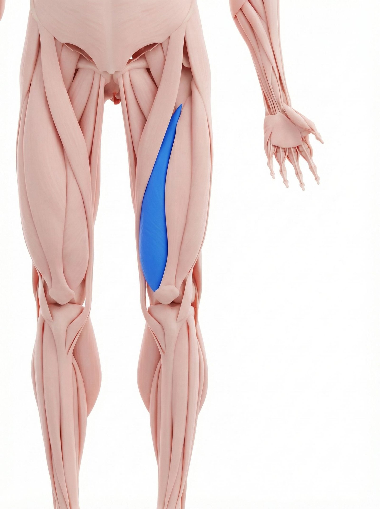
The vastus medialis is a primary dynamic stabilizer of the patella. Trigger points in this muscle frequently develop secondary to the altered gait mechanics associated with knee osteoarthritis. Clinically, these trigger points refer pain directly to the anteromedial aspect of the knee, closely mimicking the pain profile of medial compartment tibiofemoral osteoarthritis.
As the largest component of the quadriceps, the vastus lateralis is susceptible to overload when patients adopt compensatory movement strategies to offload a painful medial knee compartment. Myofascial pain here can mimic lateral compartment osteoarthritis or iliotibial band syndrome. Palpation often reveals taut bands along the lateral thigh that reproduce lateral knee pain.
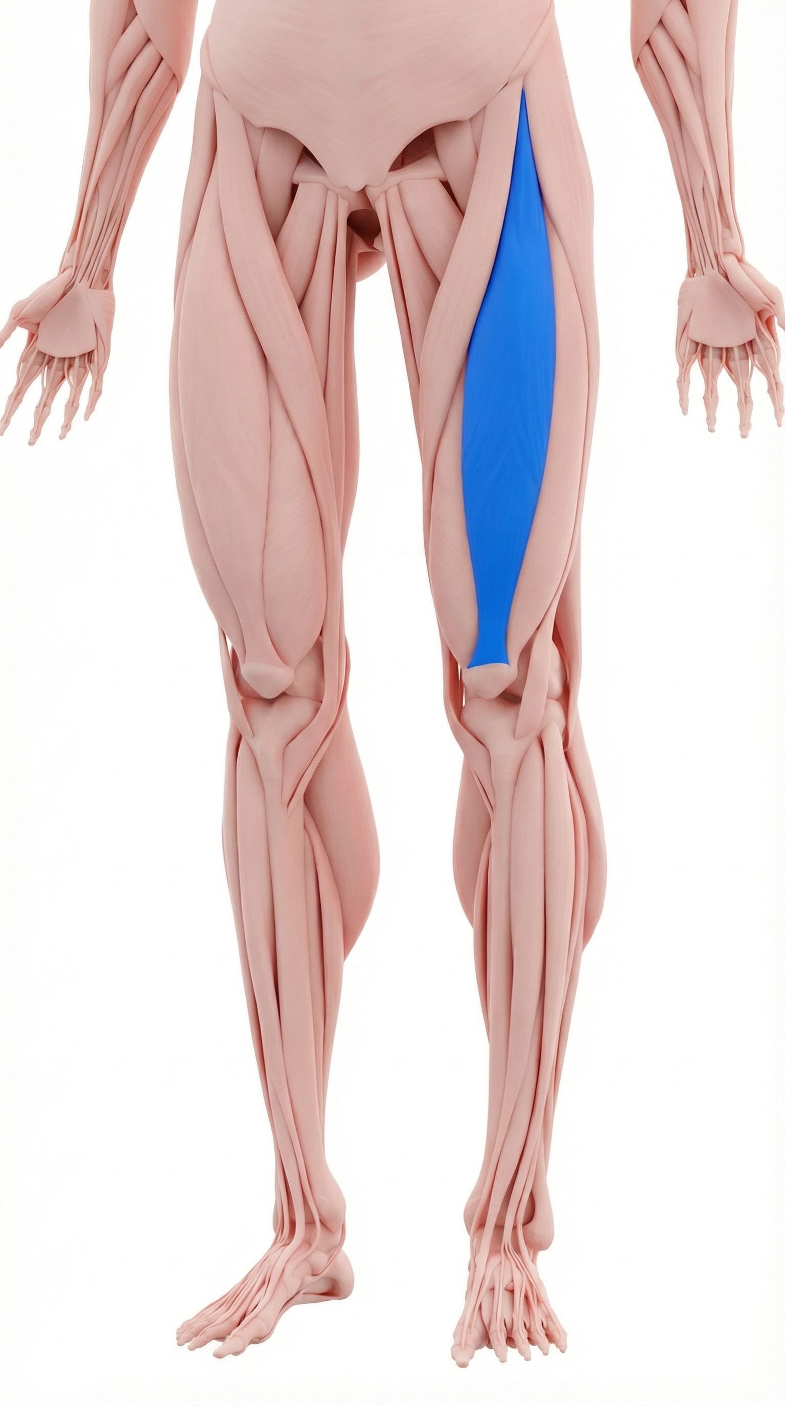
The rectus femoris crosses both the hip and the knee, making it vulnerable to dysfunction from altered biomechanics at either joint. Trigger points in the distal rectus femoris refer pain directly to the patella and deep anterior knee. This referral pattern is frequently misdiagnosed as isolated patellofemoral osteoarthritis or chondromalacia patellae.
The lateral hamstring muscle often develops increased resting tone in patients with chronic knee pain as a protective guarding mechanism. Trigger points in the biceps femoris can restrict terminal knee extension, a common finding in advanced knee osteoarthritis. The referred pain typically presents in the posterolateral knee compartment.
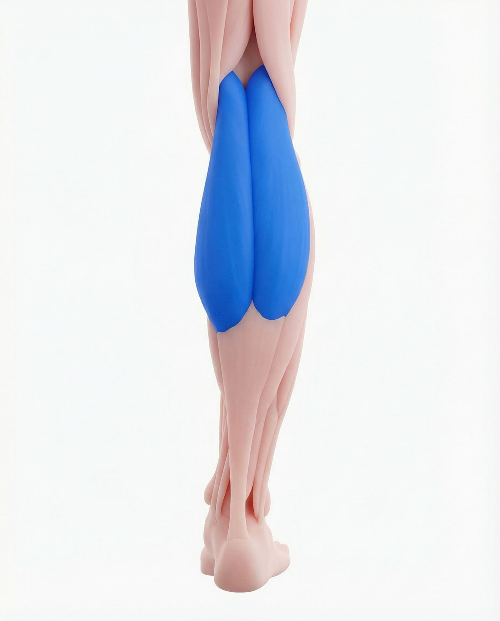
The gastrocnemius crosses the posterior knee joint and plays a crucial role in knee stability during the stance phase of gait. Trigger points in the medial or lateral heads can cause significant posterior knee pain. This presentation is frequently confused with posterior capsular pain from osteoarthritis or a symptomatic popliteal (Baker's) cyst.
The popliteus is a small but critical muscle responsible for "unlocking" the knee from terminal extension. It is frequently overloaded in the presence of knee joint effusion or instability. Popliteus trigger points produce deep posterior knee pain that is often indistinguishable from intra-articular pathology during initial clinical evaluation.
Articular Degeneration
Loss of cartilage, subchondral bone marrow lesions, and capsular inflammation directly stimulate intra-articular nociceptors.
Myofascial Referral
Active trigger points in the quadriceps and calf refer pain into the joint space, mimicking structural pathology.
Central Sensitization
Chronic peripheral nociception leads to amplified pain signaling in the central nervous system, expanding receptive fields.
Biomechanical Alteration
Pain-induced gait changes and muscle guarding increase joint contact pressures, accelerating structural wear and muscular overload.
Common Pain Referral Patterns
Myofascial trigger points in the lower extremity can project pain directly into the knee joint. Recognizing these patterns is crucial for accurate differential diagnosis.
Vastus Medialis
Rectus Femoris
Gastrocnemius
Vastus Lateralis
Clinical Differentiation
Clinical differentiation between intra-articular osteoarthritis and extra-articular myofascial pain relies on specific physical examination maneuvers. These assessments help isolate the primary pain generator.
Joint Line Tenderness Palpation
Vastus Medialis Pincer Grasp
Patellar Compression Test
Resisted Knee Extension
Evidence-Based Management
Current guidelines generally recommend a multimodal approach to knee pain, prioritizing conservative management. Effective treatment must address both the articular degeneration and the associated neuromuscular dysfunction.
Self-Care
- Weight management to reduce joint loading
- Activity modification (e.g., cycling instead of running)
- Application of thermal modalities (ice for acute flares, heat for stiffness)
- Gentle, pain-free range of motion exercises
- Use of shock-absorbing footwear or insoles
Manual Therapy
- Ischemic compression of quadriceps and calf trigger points
- Patellar mobilization to improve tracking and reduce capsular restriction
- Tibiofemoral joint mobilization to modulate pain
- Soft tissue mobilization of the iliotibial band and hamstrings
- Myofascial release techniques for the lower extremity kinetic chain
Interventional
- Diagnostic intra-articular anesthetic blocks
- Intra-articular corticosteroid injections for acute inflammatory flares
- Hyaluronic acid (viscosupplementation) injections
- Trigger point dry needling for the vastus medialis and lateralis
- Genicular nerve radiofrequency ablation for refractory cases
Rehabilitation
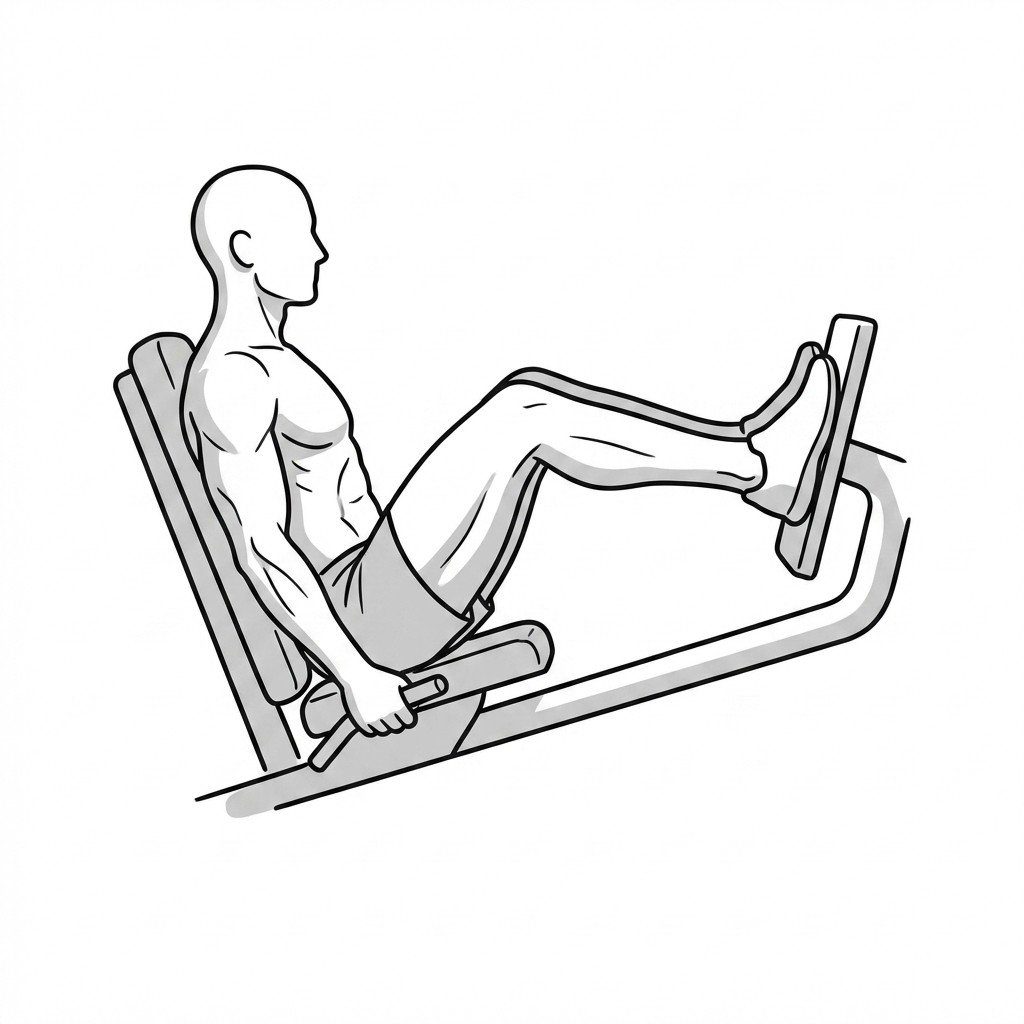
- Progressive quadriceps strengthening (especially closed-chain)
- Aquatic therapy to allow unloaded joint movement
- Proprioceptive and balance training
- Hamstring and gastrocnemius flexibility protocols
- Gait retraining to optimize lower extremity biomechanics
Red Flags — When to See a Doctor
While chronic knee pain is common and typically managed conservatively, certain signs and symptoms indicate potentially severe pathology that requires urgent medical investigation.
Radiographic severity of knee osteoarthritis often correlates poorly with the patient's actual clinical pain intensity.
Trigger points in the vastus medialis frequently mimic the exact pain distribution of medial compartment knee osteoarthritis.
Addressing myofascial pain generators can significantly improve functional outcomes and reduce pain, even when advanced structural osteoarthritis is present.
Central sensitization plays a major role in chronic knee osteoarthritis, amplifying both articular and myofascial pain signals.
Diagnostic intra-articular injections can be a valuable tool to differentiate true intra-articular pain from extra-articular myofascial pain.
Optimal management of chronic knee pain requires a comprehensive approach that addresses both joint degeneration and secondary neuromuscular dysfunction.
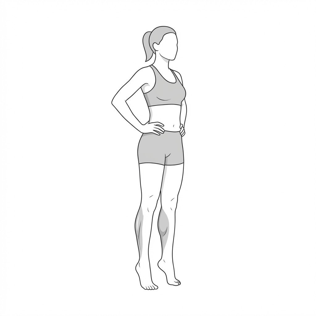
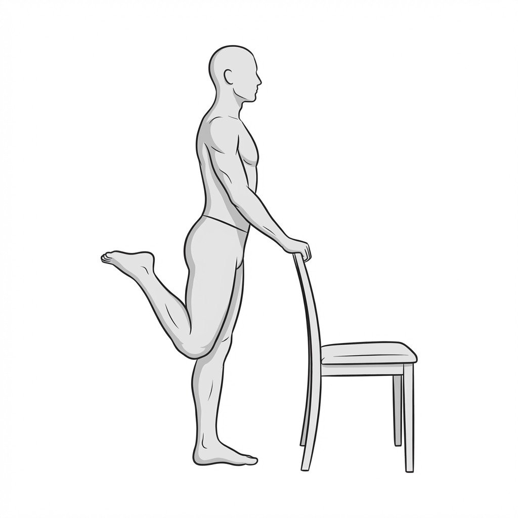
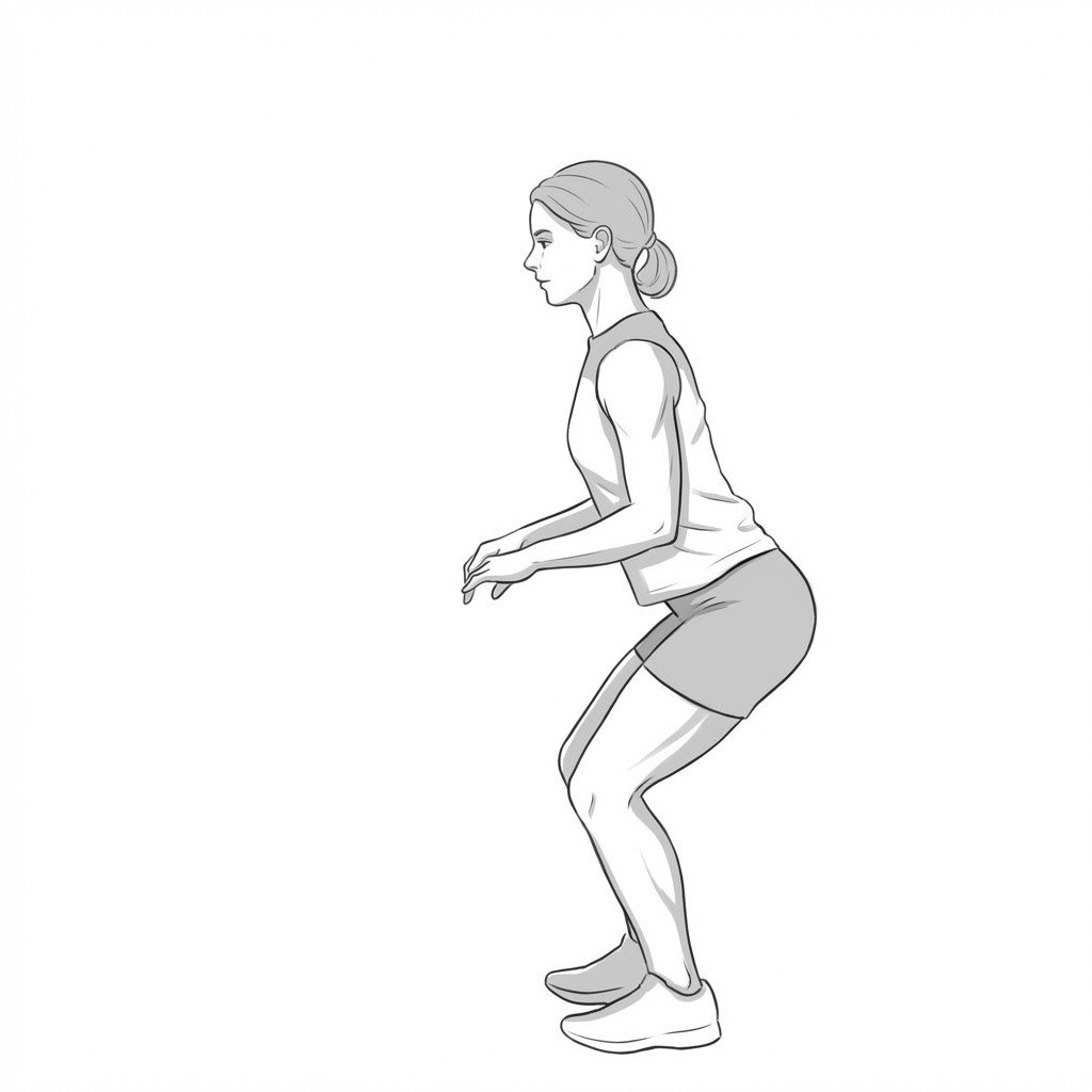
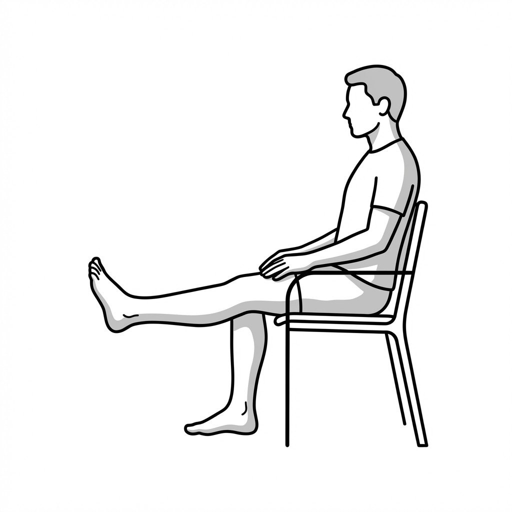
Movement therapy
Exercises for Knee Osteoarthritis
These exercises build quadriceps and calf strength around the knee joint, improve articular loading tolerance, and maintain range of motion without high-impact stress. Start with 2–3 and progress as tolerated.