Understanding the Condition
Iliotibial (IT) band syndrome is one of the most common causes of lateral knee pain, particularly among runners and cyclists. Historically described as a "friction syndrome," current evidence suggests it may be more accurately characterized by the compression of a highly innervated layer of fat and connective tissue between the IT band and the lateral femoral epicondyle. This pain typically emerges during repetitive knee flexion and extension. Differentiating true ITBS from its myofascial mimics is critical, as the treatment approach for each can differ significantly.
The IT band is not a muscle but a thick band of fascia, receiving input from the tensor fasciae latae (TFL) and gluteus maximus muscles. Consequently, myofascial pain and dysfunction in these muscles directly influence IT band mechanics. Trigger points in an overactive TFL can increase IT band tension, while trigger points in the underlying vastus lateralis muscle can refer pain directly to the lateral knee, creating a nearly identical pain presentation. Furthermore, weakness in the gluteus medius often leads to compensatory overuse of the TFL, making it a primary driver of the entire dysfunctional pattern.
Pain at the side of the knee is rarely just about the knee; a thorough investigation must always begin at the hip.
Distinguishing these overlapping conditions rests on a careful clinical evaluation. The assessment typically begins with a detailed history of training patterns and pain behavior. The physical exam focuses on locating the precise point of maximal tenderness—is it sharply localized over the bony epicondyle, or is it within the muscle belly of the thigh? Special tests, such as the Noble compression test, aim to reproduce the impingement, while functional tests like a single-leg squat can reveal the underlying biomechanical faults, such as hip weakness.
True IT Band Syndrome vs Myofascial Mimic
Pain location
Pain trigger
Key clinical sign
First-line treatment
Anatomy of the Pain
The iliotibial band is a longitudinal fibrous reinforcement of the fascia lata of the thigh. It originates from the iliac crest and inserts on Gerdy's tubercle of the tibia, with contributions from the TFL and gluteus maximus muscles.
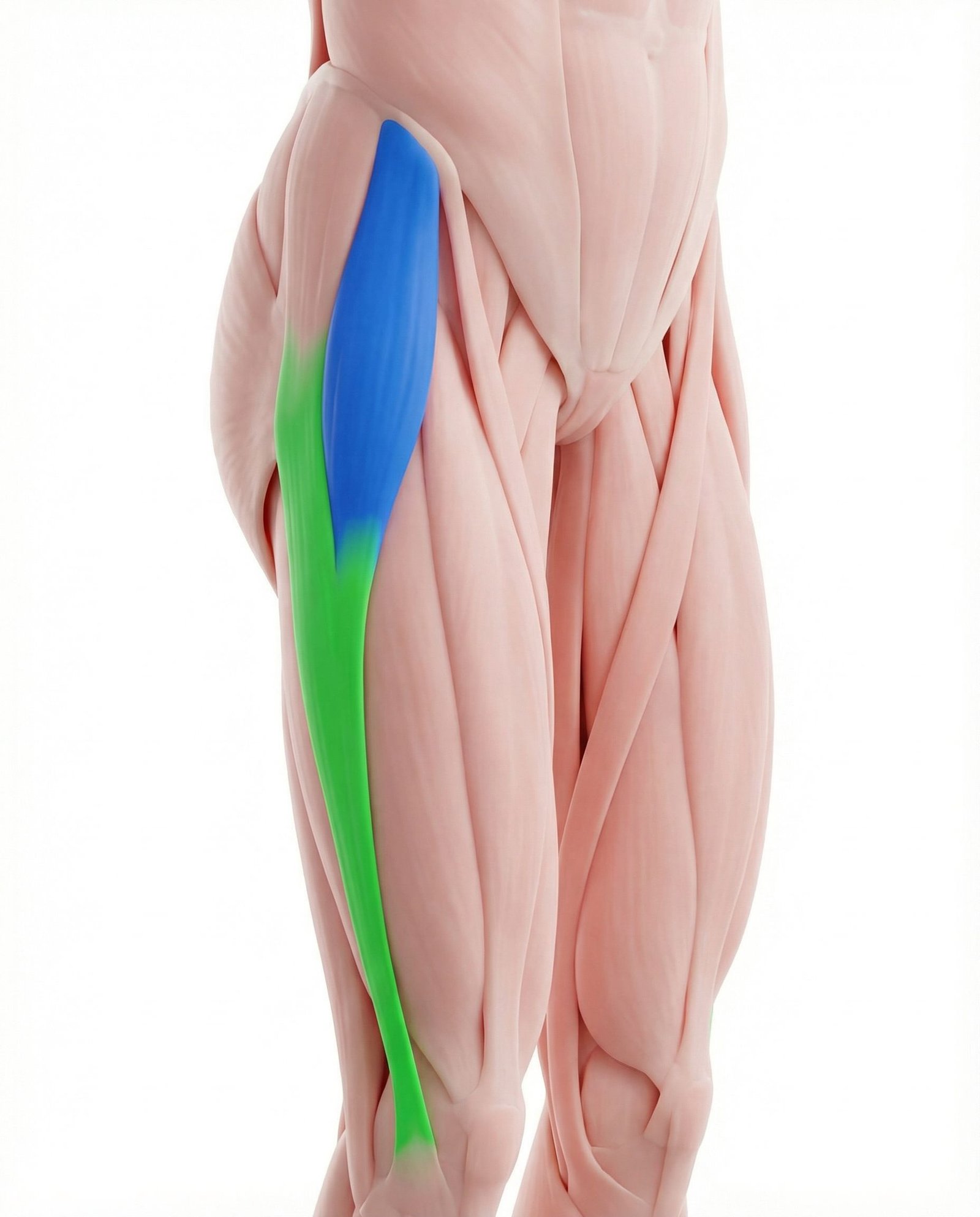
The TFL is a small muscle located at the front and side of the hip. It directly attaches to the iliotibial (IT) band and is a primary contributor to its tension. Functionally, it assists in hip flexion, abduction, and internal rotation. Chronic overuse, often due to weak gluteal muscles, can lead to the development of taut bands and trigger points within the TFL, which can both increase mechanical strain on the IT band and refer pain to the hip and thigh.
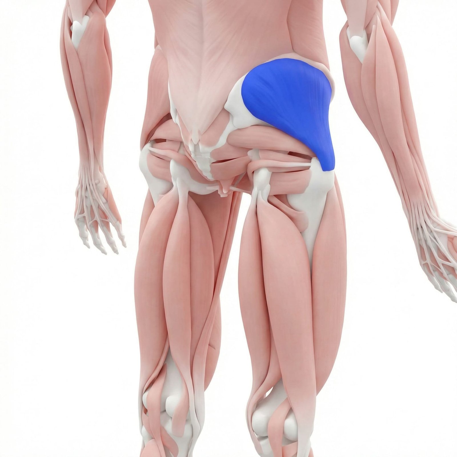
As the primary stabilizer of the pelvis during single-leg stance activities like walking and running, the gluteus medius keeps the pelvis level and directly governs lower limb alignment. Weakness or inhibition of this muscle is a well-documented contributor to numerous lower extremity injuries, including IT band syndrome. When the gluteus medius is weak, the TFL often becomes overactive to compensate for hip abduction, leading to increased IT band tension. Trigger points in the gluteus medius can also refer pain to the buttock, sacrum, and posterior thigh.
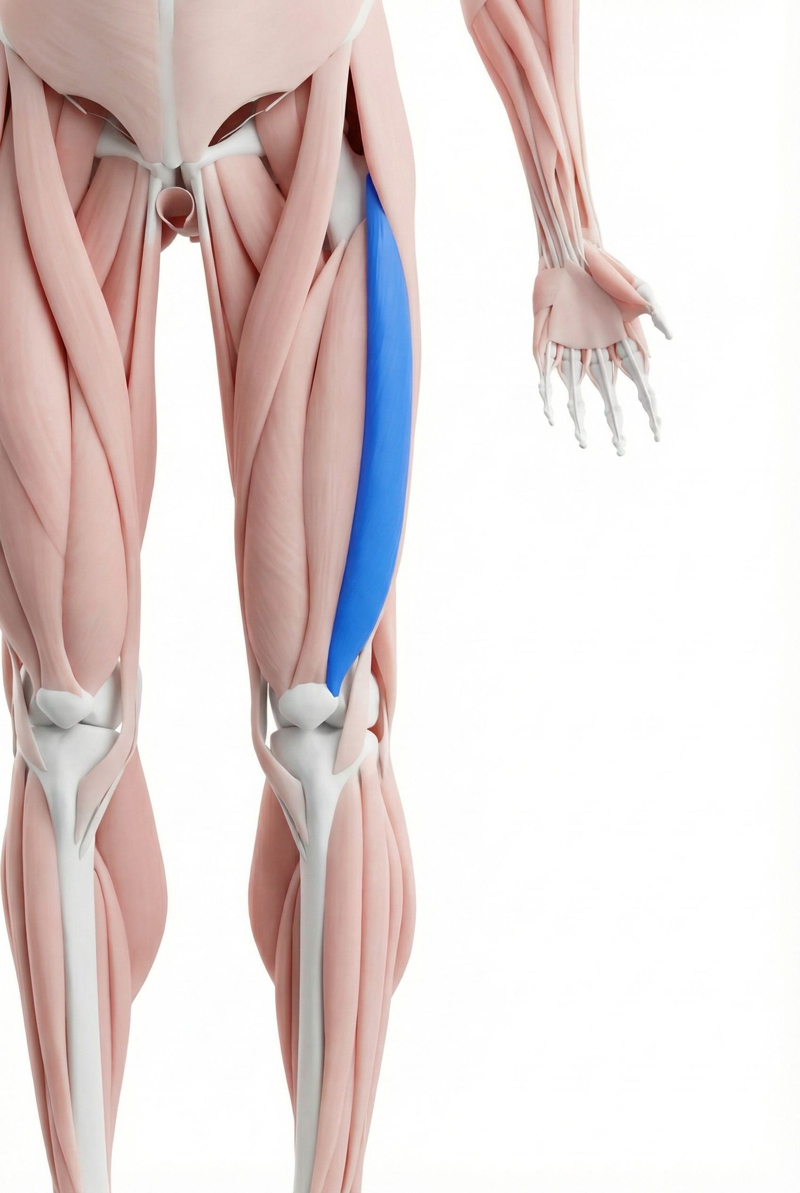
This is the largest and most powerful of the four quadriceps muscles, located on the outer side of the thigh. It lies directly deep to the IT band, and fascial adhesions can develop between the two structures. Trigger points in the vastus lateralis are a significant mimic of IT band syndrome, as they commonly refer a deep, aching pain directly to the lateral aspect of the knee, often indistinguishable from true ITBS pain without careful palpation.
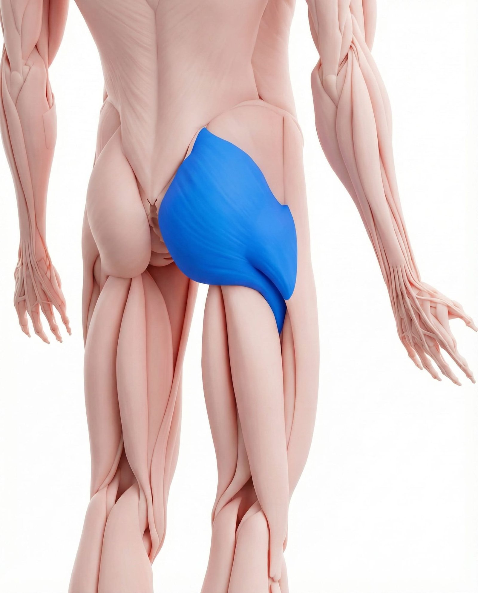
The largest muscle in the body, the gluteus maximus is a powerful hip extensor and external rotator. Its superior fibers, along with the TFL, insert into the IT band, contributing to its tension and stability. While less commonly a source of referred pain to the knee, its strength and function are integral to offloading the TFL and maintaining healthy hip mechanics. Weakness can contribute to the same compensatory patterns that overload the TFL.
As a primary muscle of the lateral hamstring, the biceps femoris tendon inserts on the head of the fibula, in close proximity to the distal insertion of the IT band. Distal biceps femoris tendinopathy can therefore present with pain in a very similar location to distal IT band syndrome. Differentiating between the two often requires precise palpation of the respective tendons and specific resistance testing.
Compression & Irritation
The IT band compresses a highly sensitive fat pad against the lateral femoral condyle, leading to inflammation and pain, especially around 30° of knee flexion.
Myofascial Trigger Points
Overloaded muscles like the TFL and vastus lateralis develop trigger points that refer pain directly to the lateral knee, mimicking ITBS.
Biomechanical Overload
Weakness in hip abductors (gluteus medius) causes poor pelvic control during gait, increasing strain on the TFL and tension in the IT band.
Training Errors
Sudden increases in training volume, intensity, or hill work can overwhelm the tissue's capacity to adapt, leading to irritation and injury.
Common Referral Patterns
Myofascial trigger points in the muscles surrounding the hip and thigh can create referred pain that complicates the diagnostic picture. Pain felt at the knee may originate from a dysfunctional muscle located much higher up the leg.
Tensor Fasciae Latae (TFL)
Vastus Lateralis
Gluteus Medius
Self-Assessment Tests
These assessments can help you explore the potential sources of your lateral knee pain. They are designed to reproduce symptoms or identify biomechanical issues commonly associated with ITBS and related myofascial pain. A positive result narrows the picture but does not confirm a diagnosis; persistent or worsening lateral knee pain warrants formal assessment.
Noble Compression Test
Ober's Test
Single-Leg Squat Assessment
Focused Palpation
Treatment Pathways
A comprehensive treatment plan for lateral knee pain generally addresses both the local symptoms and the underlying biomechanical causes. The most effective strategies often combine load management, targeted strengthening, and manual therapy to address any myofascial components.
Self-Care
- Activity modification and relative rest
- Ice application to the lateral knee post-activity
- Gentle foam rolling of TFL and gluteal muscles (avoiding the ITB itself)
- Consistent, gentle stretching of hip flexors and glutes
- Monitoring training load and avoiding sudden increases in mileage or intensity
Rehabilitation
- Targeted strengthening of hip abductors (e.g., clamshells, side-lying leg raises)
- Gluteus medius and maximus activation exercises
- Core stability and pelvic control exercises
- Neuromuscular re-education and gait analysis/retraining for runners
- Proprioceptive and balance training
Manual Therapy
- Myofascial release of the tensor fasciae latae and vastus lateralis
- Trigger point pressure release or ischemic compression
- Soft tissue mobilization to address adhesions between the IT band and underlying quadriceps
- Hip and ankle joint mobilization to improve mechanics
- Cross-friction massage at the insertion point (use to be common, evidence is mixed)
Interventional
- Dry needling of trigger points in the TFL, gluteus medius, and vastus lateralis
- Corticosteroid injection at the point of maximal tenderness (evidence for long-term benefit is debated)
- Diagnostic musculoskeletal ultrasound to assess for bursitis or tendon thickening
- Platelet-rich plasma (PRP) injections (currently considered investigational)
Red Flags — When to See a Doctor
While IT band syndrome is typically an overuse injury, it is important to be aware of signs and symptoms that could indicate a more serious condition. The following "red flags" warrant prompt or immediate evaluation by a healthcare professional.
Iliotibial band syndrome is increasingly understood as a compression syndrome, where the IT band compresses a highly innervated fat pad against the femur, rather than a simple friction issue.
Myofascial trigger points in the tensor fasciae latae (TFL) and vastus lateralis muscles are extremely common mimics and contributors to lateral knee pain.
The root cause of ITBS is often found "upstream" at the hip; weakness of the gluteus medius muscle is a primary biomechanical factor in many cases.
Effective treatment prioritizes hip abductor strengthening and motor control over aggressive stretching or foam rolling of the IT band itself, which is a non-contractile tissue.
The key diagnostic challenge is differentiating localized pain at the lateral femoral epicondyle (ITBS) from the referred pain patterns of nearby muscles.
A successful management plan almost always involves a period of load management and activity modification to allow irritated tissues to calm down before progressive reloading.
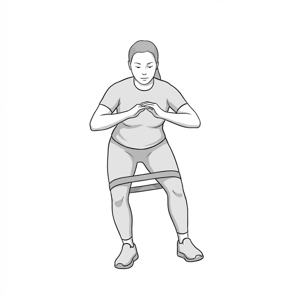
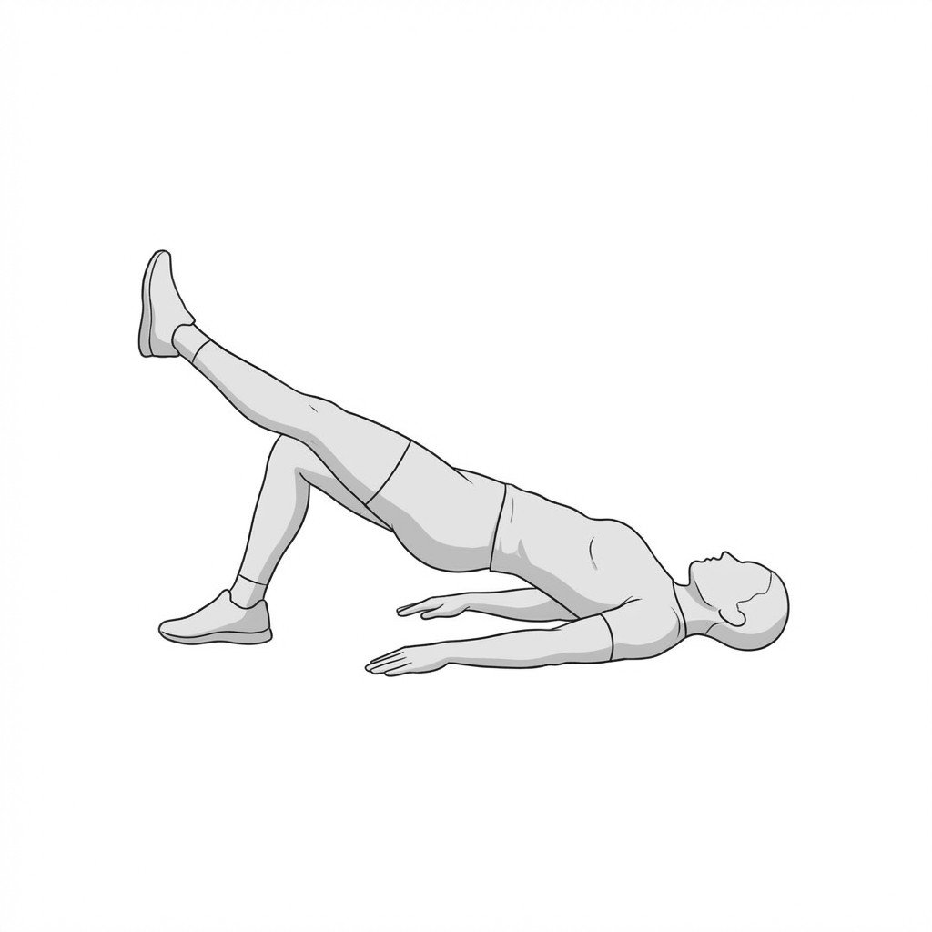
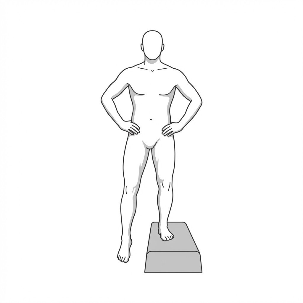
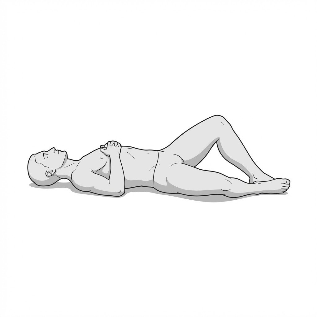
Movement therapy
Exercises for It Band Syndrome
IT band pain is driven by hip drop and contralateral pelvic shift during stance — these exercises build gluteus medius strength and TFL length to correct that pattern and reduce lateral knee compression during running and stairs. Start with 2–3 and progress as tolerated.