Two Philosophies — One Needle
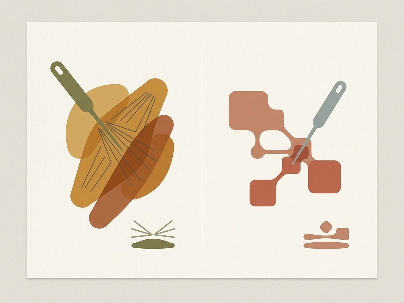
IMS vs. Dry Needling
Theoretical Model Comparison OverviewWhen a patient is referred for "dry needling," the term sounds simple — a needle, a muscle, a treatment. But behind that single needle lies a fundamental disagreement about what causes chronic myofascial pain, and that disagreement has significant consequences for what gets needled, how deep, and why. The split traces to two lineages that developed largely independently: Chan Gunn's work in Vancouver through the 1970s and 1980s, which drew on Melzack's gate control theory and Cannon's supersensitivity to develop the neuropathic model of MPS; and Janet Travell and David Simons' decades-long compilation of the trigger point atlas, which built the peripheral myofascial model that underpins Dry Needling worldwide.
Gunn's neuropathic model proposes that all chronic myofascial pain syndrome has its origin in "spondylogenic" neuropathy — subtle dysfunction at the spinal nerve root level arising from degenerative changes in the spine that do not produce the classic symptoms of frank radiculopathy. Gunn argued that even mild, subclinical nerve root compromise causes supersensitivity in all structures supplied by that root: skin, connective tissue, and especially muscle motor end plates. This supersensitivity — which follows Cannon's Law of Denervation — causes the affected muscles to become shortened, overreactive, and painful. Crucially, this is distinct from standard radiculopathy: there is no dermatomal numbness, no reflex change, no positive straight leg raise. Instead, there are subtle signs: skin that dermagraphs more easily, muscles that are diffusely shortened in a segmental pattern, and paraspinal tenderness at the relevant level.
The trigger point model, as codified by Travell and Simons and subsequently refined by researchers including David Simons, Robert Gerwin, and Jay Shah, proposes that myofascial pain arises from local contracture knots in muscle — palpable taut bands containing a central trigger point at the endplate zone. The Integrated Hypothesis (Simons, 2004) describes an energy crisis at the motor end plate: excessive acetylcholine release causes sustained sarcomere contraction that compresses local capillaries, creating local ischemia, acidosis, and the release of sensitizing substances (bradykinin, substance P, CGRP) that activate and sensitize nociceptors. Surface and intramuscular EMG studies have confirmed the presence of spontaneous electrical activity (SEA) in active trigger points — evidence of abnormal endplate activity that is the electrophysiological signature of the contracture knot.
Where the two models converge is in the reality of complex, chronic MPS that most specialist clinicians encounter daily. A patient with ten years of neck and shoulder pain, multi-segment degenerative cervical changes on MRI, diffuse scalene and upper trapezius trigger points, and subtle Gunn radiculopathic signs in C5-6 distribution cannot be cleanly assigned to either model exclusively. Most experienced needling practitioners have recognized this and work pragmatically: paraspinal IMS-style treatment to address the segmental neuropathic component, followed by peripheral trigger point Dry Needling to deactivate the peripheral contracture knots. The two techniques are not rivals but tools that address different levels of the same chronic pain system.
Key Comparison Dimensions
Five dimensions define the practical differences between IMS and Dry Needling. Understanding each dimension allows clinicians and patients to make informed decisions about which approach is appropriate and why.
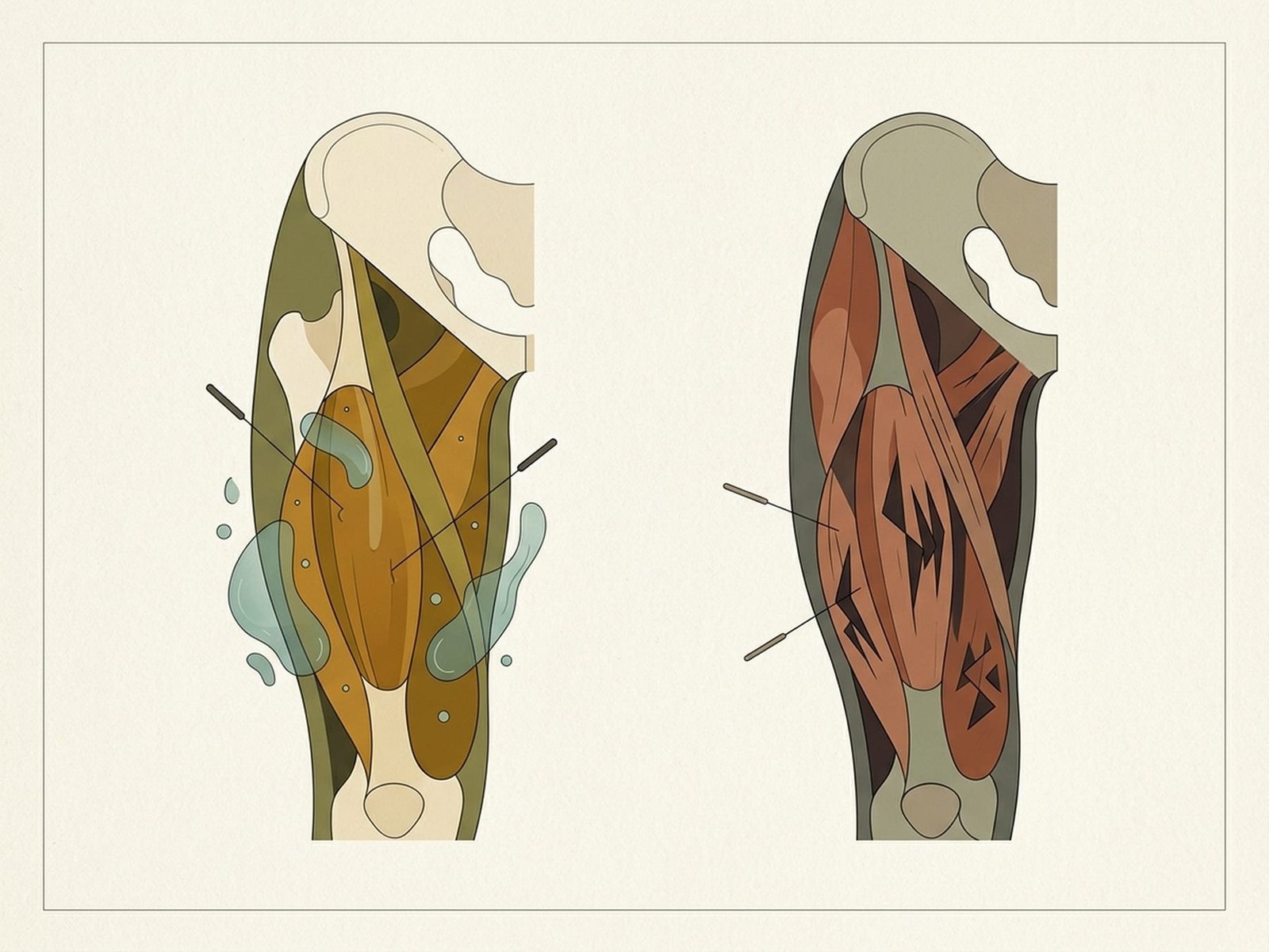
Key Comparison Dimensions
Clinical Framework DiagramTheoretical Model
Fundamentally different frameworksIMS is grounded in Chan Gunn's neuropathic model: all chronic myofascial pain syndrome is attributed to spondylogenic neuropathy — supersensitized motor end plates arising from mild, subclinical radiculopathy at the spinal root level. Dry Needling follows the Travell-Simons myofascial trigger point model: local contracture knots harboring sensitized nociceptors that develop and persist independent of spinal pathology. These divergent frameworks carry significant clinical implications — IMS mandates a spine-focused diagnostic workup before any peripheral needling, while Dry Needling directs assessment toward palpable peripheral trigger point findings. Understanding which model underlies your clinician's approach shapes how they explain your pain and why they needle where they do.
Target Tissue
What the needle is aimed atIMS targets the muscle belly at the motor point — specifically the deepest, most tender point within the "radiculopathic" segmental pattern, often deep to the surface and requiring longer needles. Dry Needling targets the palpable trigger point band, typically at mid-fiber level at the endplate zone where spontaneous electrical activity (SEA) can be recorded. This distinction produces a practical difference in needle placement guidance: IMS relies on segmental dermatomal mapping and paraspinal examination findings; Dry Needling relies on careful palpation of taut bands and jump-sign elicitation. Ultrasound guidance is gaining traction in both approaches for deep muscles, though its added benefit for routine superficial TrP needling remains debated.
Needling Depth
How deep the needle goesIMS characteristically uses deeper needling — paraspinal IMS targets deep segmental muscles (e.g., multifidus) at the level of the transverse process, and peripheral IMS targets the muscle belly at the deepest tender point, sometimes reaching periosteum. Dry Needling depth varies by target muscle but is generally aimed at the trigger point band level, which is often shallower than IMS targets. Patient experience differs accordingly: deep IMS needling in paraspinals produces an intense "needle grasp" sensation and often significant post-needling muscle soreness lasting 24-48 hours; Dry Needling at a trigger point band typically elicits a local twitch response (LTR) followed by immediate muscle relaxation. Risk profiles differ: IMS paraspinal needling carries a theoretical greater proximity to neural structures; anterior chest and scalene needling in both approaches requires meticulous depth control to avoid pneumothorax.
Local Twitch Response
Diagnostic and therapeutic responseThe local twitch response (LTR) — an involuntary spinal reflex contraction of the taut band fibers upon needle contact — holds central importance in Dry Needling. Travell-Simons placed significant weight on eliciting the LTR as both an indicator of correct needle placement within the active trigger point and as a predictor of treatment outcome; studies using EMG have confirmed LTR is a spinally mediated reflex distinct from voluntary contraction. IMS does not require LTR — Gunn's indicator of correct needle placement is the "needle grasp": mechanical resistance felt by the clinician as the needle engages a shortened, supersensitized muscle belly. The needle grasp signals that the needle is in tight, radiculopathically supersensitized muscle. To the patient, LTR feels like a brief, involuntary muscle jump or twitch; needle grasp in IMS feels like a deep ache or cramping sensation that persists while the needle is in the tissue.
Scope of Practice
Who can perform eachLegal scope of practice for needling varies significantly by jurisdiction, regulatory body, and profession. IMS training through the Gunn IMS method is primarily offered to physiotherapists through the Institute for the Study and Practice of Interdisciplinary Pain (iSPI/GUNN IMS), with formal certification requirements. Dry Needling is practiced by physical therapists, physiotherapists, chiropractors, osteopaths, some medical doctors, and in many US states, acupuncturists — with highly variable state-by-state and country-by-country legal status. The political and licensing landscape around Dry Needling remains contentious in many jurisdictions, with organized acupuncture professions challenging its practice by non-acupuncturist providers. For patients, this matters practically: always verify that your provider holds specific needling certification, not just a general professional license.
Evidence — What the Data Shows
RCT evidence comparing IMS and Dry Needling directly is limited, partly because the techniques are operationally similar enough that blinding is challenging and partly because the conditions they target — chronic MPS — are notoriously heterogeneous. Systematic reviews and meta-analyses of Dry Needling for myofascial pain generally find moderate evidence for short-term pain reduction and improved pressure pain threshold compared to sham needling or no treatment, with less certainty about long-term durability. The evidence base for IMS is smaller in terms of RCTs but is supported by a substantial case series literature and the mechanistic plausibility of Gunn's model.
Head-to-head comparison studies are rare, and where they exist (e.g., cervicogenic headache, chronic low back pain), results tend to show equivalent short-term outcomes between the approaches. This is not surprising if both techniques ultimately work through shared mechanisms: mechanical disruption of contracture knots, local tissue microtrauma triggering repair processes, and modulation of peripheral and central sensitization via needle-induced afferent input. The clinical implication is that for straightforward, acute-onset MPS without radiculopathic signs, either technique is likely to be effective. The distinction in outcomes is most apparent in chronic, treatment-resistant presentations where the presence or absence of a segmental neuropathic component determines whether peripheral needling alone will produce durable relief.
The needle is the same; the philosophy behind where you put it — and why — is what separates IMS from Dry Needling.
IMS Evidence
Strongest for chronic spine-linked MPS with radiculopathic signs
DN Evidence
Moderate RCT support for short-term pain reduction in acute/subacute MPS
Combined
Hybrid protocols show additive benefit in complex chronic presentations
Which Approach for Which Patient
Patient selection is the most important clinical decision in needling practice. These four assessment frameworks guide the choice between IMS, Dry Needling, or a combined approach — and establish the objective baseline needed to track treatment response.
Radiculopathy Pattern Assessment
IMS Patient Selection — Spine-Based Signs
Gunn's radiculopathic signs form the diagnostic basis for IMS candidacy. Assess for: skin changes (dermographia — exaggerated whealing response to light scratch), visible or measurable muscle wasting in the segmental myotome, shortened muscle with restricted range of motion at the corresponding joint, paraspinal tenderness at the relevant spinal segments, and segmental hyperalgesia (heightened skin sensitivity in the dermatome). When these signs cluster in a patient with chronic MPS, spondylogenic neuropathy is the likely driver and IMS at the paraspinal level is strongly indicated before peripheral needling. Imaging is recommended to rule out severe structural pathology but mild degenerative change is consistent with Gunn's model.
Classic Trigger Point Palpation
Dry Needling Patient Selection — TrP Signs
Classic trigger point diagnostic criteria for Dry Needling candidacy: palpable taut band within a skeletal muscle, exquisite spot tenderness on the taut band (jump sign elicitation), reproduction of the patient's familiar referred pain pattern on compression, local twitch response on snapping palpation across the band, and stretch-limited range of motion due to muscle shortening. Patients without clear Gunn radiculopathic signs but with classic TrP presentation — especially following acute overload, repetitive strain, or trauma — are ideal Dry Needling candidates. Pressure algometry (PPT measurement) is useful to quantify trigger point sensitivity and track treatment response objectively.
Neurological Examination
Distinguishing MPS from Radiculopathy
A structured neurological examination is essential before any needling to distinguish chronic MPS from active, clinically significant radiculopathy. Test dermatomal sensory distribution (light touch, pinprick, temperature), deep tendon reflexes at the relevant levels, and segmental motor testing. Red flags for serious radiculopathy requiring imaging before needling: progressive neurological deficit, bilateral lower extremity signs, bladder or bowel dysfunction, saddle anaesthesia, or severe unrelenting pain with systemic features. Positive neurological findings do not necessarily exclude needling but mandate imaging and specialist input. Mild chronic degenerative changes without active neurological deficits are consistent with Gunn's neuropathic model.
Treatment Response Tracking
Monitoring Progress with Either Approach
Objective outcome measurement is essential for both IMS and Dry Needling to guide clinical decisions about treatment continuation, technique modification, or referral. Baseline and follow-up tools: active range of motion (goniometry or inclinometry), Numeric Rating Scale (NRS) or Visual Analogue Scale (VAS) for pain intensity, pressure pain threshold (PPT) using an algometer over the primary trigger point or motor point, and condition-specific functional outcome measures (e.g., NDI for cervical, DASH for upper extremity). If a patient is not showing objective improvement by sessions 3-4, consider switching from one approach to the other or adding a combined paraspinal-peripheral protocol. Non-response to both approaches should prompt re-evaluation for central sensitization, psychosocial perpetuating factors, or missed structural pathology.
Clinical Protocols
Four clinical protocols cover the spectrum from pure IMS to pure Dry Needling to hybrid approaches. Protocol selection should follow examination findings: radiculopathic signs favor IMS; classic TrP findings without spinal signs favor Dry Needling; both together favor the combined approach. Imaging guidance is reserved for high-risk anatomical targets.
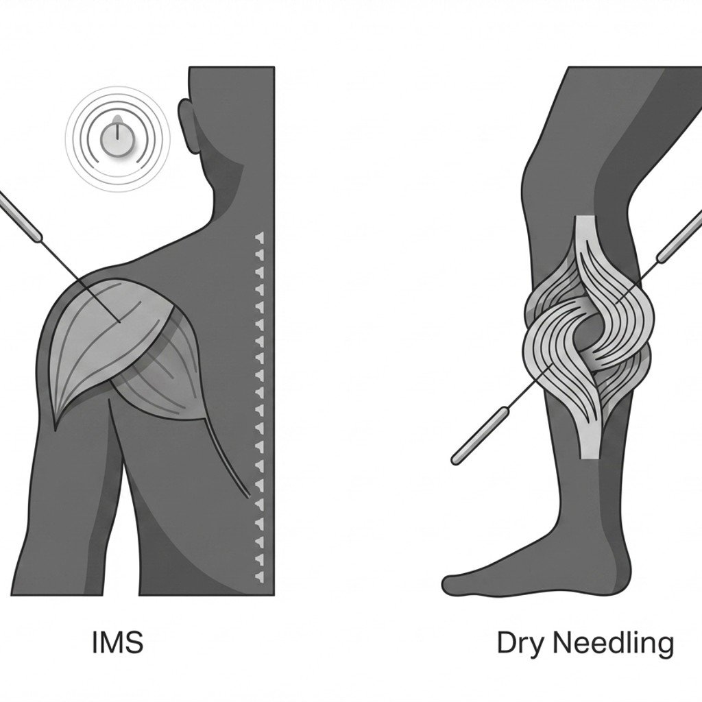
Needling Clinical Protocols
Decision and Protocol DiagramGUNN IMS Protocol
Gunn's systematic examination and treatment approach begins with a thorough segmental assessment to identify the spondylogenic component. The core principle is to treat the 'supply' (spinal nerve root via paraspinal needling) before the 'demand' (peripheral muscle trigger points) — normalizing the radiculopathic supersensitivity from which peripheral shortening derives.
- Segmental examination: identify the spinal levels contributing via paraspinal tenderness, dermatomal hyperalgesia, and muscle wasting pattern
- Paraspinal IMS first: needle deep multifidus at the relevant transverse process levels bilaterally — targets the supersensitized segmental muscle
- Peripheral muscle needling follows paraspinal treatment in the same or subsequent session, targeting motor points in radiculopathic pattern muscles
- Typical session structure: 30-45 min; 6-12 treatment series with progressive improvement expected; reassess segmental signs at each visit
- Post-needling exercise: gentle active movement and stretching of treated segments; education on posture and activity modification
Trigger Point Dry Needling
The Travell-Simons protocol for Dry Needling centers on precise trigger point identification followed by needle deactivation of the contracture knot. Eliciting the local twitch response is both the target and the confirmation of effective needle placement.
- Trigger point identification: systematic palpation for taut bands, jump sign, and referred pain reproduction — map all active TrPs before needling
- In-and-out technique (Hong): needle directed into TrP, small-amplitude pistoning until LTR elicited; multiple LTRs associated with better outcomes
- Sparrow-pecking technique: fine insertions at multiple angles within the taut band for patients unable to tolerate full pistoning
- Immediate post-needling: passive and active stretching of treated muscle to full length — consolidates the effect of TrP deactivation
- Home program: self-stretching 3x/day for treated muscles; progressive loading exercises to restore functional muscle length and strength
Combined Paraspinal + Peripheral Protocol
A hybrid approach that addresses both the segmental (spinal) component with IMS-style paraspinal needling and the peripheral trigger points with standard Dry Needling technique. Appropriate for complex, chronic MPS presentations where both radiculopathic signs and classic TrP findings coexist.
- Indication: chronic MPS (>3 months) with both Gunn radiculopathic signs and Travell-Simons TrP criteria — the most common presentation in specialist pain physiotherapy
- Session sequence: paraspinal IMS-style needling first (normalizes the segmental supersensitivity), peripheral TrP Dry Needling second in the same session or the following session
- Rationale: peripheral muscles remain supersensitized if the spinal root-level neuropathy is not addressed — treating peripherally alone is less durable
- Monitoring: expect paraspinal soreness 24-48 hours post-treatment; peripheral muscles may respond faster once paraspinal component addressed
- Typical course: 8-12 sessions; re-evaluate at session 4 for segmental sign regression as objective treatment progress marker
Ultrasound-Guided Needling
Imaging guidance improves accuracy for deep or vascular-adjacent targets where landmark-based needling carries meaningful risk. Both IMS and Dry Needling target tissues have recognizable ultrasound appearances that can guide needle placement in experienced hands.
- Indications for guidance: iliopsoas TrP (close to femoral neurovascular bundle), piriformis (close to sciatic nerve), scalenes (brachial plexus, subclavian artery), deep cervical paraspinals
- Real-time vs. static guidance: real-time (dynamic) guidance preferred for vascular-adjacent needling; static pre-needling imaging acceptable for deep paraspinal IMS
- IMS on ultrasound: tight, hypoechoic muscle belly with reduced fascial glide — needle grasp visible as tissue tenting; periosteal contact seen at transverse process
- TrP on ultrasound: hypoechoic nodule within taut band, reduced echogenicity vs. surrounding muscle; LTR visible as tissue vibration on M-mode
- Cost-benefit: US guidance adds time and cost; reserve for deep/high-risk targets or when previous landmark-guided attempts failed
Safety & Contraindications
Needling — whether IMS or Dry Needling — is a generally safe intervention in trained hands, but several patient presentations require special attention before treatment proceeds. The following contraindications and cautions apply to both techniques unless otherwise specified.
Anticoagulation — warfarin INR >3 or recent NOAC dose
Risk stratification required before any needling. INR >3 or NOAC taken within 12 hours: absolute contraindication for deep needling (paraspinal IMS, deep peripheral IMS). Superficial Dry Needling may be acceptable with patient-specific risk discussion and haematologist input for complex cases.
Pneumothorax risk — paraspinal and anterior chest needling
Thorough training in surface anatomy and needle depth control is non-negotiable before needling the cervical paraspinals, scalenes, pectoralis minor, subclavius, or thoracic paraspinals. Knowing the depth to the lung apex in each patient's body habitus is a prerequisite skill — not an optional refinement. Pneumothorax from needling is rare but serious; capnography and urgent imaging are required if suspected post-treatment.
Needling through skin lesions, active infection, or open wounds
Absolute contraindication. Active local infection, cellulitis, open skin breakdown, or undiagnosed skin lesion overlying the target area precludes needling at that site. Systemic sepsis or active bacteremia is a contraindication to any needling.
Post-needling soreness >72 hours or new neurological symptoms
Mild post-needling soreness (24-48 hours) is expected and represents a normal healing flare, particularly after IMS paraspinal treatment. Soreness persisting beyond 72 hours, or any new neurological symptom (new weakness, dermatomal numbness, bladder change) following needling, requires urgent clinical review to differentiate an exaggerated treatment flare from nerve injury. Do not rebook for further needling until assessed.
Key Takeaways
The IMS and Dry Needling models are complementary, not competing — one emphasizes the spinal (neuropathic) component and the other the peripheral (myofascial) component of the same chronic pain continuum.
Most experienced needling clinicians use elements of both approaches, adjusting their emphasis based on whether radiculopathic signs or classic trigger point criteria predominate in each patient presentation.
Outcome data from RCTs shows similar short-term efficacy for IMS and Dry Needling in pure myofascial pain syndrome — the choice of approach matters most for complex, chronic, or treatment-resistant patients.
IMS has a stronger clinical rationale for chronic spine-linked pain with Gunn's radiculopathic signs — particularly persistent cervical, lumbar, and limb pain that has not responded to peripheral trigger point treatment alone.
The distinction between the two approaches matters most when prior treatment has failed: understanding which model was applied and which was not guides the next clinical decision and prevents repeating ineffective approaches.