Why Context Matters
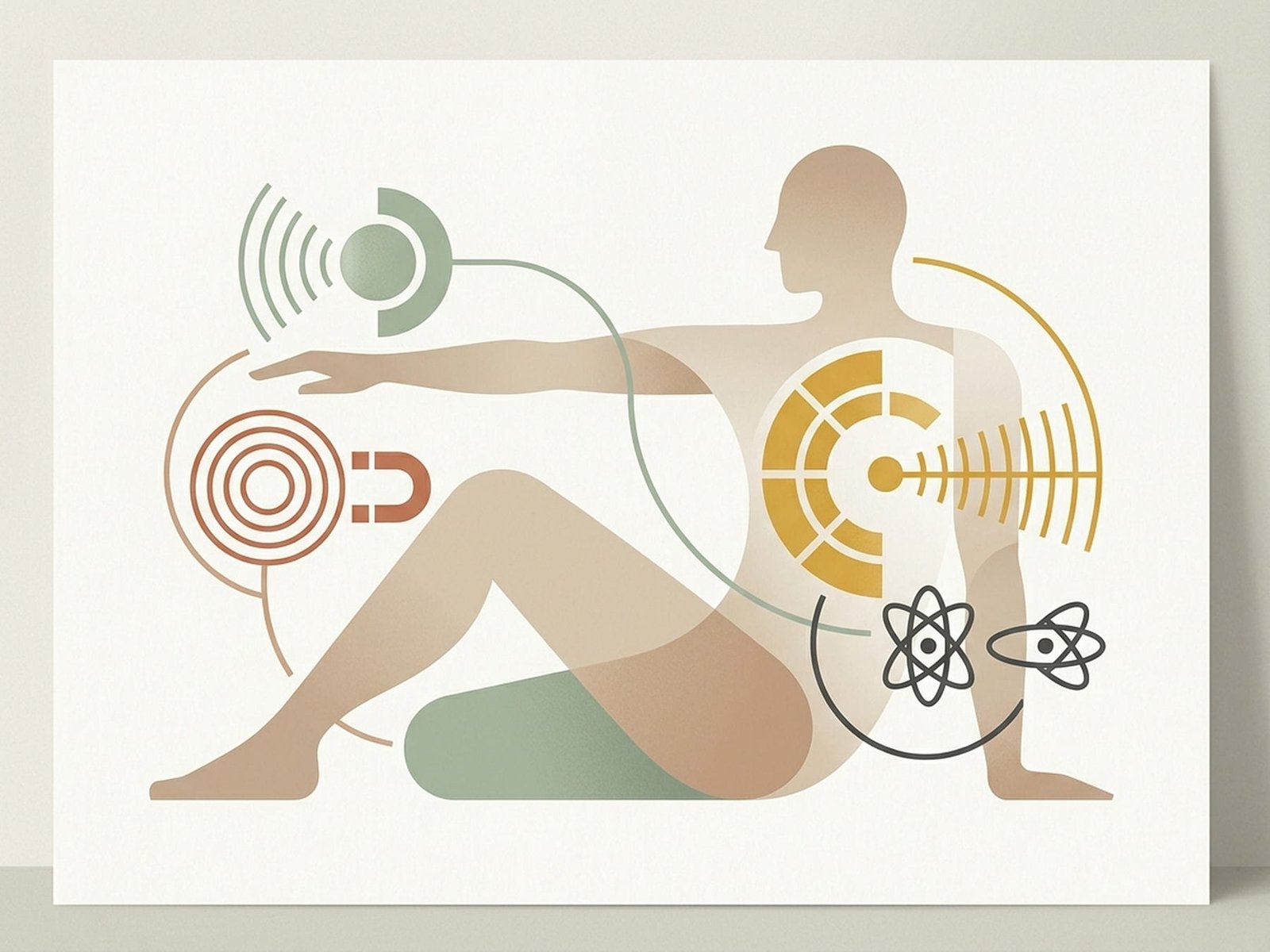
Imaging Findings
Prevalence in Asymptomatic Adults by AgeImaging technology has transformed how clinicians evaluate musculoskeletal conditions. However, imaging findings need to be interpreted in clinical context. Many findings that appear on scans — disc bulges, degenerative changes, bone spurs — are common in people who have no pain at all. The presence of a finding on a scan does not automatically mean it is the cause of your symptoms.
Research has consistently shown that many imaging abnormalities are age-related and present in a large proportion of the asymptomatic population. A landmark study (Jensen 1994, NEJM) found disc abnormalities in 64% of pain-free volunteers. A systematic review (Brinjikji 2015) confirmed that disc bulges, degeneration, and other findings are common across all age groups in people without symptoms.
Research has also shown that the language used in radiology reports can influence how patients experience their condition. Alarming terminology — even for common findings — can increase worry and may affect recovery expectations. Context on whether a finding is common for your age can keep alarming terminology from driving worry.
Some sources of pain — including muscular and soft tissue conditions — are not well visualized on standard imaging. This means that a normal scan does not rule out all possible causes of pain. When imaging is normal but symptoms persist, clinical examination can help identify contributors that scans do not capture.
Common Findings Explained
These are the imaging findings encountered most frequently. Understanding how common they are in people without symptoms can help put your own results in perspective.
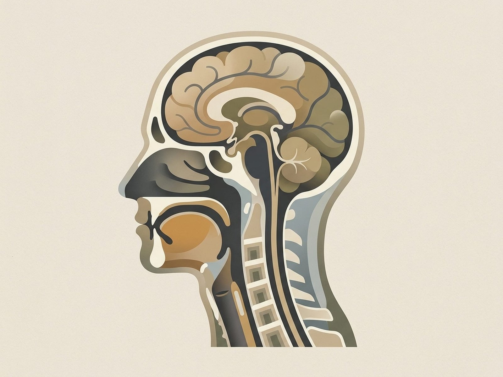
Common MRI and CT Findings
Anatomical ReferenceDisc Bulge / Herniation
Degenerative Disc Disease
Facet Arthrosis
Rotator Cuff Findings on MRI
Spinal Stenosis and Osteophytes
What the Research Shows
Research on imaging and pain has produced consistent findings over three decades: the correlation between imaging abnormalities and pain is often weaker than expected. This does not mean imaging is not useful — it means imaging is most valuable when interpreted alongside clinical assessment.
Several bodies of evidence are particularly relevant: the Brinjikji 2015 systematic review establishing how common asymptomatic imaging findings are; research showing that report language can influence pain outcomes; and the Jarvik 2003 JAMA study suggesting that early MRI for acute low back pain may increase intervention rates without improving outcomes.
A disc bulge on MRI may be as common as grey hair — it tells you about age, not necessarily about pain.
Jensen 1994
Brinjikji 2015
Jarvik 2003
Understanding Your Report
These frameworks can help you understand what your radiology report means and what questions to ask about your results.
Radiology Report Language
Report Language and Expectations
When Imaging Is Helpful
Questions to Ask About Your Scan
Working With Your Results
How you and your clinician work with imaging results depends on the clinical context. The following scenarios cover the most common situations.
Clinical Decision Framework
Imaging vs. Examination PriorityWhen Imaging Shows an Incidental Finding
Many imaging findings are common for age and may not be the source of your symptoms. When clinical assessment does not match the imaging finding, conservative approaches are usually the first step.
- Discuss the finding with your clinician in the context of your symptoms and examination
- Understand that a finding on a scan does not mean it needs treatment if it is not causing your symptoms
- Physical therapy and self-care may be appropriate regardless of what the scan shows
- Follow-up imaging is usually not needed unless symptoms change significantly
When Imaging Findings Match Clinical Assessment
When clinical examination findings and imaging point in the same direction, imaging adds value by confirming what assessment suggests and helping guide treatment decisions.
- Nerve root compression with matching clinical signs — imaging supports the clinical picture
- Pre-procedural planning: imaging defines anatomy before intervention
- Monitoring known structural conditions over time
When Imaging Is Normal but You Have Pain
Normal imaging in the presence of pain does not mean nothing is wrong. It means imaging has not identified a structural cause. Pain can come from muscles, nerves, joints, and other sources that standard imaging may not capture well. Clinical examination can help identify contributing factors that scans do not show.
- A normal scan rules out certain structural causes — that is useful information
- Musculoskeletal causes including muscle and soft tissue sources are not well visualized on standard imaging
- A clinical examination that includes musculoskeletal assessment may identify contributors that imaging cannot
- Normal imaging with persistent pain is a common clinical scenario, not a dead end
Considering Surgery
When surgery is being considered, it is reasonable to ask how well the imaging findings match your symptoms, what conservative options have been tried, and what the expected outcomes are compared to continued non-surgical care.
- Ask: "Do my symptoms match this imaging finding specifically?"
- Ask: "What conservative treatments have been tried, and for how long?"
- Ask: "What are the expected outcomes with and without surgery?"
- A second opinion is reasonable when surgery is based primarily on imaging findings
When Imaging Findings Are Significant
While most imaging findings are incidental or age-related, certain combinations of imaging and clinical findings are genuinely important and may require action.
Many imaging findings — including disc bulges, degenerative changes, and partial rotator cuff tears — are common in people without pain, especially with age.
The language used in radiology reports can influence how patients feel about their condition. Reports describe anatomy, not necessarily the cause of your pain.
Some conditions, including myofascial trigger points, are not visible on standard imaging. A normal scan in the presence of pain means imaging has not identified the cause — not that nothing is wrong.
Imaging is most useful when it answers a specific clinical question, particularly when clinical assessment and imaging findings point in the same direction.
Imaging results are best interpreted by a clinician who has also examined you — the scan and the examination together give a more complete picture than either alone.