What Is Saline Hydrodissection?
Hydrodissection Treatment
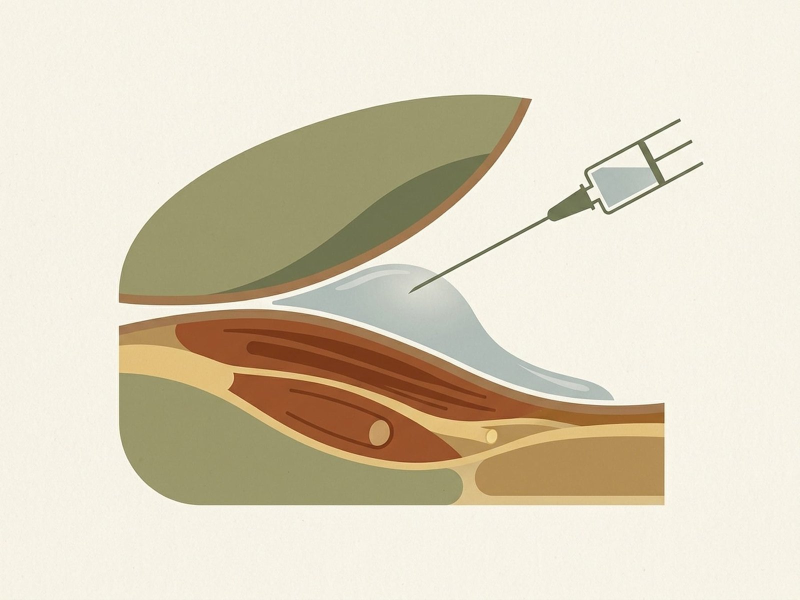
Overview DiagramSaline hydrodissection is an ultrasound-guided interventional technique in which pressurized fluid — most commonly normal saline (0.9% NaCl) or 5% dextrose in water (D5W) — is injected under real-time imaging to physically separate tissue planes, release fascial adhesions, and liberate entrapped peripheral nerves. The term "hydrodissection" literally means dissection with water: the fluid bolus acts as a hydraulic scalpel, creating space between adherent tissue layers without the need for surgical incision.
The concept of tissue plane separation is fundamental to understanding how hydrodissection works. Throughout the body, muscles, fascia, nerves, and blood vessels exist within defined tissue planes that are normally separated by thin layers of loose areolar connective tissue. This loose connective tissue functions as a gliding interface — allowing adjacent structures to slide freely relative to each other during movement. When this gliding interface is disrupted by inflammation, fibrosis, surgical scarring, or chronic mechanical stress, the tissue layers become adherent. These adhesions restrict normal tissue mobility, compress nerves, and create the structural perpetuating factors that maintain chronic myofascial trigger points.
Modern fascial research has demonstrated that fascia is not merely a passive wrapping around muscles — it is a dynamic, innervated, mechanosensitive tissue that plays a critical role in force transmission, proprioception, and nociception. When fascial layers lose their ability to glide, the resulting restriction alters biomechanics, increases intramuscular pressure, and generates nociceptive input that contributes to myofascial pain. Hydrodissection directly targets this fascial pathology by restoring the gliding interface.
Ultrasound guidance is not optional for hydrodissection — it is essential. Real-time ultrasound visualization allows the clinician to see the needle tip at all times, identify the target tissue plane or nerve, confirm correct needle positioning within the plane before injecting, and watch the tissue planes opening as the fluid is delivered. This real-time visual feedback is what distinguishes hydrodissection from blind injection techniques and is the foundation of both its safety and its efficacy. Without ultrasound, the clinician cannot confirm that the fluid is being delivered to the correct tissue plane, significantly reducing the therapeutic benefit and increasing the risk of unintended tissue injury.
Hydrodissection occupies a growing role in interventional pain medicine. Originally developed primarily for peripheral nerve entrapment (most notably carpal tunnel syndrome), the technique is increasingly applied to myofascial pain conditions where fascial adhesions, tissue plane restrictions, and nerve entrapment contribute to chronic trigger point perpetuation. It represents a paradigm shift in thinking about myofascial pain — moving beyond the isolated trigger point to address the broader fascial and neural environment in which the trigger point exists.
Hydraulic Separation
Pressurized fluid physically separates adherent tissue planes, restoring natural gliding interfaces.
Nerve Liberation
Trapped nerves are freed from surrounding fascial adhesions under real-time ultrasound guidance.
Fascial Rehydration
Injected fluid restores interstitial fluid dynamics in dehydrated, chronically adhered fascial compartments.
Inflammatory Reset
Dilution of local inflammatory mediators and nociceptive substances reduces peripheral sensitization at the injection site.
Mechanisms of Action
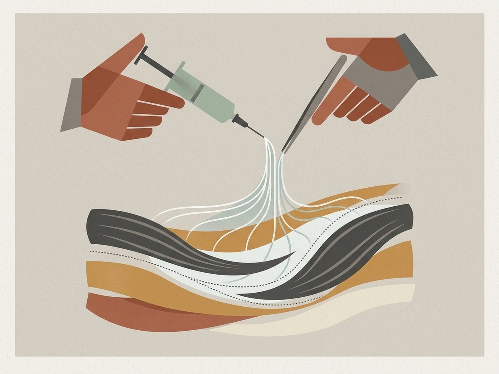
Hydrodissection physically frees trapped nerves by creating space between tissue planes using pressurized fluid injection.
Mechanisms of Action
Mechanism DiagramHydraulic Tissue Plane Separation
Nerve Decompression and Entrapment Liberation
Fascial Adhesion Disruption
Local Washout of Nociceptive Mediators
Mechanical Trigger Point Disruption via Fluid Bolus
Indications for Myofascial Pain Syndrome
Fascial Adhesions Contributing to Trigger Point Perpetuation
Peripheral Nerve Entrapment Syndromes
Chronic Fibrotic Trigger Points Unresponsive to Dry Needling
Post-Surgical Adhesions Restricting Muscle Function
Thoracolumbar Fascia Stiffness and Dysfunction
Solutions Used in Hydrodissection
Normal Saline (0.9% NaCl)
Isotonic, pH 5.5, widely available
5% Dextrose in Water (D5W)
Hypotonic when dextrose metabolized, pH 4.0
Local Anesthetic + Saline Combination
Typically 0.5–1% lidocaine diluted in normal saline
PRP + Saline (Regenerative Hydrodissection)
Autologous PRP diluted with saline for volume
Technique — Step by Step
Technique
Step by StepUltrasound Probe Positioning and Target Identification
Needle Approach Selection — In-Plane vs Out-of-Plane
Needle Insertion and Tissue Plane Access
Test Injection and Tissue Plane Confirmation
Controlled Volume Injection
Dynamic Assessment of Plane Separation
Key Technical Parameters
Ultrasound Probe
Needle Approach
Needle Gauge and Length
Volume per Site
Key Ultrasound Sign
Injection Speed
Synergistic Treatment Protocols
Hydrodissection is most powerful when integrated into a multimodal treatment strategy. The following combinations leverage the structural release achieved by hydrodissection as a foundation for complementary therapies that address different aspects of the myofascial pain cycle.
Hydrodissection + Extracorporeal Shockwave Therapy (ESWT)
Hydrodissection first separates adherent fascial planes and liberates entrapped nerves, restoring tissue mobility. ESWT is then applied to the newly mobile tissue to stimulate mechanotransduction-driven tissue remodeling, neovascularization, and stem cell activation. The sequential approach addresses the structural pathology (adhesions) with hydrodissection and the biological healing response (regeneration of healthy tissue within the opened plane) with ESWT. This combination is particularly powerful for chronic fascial adhesions where both mechanical restriction and tissue degeneration coexist.
Learn about Shockwave TherapyHydrodissection + Dry Needling
When myofascial trigger points coexist with fascial adhesions (which is common in chronic Myofascial Pain Syndrome), hydrodissection can first release the fascial adhesion that is perpetuating the trigger point, followed by dry needling of the trigger point itself. With the adhesion released, the local twitch response to dry needling is often more robust and complete, because the muscle fiber is no longer tethered by the fascial restriction. This combination addresses both the structural (fascial adhesion) and neuromuscular (trigger point) components of the pain generator.
Learn about Dry NeedlingHydrodissection + Platelet-Rich Plasma (PRP)
Following hydrodissection to create a tissue plane, PRP can be injected into the newly opened space to deliver concentrated growth factors directly to the site of previous adhesion. The growth factors promote healthy tissue remodeling within the plane, theoretically reducing the likelihood of re-adhesion. PDGF, TGF-beta, and VEGF stimulate angiogenesis and collagen remodeling, creating healthy loose areolar connective tissue rather than dense scar. This "regenerative hydrodissection" approach is particularly relevant for patients with recurrent adhesions that reform after conventional hydrodissection alone.
Learn about PRP TherapyHydrodissection + Motor Control Rehabilitation
Hydrodissection restores tissue plane mobility, but the motor control deficits that developed as a consequence of chronic fascial restriction persist beyond the procedure. After hydrodissection releases the structural restriction, targeted motor control rehabilitation retrains the neuromuscular system to utilize the restored range of motion. This includes progressive loading of the previously restricted muscles, proprioceptive retraining, and movement pattern correction. Without rehabilitation, compensatory movement patterns persist and mechanical forces may cause the adhesions to reform.
Learn about NeuromodulationEvidence Base
Wu et al. (2019) — Muscle Nerve
Wu et al. (2022) — BioMed Research International
Cass (2016) — Curr Sports Med Rep
Emerging Myofascial-Pain Evidence — Fascial Hydrodissection
Hydrodissection vs Other Treatments
Primary Target
Mechanism
Imaging Guidance
Injection Volume
Depth Range
Evidence Level (myofascial pain)
Key Advantage
Safety Profile
Complications and Expected Effects
Very Safe ProfileGenerally Very Safe Procedure
Infection (Very Rare)
Nerve Injury from Needle Contact (Very Rare with Ultrasound)
Post-Procedure Soreness (Expected, 24–48 Hours)
Transient Numbness (With Local Anesthetic Use)
Hydrodissection uses pressurized fluid injection under ultrasound guidance to physically separate adherent tissue planes and liberate entrapped nerves — directly addressing structural perpetuating factors in chronic myofascial pain.
Ultrasound guidance is essential, not optional. Real-time visualization of the needle tip, fluid spread, and tissue planes opening is what makes hydrodissection safe and effective. This is a precision technique that requires image guidance.
The primary indications for myofascial pain are fascial adhesions perpetuating trigger points, peripheral nerve entrapment syndromes mimicking myofascial pain, and chronic fibrotic trigger points unresponsive to conventional dry needling.
Normal saline and D5W are the most commonly used solutions. D5W has emerging evidence for neuroprotective properties and reduced post-procedure pain, making it the preferred injectate for perineural hydrodissection.
The strongest evidence base exists for nerve entrapment (particularly carpal tunnel syndrome), with growing application to myofascial fascial plane pathology. The myofascial pain-specific evidence is emerging but the mechanistic rationale is compelling.
Hydrodissection is most powerful when combined with other therapies — ESWT for tissue regeneration, dry needling for trigger point deactivation, PRP for regenerative plane filling, and motor control rehabilitation for functional retraining.
The procedure is generally very safe. Serious complications are rare when performed with proper ultrasound guidance and aseptic technique. Post-procedure soreness for 24 to 48 hours is expected and self-limiting.
Think of hydrodissection as addressing the "hardware" problem (structural adhesions and entrapment) while other therapies address the "software" problem (neuromuscular dysfunction and central sensitization).
Bottom line:Saline hydrodissection is a precision ultrasound-guided technique that addresses the structural fascial and neural environment surrounding chronic myofascial trigger points. It is not a replacement for dry needling or trigger point injection — it is a complementary tool that targets a different layer of pathology. When fascial adhesions and nerve entrapment are contributing to chronic, treatment-resistant myofascial pain, hydrodissection offers a targeted, minimally invasive solution with an excellent safety profile and growing evidence base.