Understanding the Differential
Hip osteoarthritis (OA) is a prevalent degenerative joint condition characterized by the progressive loss of articular cartilage, subchondral bone changes, and synovial inflammation. Clinical diagnosis relies on a combination of patient history and physical examination findings, classically presenting as a triad of groin, buttock, and anterior thigh pain. However, systematic reviews consistently note a significant discordance between radiographic severity, such as the Kellgren-Lawrence grading, and the patient's reported pain levels. This discrepancy highlights the necessity of a comprehensive differential diagnosis, as extra-articular structures frequently contribute to the clinical presentation.
Myofascial pain syndrome (MPS) frequently coexists with hip osteoarthritis, complicating the clinical picture. As the hip joint degenerates and loses its normal range of motion—particularly internal rotation and extension—the surrounding musculature engages in chronic protective splinting. This sustained tension often activates secondary myofascial trigger points in the iliopsoas, gluteus medius, and adductor complex. Current clinical observations suggest that in many patients, the pain generated by these hyperirritable muscular nodules can equal or even exceed the nociceptive input from the arthritic joint itself.
Treating the surrounding myofascial dysfunction can often yield significant symptomatic relief, even when radiographic evidence of joint degeneration remains unchanged.
Differentiating true intra-articular hip pain from myofascial pain requires careful clinical correlation. Provocative maneuvers like the FADIR and FABER tests can help localize the pathology, while assessing the joint's capsular end-feel provides clues about structural restriction. In younger patients, femoroacetabular impingement (FAI) or labral tears must be considered, whereas older adults may present with concurrent L3/L4 radiculopathy mimicking hip pain. When conservative differentiation remains ambiguous, a fluoroscopically guided intra-articular injection of local anesthetic serves as a reliable diagnostic tool to isolate the articular contribution.
True Hip OA vs Myofascial Mimic
Pain location
Pain behavior
Hallmark exam finding
Imaging correlation
Diagnostic gold standard
Key Muscular Contributors
The hip joint is deeply invested by powerful musculature. Understanding the relationship between these muscles and the joint capsule is essential for differentiating articular from extra-articular pain.
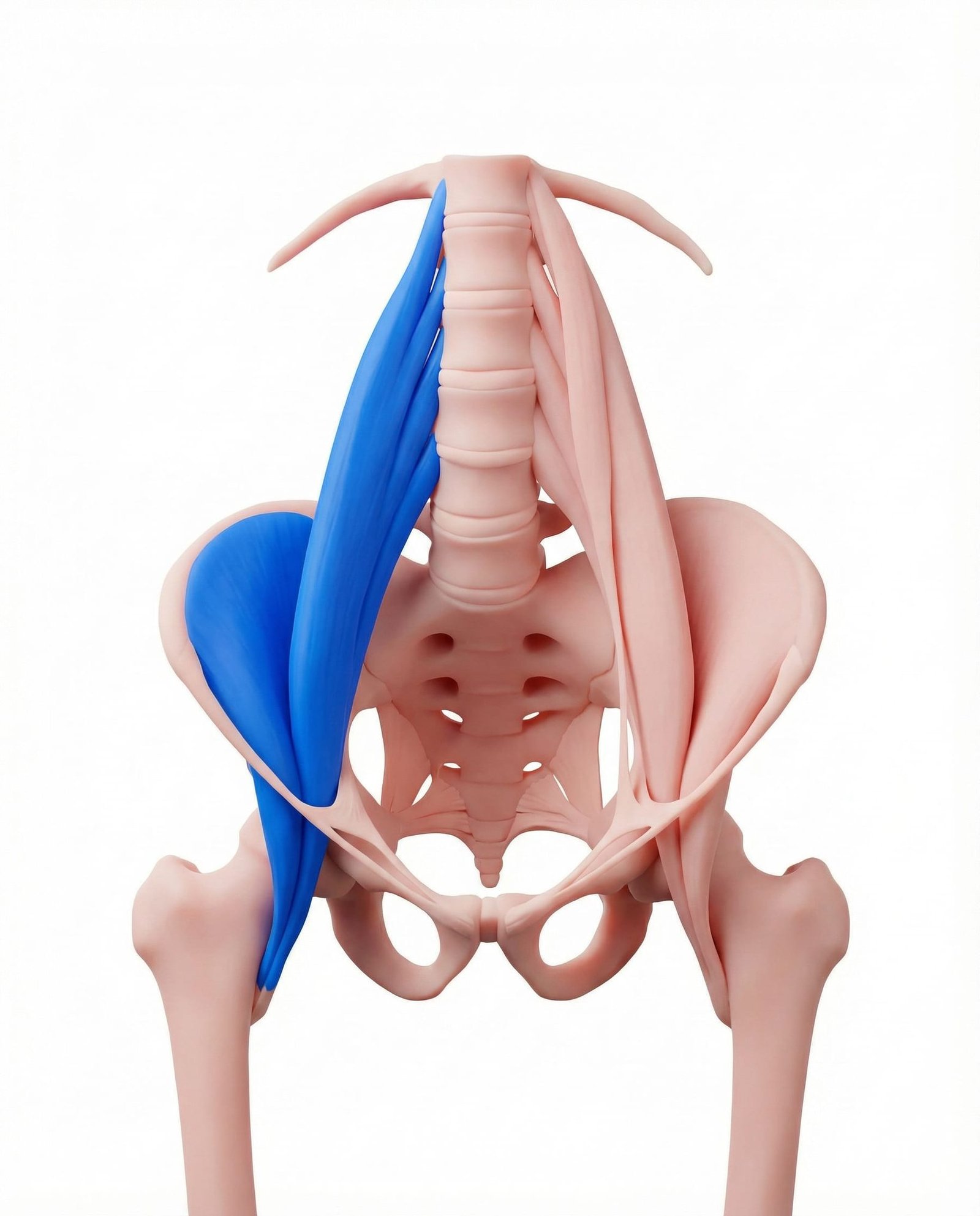
The iliopsoas is the primary hip flexor and lies directly anterior to the hip joint capsule. In the presence of hip osteoarthritis, this muscle frequently develops secondary myofascial trigger points due to chronic protective splinting. Its pain referral pattern strongly mimics true intra-articular hip pathology, often presenting as deep, aching groin pain that worsens with weight-bearing or hip extension.
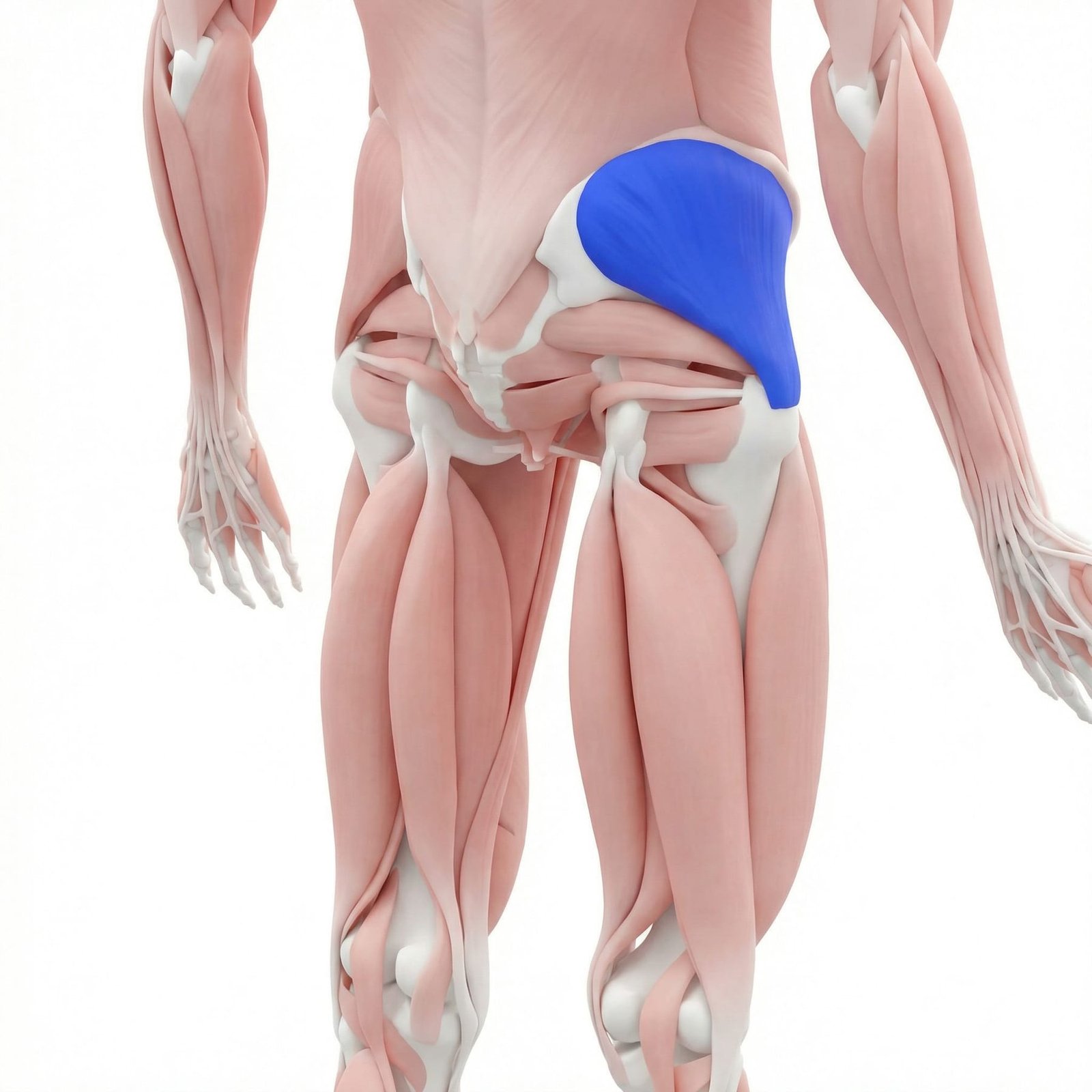
Serving as the primary hip abductor and pelvic stabilizer, the gluteus medius is subjected to altered biomechanical loads when hip joint mechanics are compromised by osteoarthritis. Patients often develop a compensatory Trendelenburg gait, which chronically overloads the muscle fibers. Trigger points here are a primary source of lateral hip and buttock pain, frequently misdiagnosed as greater trochanteric pain syndrome or isolated joint pain.
The adductor longus assists in hip adduction and flexion, and is often recruited to stabilize a degenerating hip joint. Restricted hip abduction, a hallmark clinical sign of hip OA, places chronic tension on the adductor compartment. Myofascial pain in this muscle contributes significantly to the medial thigh and groin pain commonly reported by patients with hip joint pathology.
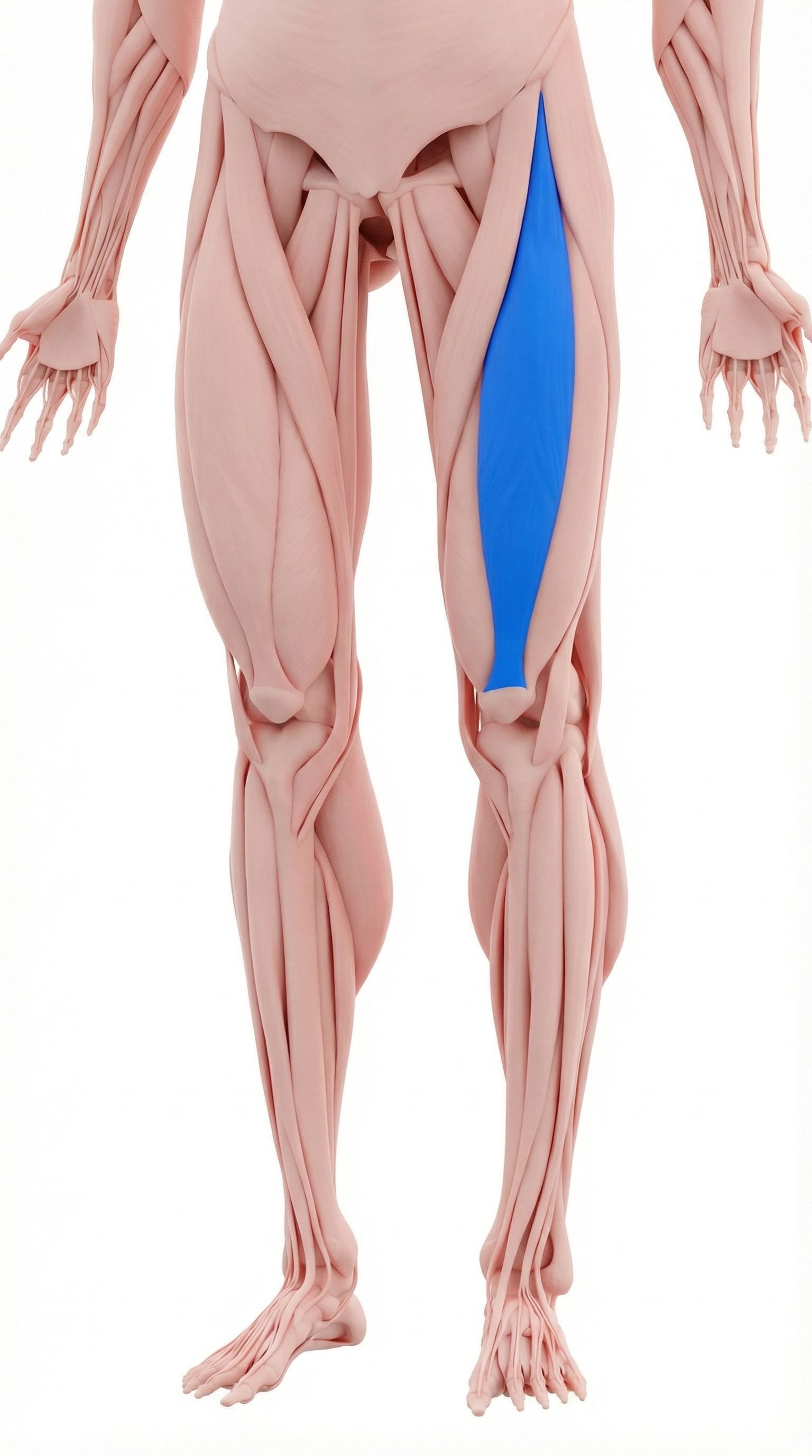
As a biarticular muscle crossing both the hip and the knee, the rectus femoris is vulnerable to tension from altered gait mechanics associated with hip OA. Trigger points in the proximal rectus femoris can mimic the anterior thigh pain characteristic of hip joint degeneration. Clinical evaluation often reveals restricted hip extension and knee flexion when this muscle is involved.
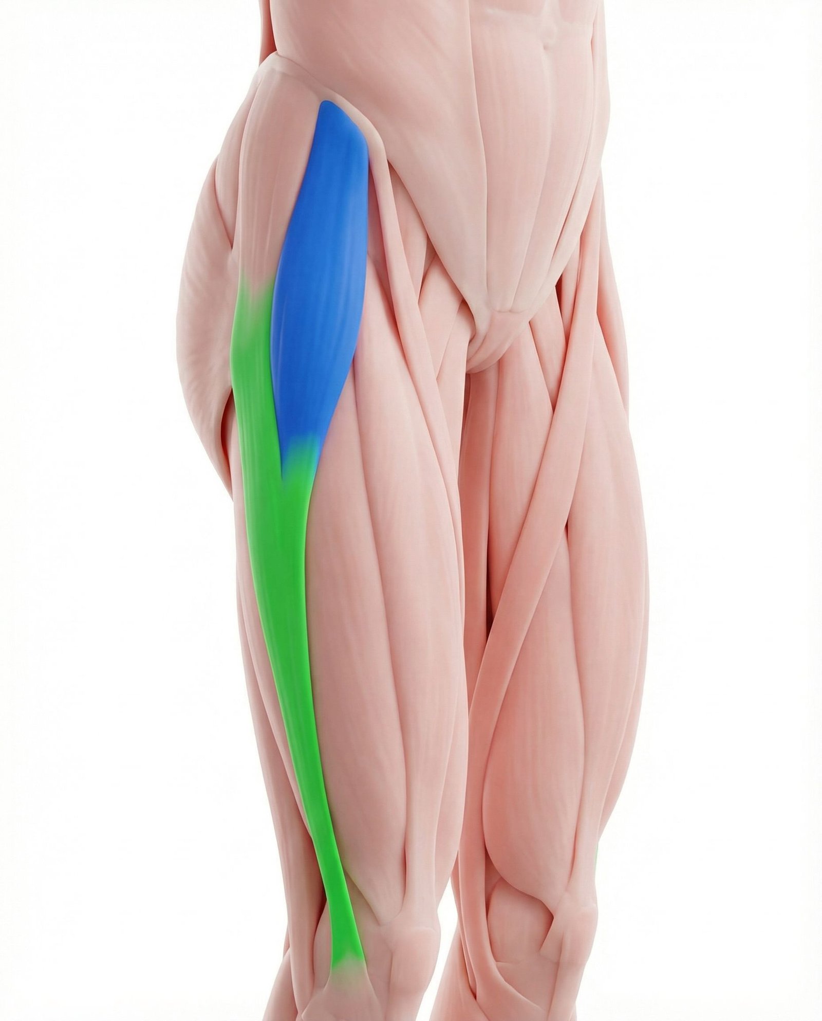
The TFL works synergistically with the gluteus medius to stabilize the pelvis during the stance phase of gait. In patients with hip OA, the TFL often becomes hyperactive to compensate for gluteal weakness or joint instability. This chronic overactivity frequently leads to myofascial pain that presents as lateral thigh aching, complicating the clinical picture.
The piriformis functions as an external rotator of the hip and provides posterior joint stability. Hip joint capsular restriction, particularly the loss of internal rotation seen in early OA, alters the resting length and tension of the piriformis. Trigger points here can produce deep gluteal pain that mimics both posterior hip joint pain and lumbar radiculopathy.
Articular Degeneration
Progressive loss of cartilage, subchondral sclerosis, and osteophyte formation leading to mechanical joint pain and capsular restriction.
Myofascial Splinting
Chronic protective contraction of the iliopsoas, gluteals, and adductors in response to joint instability or pain, leading to trigger point formation.
Altered Biomechanics
Compensatory gait patterns, such as a Trendelenburg gait, which abnormally load the lateral hip musculature and lumbar spine.
Central Sensitization
Prolonged nociceptive input from the degenerating joint and surrounding muscles can lead to an amplified pain response in the central nervous system.
Pain Referral Patterns
Myofascial trigger points around the hip produce distinct referral patterns that frequently mimic the groin, thigh, and buttock pain classically attributed to hip osteoarthritis.
Iliopsoas
Gluteus Medius
Adductor Longus
Piriformis
Clinical Assessment Maneuvers
Clinicians utilize specific orthopedic maneuvers to stress the hip joint capsule and surrounding musculature, aiding in the differentiation of pain generators.
FABER (Patrick's) Test
FADIR Test
Modified Thomas Test
Trendelenburg Sign
Evidence-Based Pathways
Current guidelines generally recommend a multimodal approach to hip pain, addressing both the articular degeneration and the secondary myofascial adaptations to optimize function.
Self-Care
- Activity modification to reduce high-impact joint loading
- Weight management to decrease biomechanical stress on the hip
- Gentle, pain-free range of motion exercises daily
- Use of a cane or walking aid on the contralateral side
- Application of heat to surrounding musculature to reduce spasm
Manual Therapy
- Ischemic compression of iliopsoas and gluteal trigger points
- Gentle joint mobilization to improve capsular mobility
- Soft tissue mobilization of the adductor compartment
- Passive stretching of the hip flexors and external rotators
- Myofascial release targeting the tensor fasciae latae
Interventional
- Fluoroscopically guided intra-articular diagnostic block
- Intra-articular corticosteroid or hyaluronic acid injection
- Trigger point injections for refractory iliopsoas or gluteal pain
- Ultrasound-guided greater trochanteric bursa injection
- Radiofrequency ablation of articular branches (in select cases)
Rehabilitation
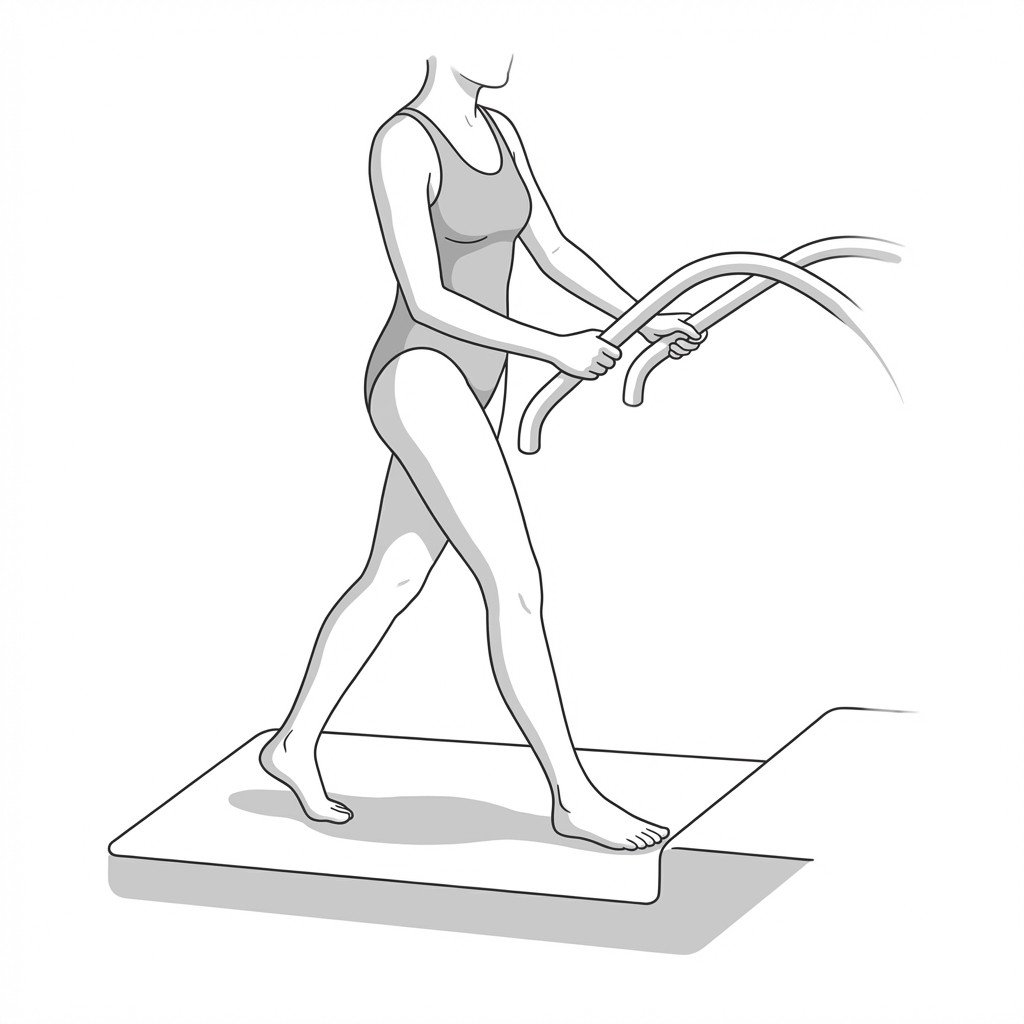
- Aquatic therapy to allow unloaded joint movement
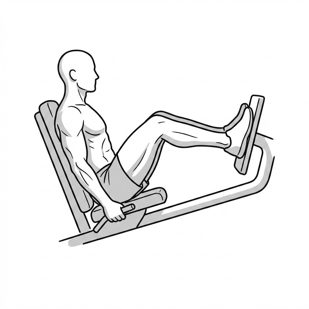
- Progressive strengthening of the gluteus medius and minimus
- Core stabilization exercises to improve pelvic control
- Gait retraining to address compensatory movement patterns
- Stationary cycling with minimal resistance to promote synovial fluid circulation
Red Flags — When to See a Doctor
While chronic hip pain is common, certain signs and symptoms warrant prompt medical investigation to rule out severe or life-threatening conditions.
Radiographic evidence of hip osteoarthritis frequently correlates poorly with the severity of a patient's clinical pain.
Myofascial trigger points in the iliopsoas, gluteals, and adductors routinely coexist with hip OA due to chronic protective muscle splinting.
Pain from secondary myofascial trigger points can sometimes exceed the pain generated by the degenerating joint itself.
A fluoroscopically guided intra-articular injection of local anesthetic is considered the gold standard for differentiating true articular pain from extra-articular sources.
Addressing coexisting myofascial pain through conservative measures may significantly improve function and potentially delay the need for arthroplasty.
Restricted internal rotation is typically the earliest physical examination finding in true hip joint capsular restriction.
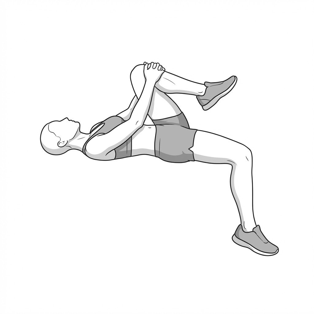
Movement therapy
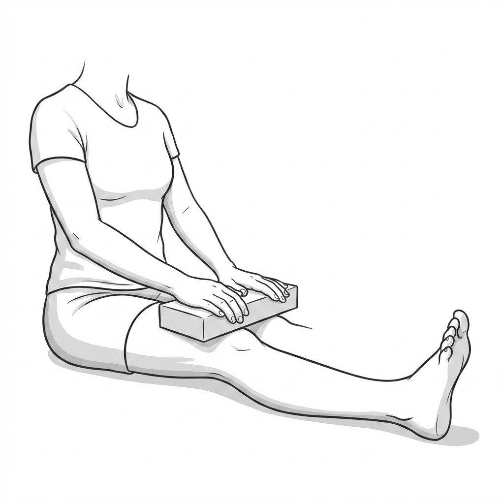
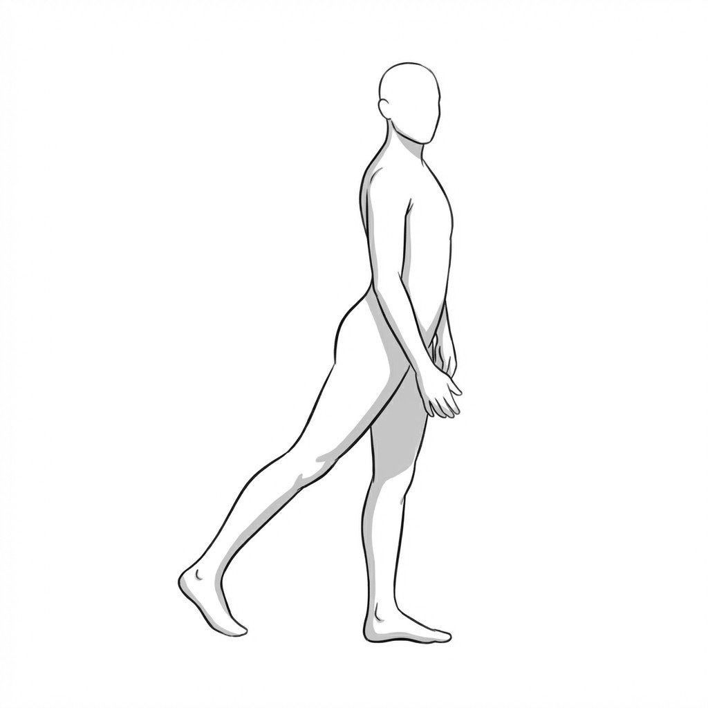
Exercises for Hip Osteoarthritis
Hip OA responds to low-load movement that preserves joint nutrition and strengthens the surrounding musculature without compressing an already-narrowed joint space. Start with 2–3 and progress as tolerated.